Circadian Clock in a Mouse Colon Tumor Regulates ... · Moreover, growth in colon-26( pendent on...
13
Circadian Clock in a Mouse Colon Tumor Regulates Intracellular Iron Levels to Promote Tumor Progression * Received for publication, December 30, 2015, and in revised form, January 21, 2016 Published, JBC Papers in Press, January 21, 2016, DOI 10.1074/jbc.M115.713412 Fumiyasu Okazaki ‡§1 , Naoya Matsunaga §1 , Hiroyuki Okazaki ¶1 , Hiroki Azuma §1 , Kengo Hamamura ¶ , Akito Tsuruta § , Yuya Tsurudome § , Takashi Ogino § , Yukinori Hara § , Takuya Suzuki , Kenji Hyodo , Hiroshi Ishihara , Hiroshi Kikuchi**, Hideto To ‡ , Hironori Aramaki ¶‡‡ , Satoru Koyanagi §§ , and Shigehiro Ohdo §2 From the ‡ Department of Medical Pharmaceutics, Graduate School of Medicine and Pharmaceutical Sciences for Research, University of Toyama, Toyama 930-0194, the § Department of Pharmaceutics and the §§ Department of Glocal Healthcare Science, Faculty of Pharmaceutical Sciences, Kyushu University, Fukuoka 812-8582, the ¶ Department of Molecular Biology and ‡‡ Drug Innovation Research Center, Daiichi University of Pharmacy, Fukuoka 815-8511, and the Formulation Research, Pharmaceutical Science & Technology Core Function Unit and **Chief Innovation Officer Group, Eisai Product Creation Systems, Eisai Co., Ltd., Ibaraki 300-2635, Japan Iron is an important biological catalyst and is critical for DNA synthesis during cell proliferation. Cellular iron uptake is enhanced in tumor cells to support increased DNA synthesis. Circadian variations in DNA synthesis and proliferation have been identified in tumor cells, but their relationship with intra- cellular iron levels is unclear. In this study, we identified a 24-h rhythm in iron regulatory protein 2 (IRP2) levels in colon-26 tumors implanted in mice. Our findings suggest that IRP2 reg- ulates the 24-h rhythm of transferrin receptor 1 (Tfr1) mRNA expression post-transcriptionally, by binding to RNA stem-loop structures known as iron-response elements. We also found that Irp2 mRNA transcription is promoted by circadian clock genes, including brain and muscle Arnt-like 1 (BMAL1) and the circa- dian locomotor output cycles kaput (CLOCK) heterodimer. Moreover, growth in colon-26(19) tumors expressing the clock-mutant protein (CLOCK 19 ) was low compared with that in wild-type colon-26 tumor. The time-dependent variation of cellular iron levels, and the proliferation rate in wild-type colon-26 tumor was decreased by CLOCK 19 expression. Our findings suggest that circadian organization contributes to tumor cell proliferation by regulating iron metabolism in the tumor. Circadian rhythms affect blood pressure, locomotor activity, core body temperature, and the sleep-wake cycle. These circa- dian controls of physiology and behavior are driven by a master pacemaker located in the suprachiasmatic nucleus of the hypo- thalamus, which relies on the interplay of interconnected tran- scriptional and translational feedback loops (1). The brain and muscle Arnt-like 1 (BMAL1) 3 and circadian locomotor output cycles kaput (CLOCK) heterodimer drive the transcription of cryptochrome (CRY) and period (PER) genes by binding to the E-box in their promoter regions. CRY and PER homodimers and heterodimers inhibit the function of BMAL1/CLOCK, thus decreasing their own expression (2–5). This core loop creates a 24-h rhythmic oscillation of clock-controlled gene expression in normal and tumor cells. Iron is an important metal for cell proliferation, metabolism, respiration, and DNA synthesis (6 – 8). Iron homeostasis is de- pendent on the expression of transferrin receptor 1 (TfR1) (9, 10). TfR1 is a membrane receptor responsible for iron uptake and ferritin is an intracellular protein that stores iron. When present in excess, cellular iron is toxic (11–13). Therefore, iron concentration must be tightly regulated. In general, the expres- sion of TfR1 is regulated by iron-regulatory proteins (IRPs), which are sensors of intracellular iron levels. IRPs control TfR1 and divalent metal transporter 1 (Dmt1) at the post-transcrip- tional level by binding to RNA stem-loop structures known as iron-responsive elements (IREs) in the 3-untranslated region (UTR) of mRNA. IRP binding stabilizes transcripts in these genes (14 –16). Iron metabolism is critical for the rapid growth of tumors and iron is also required for DNA synthesis in tumor cells. Therefore, the cellular iron level is higher in tumor cells. TfR1 expression in tumor cells is higher than that in normal cells (17). Additionally, recent studies have demonstrated diurnal variation in DNA synthesis and cell proliferation in tumor masses and that such variation is important for tumor growth (18, 19). However, the mechanism by which iron metabolism relates to these rhythms is unclear. * This work was supported in part by Grants-in-Aid for Scientific Research (A; 25253038) (to S. O.), Scientific Research on Innovative Areas (25136716) (to S. O.), and Challenging Exploratory Research (25670079) (to S. O.), the Uehara Memorial Foundation (to S. O.), Grant-in-Aid for Scientific Research (C; 15K08098) (to N. M.) and Grant-in-Aid for Young Scientists (B; 26860097) (to F. O.), Grant-in-Aid for Young Scientists (B; 15K19167) (to H. O.), and Grant-in-Aid for Young Scientists (Start-up; 15H06795K) (to K. H.), the Japan Research Foundation for Clinical Pharmacology (to N. M.), a Grant-in-Aid for JSPS Fellows (25-4175) (to K. H.) from the Japan Society for the Promotion of Science (JSPS), the Fukuoka Foundation for Sound Health, and the Platform for Drug Discovery, Informatics, and Structural Life Science of the Ministry of Education, Culture, Sports, Science and Tech- nology, Japan. The authors declare that they have no conflicts of interest with the contents of this article. 1 These authors contributed equally to this work. 2 To whom correspondence should be addressed: 3-1-1 Maidashi Higashi-ku, Fukuoka 812-8582, Japan. Tel.: 81-92-642-6610; Fax: 81-92-642-6614; E-mail: [email protected]. 3 The abbreviations used are: BMAL1, brain and muscle Arnt-like 1; CLOCK, circadian locomotor output cycles kaput; 5-BrU, 5-bromouridine; CRY, cryptochrome; DFO, deferoxamine; Dmt1, divalent metal transporter 1; IRE, iron-responsive element; IRP, iron-regulatory protein; PER, period; RIPA, RNA immunoprecipitation assay; TBS, BSA containing Tween; TfR1, transferrin receptor 1; ANOVA, analysis of variance. crossmark THE JOURNAL OF BIOLOGICAL CHEMISTRY VOL. 291, NO. 13, pp. 7017–7028, March 25, 2016 © 2016 by The American Society for Biochemistry and Molecular Biology, Inc. Published in the U.S.A. MARCH 25, 2016 • VOLUME 291 • NUMBER 13 JOURNAL OF BIOLOGICAL CHEMISTRY 7017 by guest on February 16, 2019 http://www.jbc.org/ Downloaded from
Transcript of Circadian Clock in a Mouse Colon Tumor Regulates ... · Moreover, growth in colon-26( pendent on...
Circadian Clock in a Mouse Colon Tumor RegulatesIntracellular Iron Levels to Promote Tumor Progression*
Received for publication, December 30, 2015, and in revised form, January 21, 2016 Published, JBC Papers in Press, January 21, 2016, DOI 10.1074/jbc.M115.713412
Fumiyasu Okazaki‡§1, Naoya Matsunaga§1, Hiroyuki Okazaki¶1, Hiroki Azuma§1, Kengo Hamamura¶, Akito Tsuruta§,Yuya Tsurudome§, Takashi Ogino§, Yukinori Hara§, Takuya Suzuki�, Kenji Hyodo�, Hiroshi Ishihara�,Hiroshi Kikuchi**, Hideto To‡, Hironori Aramaki¶‡‡, Satoru Koyanagi§§, and Shigehiro Ohdo§2
From the ‡Department of Medical Pharmaceutics, Graduate School of Medicine and Pharmaceutical Sciences for Research,University of Toyama, Toyama 930-0194, the §Department of Pharmaceutics and the §§Department of Glocal Healthcare Science,Faculty of Pharmaceutical Sciences, Kyushu University, Fukuoka 812-8582, the ¶Department of Molecular Biology and ‡‡DrugInnovation Research Center, Daiichi University of Pharmacy, Fukuoka 815-8511, and the �Formulation Research, PharmaceuticalScience & Technology Core Function Unit and **Chief Innovation Officer Group, Eisai Product Creation Systems, Eisai Co., Ltd.,Ibaraki 300-2635, Japan
Iron is an important biological catalyst and is critical for DNAsynthesis during cell proliferation. Cellular iron uptake isenhanced in tumor cells to support increased DNA synthesis.Circadian variations in DNA synthesis and proliferation havebeen identified in tumor cells, but their relationship with intra-cellular iron levels is unclear. In this study, we identified a 24-hrhythm in iron regulatory protein 2 (IRP2) levels in colon-26tumors implanted in mice. Our findings suggest that IRP2 reg-ulates the 24-h rhythm of transferrin receptor 1 (Tfr1) mRNAexpression post-transcriptionally, by binding to RNA stem-loopstructures known as iron-response elements. We also found thatIrp2 mRNA transcription is promoted by circadian clock genes,including brain and muscle Arnt-like 1 (BMAL1) and the circa-dian locomotor output cycles kaput (CLOCK) heterodimer.Moreover, growth in colon-26(�19) tumors expressing theclock-mutant protein (CLOCK�19) was low compared with thatin wild-type colon-26 tumor. The time-dependent variation ofcellular iron levels, and the proliferation rate in wild-typecolon-26 tumor was decreased by CLOCK�19 expression. Ourfindings suggest that circadian organization contributes totumor cell proliferation by regulating iron metabolism in thetumor.
Circadian rhythms affect blood pressure, locomotor activity,core body temperature, and the sleep-wake cycle. These circa-
dian controls of physiology and behavior are driven by a masterpacemaker located in the suprachiasmatic nucleus of the hypo-thalamus, which relies on the interplay of interconnected tran-scriptional and translational feedback loops (1). The brain andmuscle Arnt-like 1 (BMAL1)3 and circadian locomotor outputcycles kaput (CLOCK) heterodimer drive the transcription ofcryptochrome (CRY) and period (PER) genes by binding to theE-box in their promoter regions. CRY and PER homodimersand heterodimers inhibit the function of BMAL1/CLOCK, thusdecreasing their own expression (2–5). This core loop creates a24-h rhythmic oscillation of clock-controlled gene expressionin normal and tumor cells.
Iron is an important metal for cell proliferation, metabolism,respiration, and DNA synthesis (6 – 8). Iron homeostasis is de-pendent on the expression of transferrin receptor 1 (TfR1) (9,10). TfR1 is a membrane receptor responsible for iron uptakeand ferritin is an intracellular protein that stores iron. Whenpresent in excess, cellular iron is toxic (11–13). Therefore, ironconcentration must be tightly regulated. In general, the expres-sion of TfR1 is regulated by iron-regulatory proteins (IRPs),which are sensors of intracellular iron levels. IRPs control TfR1and divalent metal transporter 1 (Dmt1) at the post-transcrip-tional level by binding to RNA stem-loop structures known asiron-responsive elements (IREs) in the 3�-untranslated region(UTR) of mRNA. IRP binding stabilizes transcripts in thesegenes (14 –16).
Iron metabolism is critical for the rapid growth of tumorsand iron is also required for DNA synthesis in tumor cells.Therefore, the cellular iron level is higher in tumor cells. TfR1expression in tumor cells is higher than that in normal cells(17). Additionally, recent studies have demonstrated diurnalvariation in DNA synthesis and cell proliferation in tumormasses and that such variation is important for tumor growth(18, 19). However, the mechanism by which iron metabolismrelates to these rhythms is unclear.
* This work was supported in part by Grants-in-Aid for Scientific Research (A;25253038) (to S. O.), Scientific Research on Innovative Areas (25136716) (toS. O.), and Challenging Exploratory Research (25670079) (to S. O.), theUehara Memorial Foundation (to S. O.), Grant-in-Aid for Scientific Research(C; 15K08098) (to N. M.) and Grant-in-Aid for Young Scientists (B;26860097) (to F. O.), Grant-in-Aid for Young Scientists (B; 15K19167) (toH. O.), and Grant-in-Aid for Young Scientists (Start-up; 15H06795K) (toK. H.), the Japan Research Foundation for Clinical Pharmacology (to N. M.),a Grant-in-Aid for JSPS Fellows (25-4175) (to K. H.) from the Japan Societyfor the Promotion of Science (JSPS), the Fukuoka Foundation for SoundHealth, and the Platform for Drug Discovery, Informatics, and StructuralLife Science of the Ministry of Education, Culture, Sports, Science and Tech-nology, Japan. The authors declare that they have no conflicts of interestwith the contents of this article.
1 These authors contributed equally to this work.2 To whom correspondence should be addressed: 3-1-1 Maidashi Higashi-ku,
Fukuoka 812-8582, Japan. Tel.: 81-92-642-6610; Fax: 81-92-642-6614;E-mail: [email protected].
3 The abbreviations used are: BMAL1, brain and muscle Arnt-like 1; CLOCK,circadian locomotor output cycles kaput; 5-BrU, 5-bromouridine; CRY,cryptochrome; DFO, deferoxamine; Dmt1, divalent metal transporter 1;IRE, iron-responsive element; IRP, iron-regulatory protein; PER, period;RIPA, RNA immunoprecipitation assay; TBS, BSA containing Tween; TfR1,transferrin receptor 1; ANOVA, analysis of variance.
crossmarkTHE JOURNAL OF BIOLOGICAL CHEMISTRY VOL. 291, NO. 13, pp. 7017–7028, March 25, 2016
© 2016 by The American Society for Biochemistry and Molecular Biology, Inc. Published in the U.S.A.
MARCH 25, 2016 • VOLUME 291 • NUMBER 13 JOURNAL OF BIOLOGICAL CHEMISTRY 7017
by guest on February 16, 2019http://w
ww
.jbc.org/D
ownloaded from
We previously reported that TfR1 mRNA expression intumors exhibits a circadian rhythm (20). Therefore, we hypoth-esized that iron levels in tumor masses also follow a circadianrhythm. Moreover, because cellular iron metabolism is con-trolled by IRPs (14 –16), IRP mRNA expression may also exhibita circadian rhythm in tumor masses.
In this study, we identified a 24-h cycle in IRP2 expression intumor masses. In addition, we found that circadian clock genescontrolled the 24-h oscillation of IRP2 mRNA. Furthermore,circadian expression of IRP2 affected the stability of mRNAincluding that of IRE in colon-26 tumors. Our findings suggestthat circadian organization contributes to cell proliferation byregulating iron metabolism.
Experimental Procedures
Animals and Cells—Seven-week-old male BALB/c mice(Charles River Japan) were housed with lights on from 7:00 a.m.to 7:00 p.m. at an ambient temperature of 24 � 1 °C and ahumidity of 60 � 10%, with food and water provided ad libitum.The colon-26 cell line was purchased from Cell Bank RikenBioResource Center. Cells were maintained in RPMI 1640 sup-plemented with 10% fetal bovine serum (FBS) at 37 °C in ahumidified 5% CO2 atmosphere. Colon-26(�19) expressing thestable clock mutant protein was generated by transfection ofthe clock mutant protein (CLOCK�19) expression vector andthe cells were clonally selected by treatment of G418 (Wako).Expression of CLOCK�19 was confirmed by polymerase chainreaction (PCR) products (Fig. 3c). Colon-26 (irp2 knock down)expressing stable Irp2 microRNA was generated by transfec-tion of Irp2 microRNA expression vector (BLOCK-iT Pol IImiR RNAi Expression Vector, Invitrogen) and the cells wasclonally selected by treatment of G418 (Wako). We confirmedthat the cell line was authenticated by the cell bank using shorttandem repeat-PCR analysis, and used within 3 months fromfrozen stock. Tumor model mice were euthanized after thetumor size reached �200 mm3. The tumor volume was esti-mated using the formula: tumor volume (mm3) � 4�xyz/3,where 2x, 2y, and 2z are the three perpendicular diameters ofthe tumor. All experiments were performed in accordance withthe Guide for the Care and Use of Laboratory Animals distrib-uted by the United States National Institutes of Health.
Experimental Design—To assess the temporal IRP2 mRNAand protein expression profiles in tumor cells and normal cells,the right tumor mass or left normal footpad was removed fromindividual tumor-bearing mice at 6 different time points (09:00,13:00, 17:00, 21:00, 01:00, and 05:00 h) on day 7 post-implanta-tion. The levels of IRP2 mRNA and protein were measured byreal-time reverse transcriptase (RT)-PCR and Western blottinganalysis, respectively. To determine whether the molecularcomponents of the circadian clock regulate the expression ofIRP2, the effect of clock gene products on the transcriptionalactivity of IRP2 was assessed using a luciferase reporter con-taining lengths of the IRP2 5�-flanking region. To assess thetemporal CLOCK and BMAL1 protein expression profiles intumor cells, the levels of each protein were measured by West-ern blotting analysis. To analyze the temporal binding ofendogenous BMAL1/CLOCK to the Irp2 promoter in colon-26tumors, chromatin immunoprecipitation (ChIP) assays were
performed using samples isolated at 09:00 and 21:00 h. Toassess the relationship between oscillations in IRP2 and clockgene expression, colon-26(�19) mutant cells, which overex-press a CLOCK mutant that lacks transcriptional activity(CLOCK�19), were used. To assess the temporal Per2 and Irp2mRNA expression profiles in wild-type colon-26 and colon-26(�19) tumor cells, the levels of each mRNA were measuredby real-time RT-PCR.
To determine the effect of IRP2 abundance on TfR1 mRNAstability, mRNA was extracted from colon-26 cells treated withactinomycin D (Act D) to inhibit transcription 24 h after IRP2expression had been induced by deferoxamine (DFO) treat-ment. The levels of TfR1 mRNA were measured by real-timeRT-PCR. Wild-type colon-26 or colon-26(�19) tumor masseswere removed at 6 different time points and the temporal Tfr1and Dmt1 mRNA expression profiles were assessed. To assessthe time-dependent changes in TfR1 mRNA stability, tumormasses were removed at 09:00 and 21:00 h. The temporal bind-ing of endogenous IRP2 to the IREs of murine TfR1 mRNA inindividual tumor masses at 09:00 and 21:00 h was assessed byimmunoprecipitation analysis.
To assess the importance of clock genes in tumor cell prolif-eration, tumor volumes were measured on day 15 post-implan-tation. To assess the temporal iron concentration profiles intumor cells, tumor masses were removed from individual wild-type colon-26 or colon-26(�19) tumor-bearing mice at 6 differ-ent time points on day 7 post-implantation. The iron levelswere determined using an atomic absorption photometer. Toinvestigate the influence of iron on cell growth, cell viability ofcolon-26 cells, and colon-26(�19) cells after treatment of apo-transferrin was analyzed with an ATP assay.
Atomic Absorption Spectrophotometry—The samples wereadded to �2 ml of HNO3 (60%) and then heated for 1 h at 70 °C.Organic compounds were completely oxidized and iron wasionized. Samples were condensed by further heating at 150 °Cfor 3 h. Finally, sample volumes were adjusted to 4 ml usingMilli-Q water. Iron levels were quantified by inductively cou-pled plasma mass spectrometry using the Agilent 7500c system(Agilent, Tokyo, Japan).
RT-PCR Analysis—Total RNA was extracted using RNAiso(TaKaRa, Otsu, Japan). Mouse IRP1 (NM_007386), IRP2(NM_022655), TfR1 (NM_011638), Dmt1 (NM_008732), Per2(NM_011066), and �-actin (NM_007393) cDNAs were synthe-sized using the ReverTra Ace quantitative RT-PCR Kit(Toyobo, Kita, Japan). Real-time PCR analysis was performedwith diluted cDNA samples using THUNDERBIRD SYBRqPCR Mix (Toyobo) with the 7500 Real-Time PCR System(Applied Biosystems). Primer sequences are shown in Tables1–3.
Western Blotting Analysis—Cytoplasmic proteins in tumormasses were extracted using NE-PER Nuclear and CytoplasmicExtraction Reagents (Pierce). The protein concentrations weredetermined using the BCA Protein Assay kit (Pierce Biotech-nology). Lysates were separated using 6 or 10% SDS-PAGE andtransferred to polyvinylidene difluoride membranes. Themembranes were incubated with antibodies against IRP2(Santa Cruz Biotechnology, Dallas, TX; SC-33682) and �-AC-TIN (Santa Cruz Biotechnology; SC-4778HRP). The immuno-
Circadian Rhythm of IRP2 in Tumors
7018 JOURNAL OF BIOLOGICAL CHEMISTRY VOLUME 291 • NUMBER 13 • MARCH 25, 2016
by guest on February 16, 2019http://w
ww
.jbc.org/D
ownloaded from
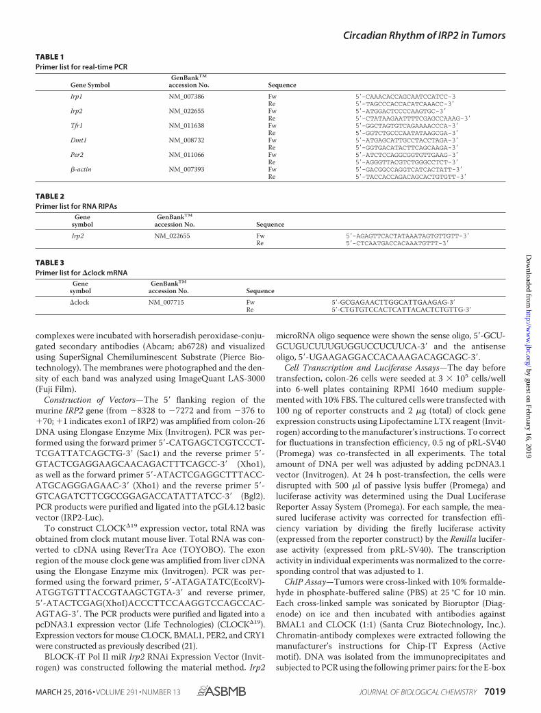
complexes were incubated with horseradish peroxidase-conju-gated secondary antibodies (Abcam; ab6728) and visualizedusing SuperSignal Chemiluminescent Substrate (Pierce Bio-technology). The membranes were photographed and the den-sity of each band was analyzed using ImageQuant LAS-3000(Fuji Film).
Construction of Vectors—The 5� flanking region of themurine IRP2 gene (from �8328 to �7272 and from �376 to�70; �1 indicates exon1 of IRP2) was amplified from colon-26DNA using Elongase Enzyme Mix (Invitrogen). PCR was per-formed using the forward primer 5�-CATGAGCTCGTCCCT-TCGATTATCAGCTG-3� (Sac1) and the reverse primer 5�-GTACTCGAGGAAGCAACAGACTTTCAGCC-3� (Xho1),as well as the forward primer 5�-ATACTCGAGGCTTTACC-ATGCAGGGAGAAC-3� (Xho1) and the reverse primer 5�-GTCAGATCTTCGCCGGAGACCATATTATCC-3� (Bgl2).PCR products were purified and ligated into the pGL4.12 basicvector (IRP2-Luc).
To construct CLOCK�19 expression vector, total RNA wasobtained from clock mutant mouse liver. Total RNA was con-verted to cDNA using ReverTra Ace (TOYOBO). The exonregion of the mouse clock gene was amplified from liver cDNAusing the Elongase Enzyme mix (Invitrogen). PCR was per-formed using the forward primer, 5�-ATAGATATC(EcoRV)-ATGGTGTTTACCGTAAGCTGTA-3� and reverse primer,5�-ATACTCGAG(XhoI)ACCCTTCCAAGGTCCAGCCAC-AGTAG-3�. The PCR products were purified and ligated into apcDNA3.1 expression vector (Life Technologies) (CLOCK�19).Expression vectors for mouse CLOCK, BMAL1, PER2, and CRY1were constructed as previously described (21).
BLOCK-iT Pol II miR Irp2 RNAi Expression Vector (Invit-rogen) was constructed following the material method. Irp2
microRNA oligo sequence were shown the sense oligo, 5�-GCU-GCUGUCUUUGUGGUCCUCUUCA-3� and the antisenseoligo, 5�-UGAAGAGGACCACAAAGACAGCAGC-3�.
Cell Transcription and Luciferase Assays—The day beforetransfection, colon-26 cells were seeded at 3 105 cells/wellinto 6-well plates containing RPMI 1640 medium supple-mented with 10% FBS. The cultured cells were transfected with100 ng of reporter constructs and 2 �g (total) of clock geneexpression constructs using Lipofectamine LTX reagent (Invit-rogen) according to the manufacturer’s instructions. To correctfor fluctuations in transfection efficiency, 0.5 ng of pRL-SV40(Promega) was co-transfected in all experiments. The totalamount of DNA per well was adjusted by adding pcDNA3.1vector (Invitrogen). At 24 h post-transfection, the cells weredisrupted with 500 �l of passive lysis buffer (Promega) andluciferase activity was determined using the Dual LuciferaseReporter Assay System (Promega). For each sample, the mea-sured luciferase activity was corrected for transfection effi-ciency variation by dividing the firefly luciferase activity(expressed from the reporter construct) by the Renilla lucifer-ase activity (expressed from pRL-SV40). The transcriptionactivity in individual experiments was normalized to the corre-sponding control that was adjusted to 1.
ChIP Assay—Tumors were cross-linked with 10% formalde-hyde in phosphate-buffered saline (PBS) at 25 °C for 10 min.Each cross-linked sample was sonicated by Bioruptor (Diag-enode) on ice and then incubated with antibodies againstBMAL1 and CLOCK (1:1) (Santa Cruz Biotechnology, Inc.).Chromatin-antibody complexes were extracted following themanufacturer’s instructions for Chip-IT Express (Activemotif). DNA was isolated from the immunoprecipitates andsubjected to PCR using the following primer pairs: for the E-box
TABLE 1Primer list for real-time PCR
Gene SymbolGenBankTM
accession No. Sequence
Irp1 NM_007386 Fw 5�-CAAACACCAGCAATCCATCC-3Re 5�-TAGCCCACCACATCAAACC-3�
Irp2 NM_022655 Fw 5�-ATGGACTCCCCAAGTGC-3�Re 5�-CTATAAGAATTTTCGAGCCAAAG-3�
Tfr1 NM_011638 Fw 5�-GGCTAGTGTCAGAAAACCCA-3�Re 5�-GGTCTGCCCAATATAAGCGA-3�
Dmt1 NM_008732 Fw 5�-ATGAGCATTGCCTACCTAGA-3�Re 5�-GGTGACATACTTCAGCAAGA-3�
Per2 NM_011066 Fw 5�-ATCTCCAGGCGGTGTTGAAG-3�Re 5�-AGGGTTACGTCTGGGCCTCT-3�
�-actin NM_007393 Fw 5�-GACGGCCAGGTCATCACTATT-3�Re 5�-TACCACCAGACAGCACTGTGTT-3�
TABLE 2Primer list for RNA RIPAs
Genesymbol
GenBankTM
accession No. Sequence
Irp2 NM_022655 Fw 5�-AGAGTTCACTATAAATAGTGTTGTT-3�Re 5�-CTCAATGACCACAAATGTTT-3�
TABLE 3Primer list for �clock mRNA
Genesymbol
GenBankTM
accession No. Sequence
�clock NM_007715 Fw 5�-GCGAGAACTTGGCATTGAAGAG-3�Re 5�-CTGTGTCCACTCATTACACTCTGTTG-3�
Circadian Rhythm of IRP2 in Tumors
MARCH 25, 2016 • VOLUME 291 • NUMBER 13 JOURNAL OF BIOLOGICAL CHEMISTRY 7019
by guest on February 16, 2019http://w
ww
.jbc.org/D
ownloaded from
in the IRP2 promoter region, 5�-GTTCTGAACTGCTCAGG-AAG-3� and 5�-CTGAACTGGAGAGAATGTCC-3�, and forthe E-box in the IRP2 intron region, 5�-GGCACAGTAACCC-TAAGTTC-3� and 5�-CAAGGAGCTGGAGAATGTAC-3�.Products were separated on 3% agarose gel electrophoresis,stained with ethidium bromide, and photographed withImageQuant LAS 3000 mini (Fuji film).
RNA Immunoprecipitation Assays (RIPA)—RIPA was per-formed as previously described (22, 23), using IRP2 or IgG(Santa Cruz Biotechnology) antibody. Each experiment wasindependently performed 3 times. PCR was performed withdiluted cDNA samples using the Gotaq Green Master Mix(Promega). PCR products were run on 2% agarose gels. Afterstaining with ethidium bromide, the gels were photographedusing Polaroid-type film or ImageQuant LAS-3000 (Fujifilm)and the density of each band was analyzed using ImageJ soft-ware (National Institutes of Health, Bethesda, MD). Primersequences are shown in Tables 1–3.
Cell Viability Assays—Colon-26 and colon-26(�19) cellswere seeded at a density of 1 105 cells/well in a 6-well cultureplate. After 48 and 72 h, intracellular ATP was measured as anindicator of cell viability using the CellTiter-Glo LuminescentCell Viability Assay kit (Promega).
Incorporation of 5-Bromouridine into DNA—Tumor masseswere collected from wild-type or colon-26(�19) cell-bearingmice at 09:00 or 21:00 h. Tumor mass was chopped by scissors.The tumor pieces were treated with 5-bromouridine (5-BrU)/DMEM solution (100 �g/ml) for 60 min at 37 °C and later fixedin 8% paraformaldehyde in PBS for 15 min at room tempera-ture. The tumor pieces were embedded into gelatin. Thetumor-gelatin masses were cut in 20-�m thick slices by LeicaCM1100 (Leica). Slices were blocked in 3% bovine serum albu-min (BSA), 0.2% Tween (TBS) for 1 h. Then, slices were incu-bated in anti-5-bromodeoxyuridine-fluorescein isothiocyanate(anti-BrdU-FITC) antibody (Santa Cruz Biotechnology)/T-TBS (1:2000) for 24 h. The stained cells were observed using afluorescence microscope (BZ-9000, KEYENCE). The imageswere taken in 3 regions of culture sections at a magnificationof 100.
Statistical Analysis—Analysis of variance (ANOVA) wasused for multiple comparisons, and Scheffé’s test was used forcomparison between two groups. A p value of 0.05 was con-sidered significant.
Results
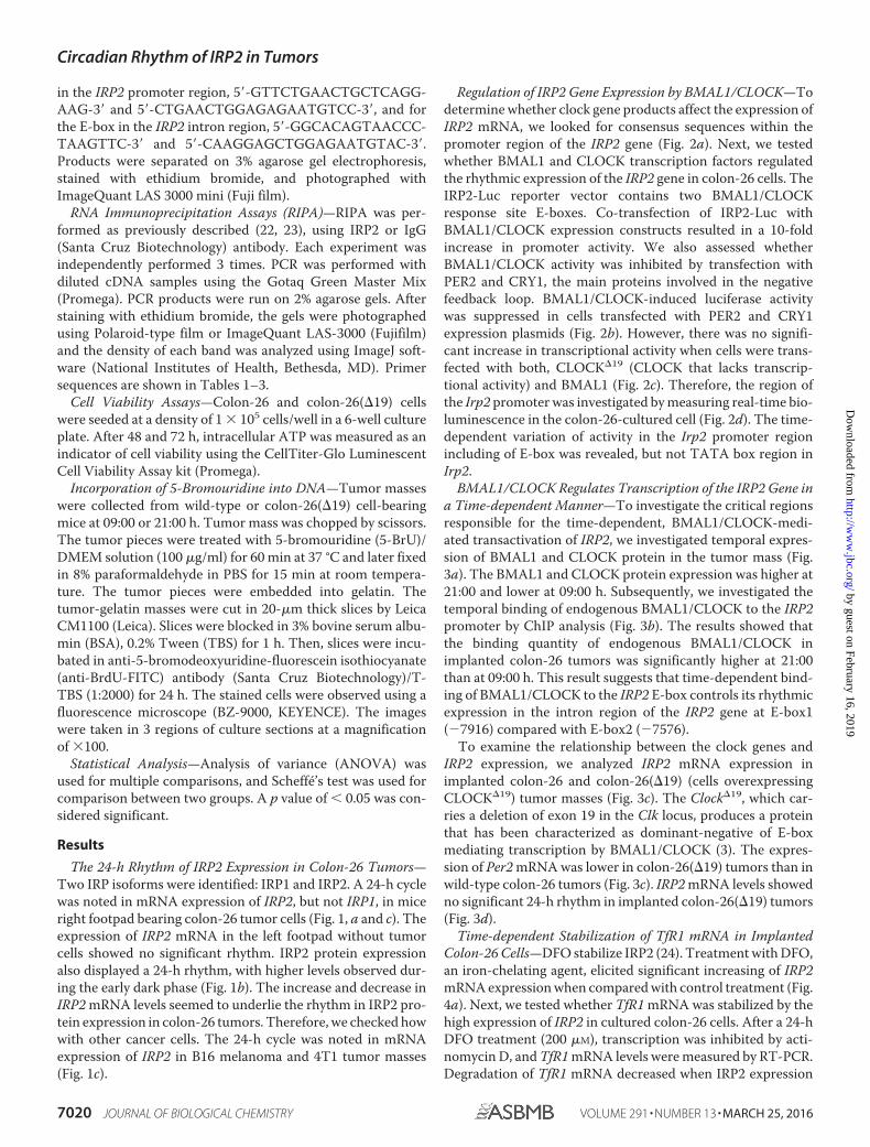
The 24-h Rhythm of IRP2 Expression in Colon-26 Tumors—Two IRP isoforms were identified: IRP1 and IRP2. A 24-h cyclewas noted in mRNA expression of IRP2, but not IRP1, in miceright footpad bearing colon-26 tumor cells (Fig. 1, a and c). Theexpression of IRP2 mRNA in the left footpad without tumorcells showed no significant rhythm. IRP2 protein expressionalso displayed a 24-h rhythm, with higher levels observed dur-ing the early dark phase (Fig. 1b). The increase and decrease inIRP2 mRNA levels seemed to underlie the rhythm in IRP2 pro-tein expression in colon-26 tumors. Therefore, we checked howwith other cancer cells. The 24-h cycle was noted in mRNAexpression of IRP2 in B16 melanoma and 4T1 tumor masses(Fig. 1c).
Regulation of IRP2 Gene Expression by BMAL1/CLOCK—Todetermine whether clock gene products affect the expression ofIRP2 mRNA, we looked for consensus sequences within thepromoter region of the IRP2 gene (Fig. 2a). Next, we testedwhether BMAL1 and CLOCK transcription factors regulatedthe rhythmic expression of the IRP2 gene in colon-26 cells. TheIRP2-Luc reporter vector contains two BMAL1/CLOCKresponse site E-boxes. Co-transfection of IRP2-Luc withBMAL1/CLOCK expression constructs resulted in a 10-foldincrease in promoter activity. We also assessed whetherBMAL1/CLOCK activity was inhibited by transfection withPER2 and CRY1, the main proteins involved in the negativefeedback loop. BMAL1/CLOCK-induced luciferase activitywas suppressed in cells transfected with PER2 and CRY1expression plasmids (Fig. 2b). However, there was no signifi-cant increase in transcriptional activity when cells were trans-fected with both, CLOCK�19 (CLOCK that lacks transcrip-tional activity) and BMAL1 (Fig. 2c). Therefore, the region ofthe Irp2 promoter was investigated by measuring real-time bio-luminescence in the colon-26-cultured cell (Fig. 2d). The time-dependent variation of activity in the Irp2 promoter regionincluding of E-box was revealed, but not TATA box region inIrp2.
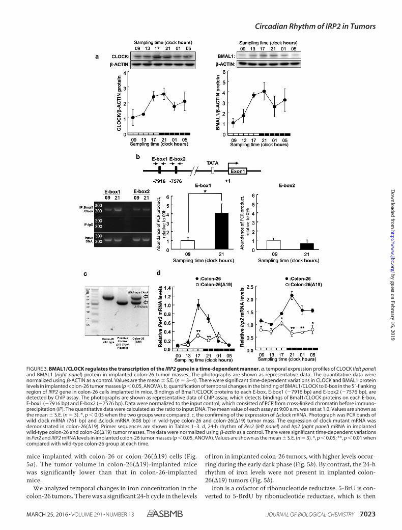
BMAL1/CLOCK Regulates Transcription of the IRP2 Gene ina Time-dependent Manner—To investigate the critical regionsresponsible for the time-dependent, BMAL1/CLOCK-medi-ated transactivation of IRP2, we investigated temporal expres-sion of BMAL1 and CLOCK protein in the tumor mass (Fig.3a). The BMAL1 and CLOCK protein expression was higher at21:00 and lower at 09:00 h. Subsequently, we investigated thetemporal binding of endogenous BMAL1/CLOCK to the IRP2promoter by ChIP analysis (Fig. 3b). The results showed thatthe binding quantity of endogenous BMAL1/CLOCK inimplanted colon-26 tumors was significantly higher at 21:00than at 09:00 h. This result suggests that time-dependent bind-ing of BMAL1/CLOCK to the IRP2 E-box controls its rhythmicexpression in the intron region of the IRP2 gene at E-box1(�7916) compared with E-box2 (�7576).
To examine the relationship between the clock genes andIRP2 expression, we analyzed IRP2 mRNA expression inimplanted colon-26 and colon-26(�19) (cells overexpressingCLOCK�19) tumor masses (Fig. 3c). The Clock�19, which car-ries a deletion of exon 19 in the Clk locus, produces a proteinthat has been characterized as dominant-negative of E-boxmediating transcription by BMAL1/CLOCK (3). The expres-sion of Per2 mRNA was lower in colon-26(�19) tumors than inwild-type colon-26 tumors (Fig. 3c). IRP2 mRNA levels showedno significant 24-h rhythm in implanted colon-26(�19) tumors(Fig. 3d).
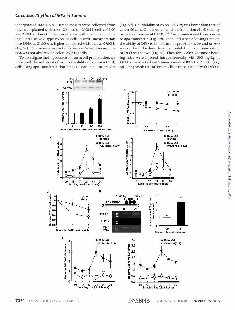
Time-dependent Stabilization of TfR1 mRNA in ImplantedColon-26 Cells—DFO stabilize IRP2 (24). Treatment with DFO,an iron-chelating agent, elicited significant increasing of IRP2mRNA expression when compared with control treatment (Fig.4a). Next, we tested whether TfR1 mRNA was stabilized by thehigh expression of IRP2 in cultured colon-26 cells. After a 24-hDFO treatment (200 �M), transcription was inhibited by acti-nomycin D, and TfR1 mRNA levels were measured by RT-PCR.Degradation of TfR1 mRNA decreased when IRP2 expression
Circadian Rhythm of IRP2 in Tumors
7020 JOURNAL OF BIOLOGICAL CHEMISTRY VOLUME 291 • NUMBER 13 • MARCH 25, 2016
by guest on February 16, 2019http://w
ww
.jbc.org/D
ownloaded from
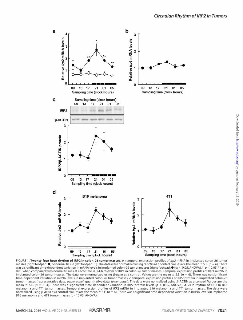
FIGURE 1. Twenty-four hour rhythm of IRP2 in colon-26 tumor masses. a, temporal expression profiles of Irp2 mRNA in implanted colon-26 tumormasses (right footpad: ●) or normal tissue (left footpad:E). The data were normalized using �-actin as a control. Values are the mean � S.E. (n � 6). Therewas a significant time-dependent variation in mRNA levels in implanted colon-26 tumor masses (right footpad: ●) (p 0.05, ANOVA). *, p 0.05; **, p 0.01 when compared with normal tissues at each time. b, 24-h rhythm of IRP1 in colon-26 tumor masses. Temporal expression profiles of IRP1 mRNA inimplanted colon-26 tumor masses. The data were normalized using �-actin as a control. Values are the mean � S.E. (n � 6). There was no significanttime-dependent variation in mRNA levels in implanted colon-26 tumor masses. c, temporal expression profiles of IRP2 protein in implanted colon-26tumor masses (representative data, upper panel, quantitative data, lower panel). The data were normalized using �-ACTIN as a control. Values are themean � S.E. (n � 3– 4). There was a significant time-dependent variation in IRP2 protein levels (p 0.05, ANOVA). d, 24-h rhythm of IRP2 in B16melanoma and 4T1 tumor masses. Temporal expression profiles of IRP2 mRNA in implanted B16 melanoma and 4T1 tumor masses. The data werenormalized using �-actin as a control. Values are the mean � S.E. (n � 6). There was a significant time-dependent variation in mRNA levels in implantedB16 melanoma and 4T1 tumor masses (p 0.05, ANOVA).
Circadian Rhythm of IRP2 in Tumors
MARCH 25, 2016 • VOLUME 291 • NUMBER 13 JOURNAL OF BIOLOGICAL CHEMISTRY 7021
by guest on February 16, 2019http://w
ww
.jbc.org/D
ownloaded from
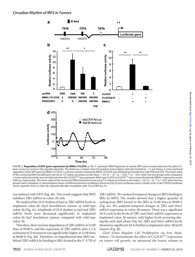
was induced with DFO (Fig. 4b). This result suggests that IRP2stabilizes TfR1 mRNA in colon-26 cells.
We analyzed the 24-h rhythm of Irp2 or TfR1 mRNA levels inimplanted colon-26 (Irp2 knockdown) tumors or wild-typecolon-26 (Fig. 4c). Amplitude of 24-h rhythm in Irp2 and TfR1mRNA levels were decreased significantly in implantedcolon-26 Irp2 knockdown tumors compared with wild-typecolon-26.
Therefore, there was less degradation of TfR1 mRNA at 21:00than at 09:00 h, and the expression of TfR1 mRNA after a 2-hactinomycin D treatment was significantly higher at 21:00 thanat 09:00 h (Fig. 4d). Therefore, we hypothesized that IRP2 sta-bilized TfR1 mRNA by binding to IREs located in the 3�-UTR of
TfR1 mRNA. We analyzed temporal changes in IRP2 binding toIREs by RIPA. The results showed that a higher quantity ofendogenous IRP2 bound to the IREs at 21:00 than at 09:00 h(Fig. 4e). We analyzed temporal changes in TfR1 and Dmt1mRNA expression in colon-26 tumors. There was a significant24-h cycle in the levels of TfR1 and Dmt1 mRNA expression inimplanted colon-26 tumors, with higher levels occurring dur-ing the early dark phase (Fig. 4c). TfR1 and Dmt1 mRNA levelsshowed no significant 24-h rhythm in implanted colon-26(�19)tumors (Fig. 4f).
Clock Genes Regulate Cell Proliferation via Iron Meta-bolism—To demonstrate the effect of CLOCK�19 expressionon tumor cell growth, we measured the tumor volume in
FIGURE 2. Regulation of IRP2 gene expression by BMAL1/CLOCK. a, the 5�-upstream DNA fragments of murine IRP2 were incorporated into the pGL4.12-basic vector to construct the reporter plasmids. The distances, in bases, from the putative transcription start site (marked as �1) are shown. b, transcriptionalregulation of the IRP2 gene by BMAL1/CLOCK. Luciferase activity induced by BMAL1/CLOCK was inhibited by transfection with PER and CRY. The mean valueof the control (pcDNA transfection) was set at 1.0. Values are shown as the mean � S.E. (n � 3). *, p 0.05; **, p 0.01 when the two groups were compared.c, transcriptional activity was not induced when the CLOCK�19 was expressed. Wild-type CLOCK or CLOCK�19 were cotransfected with BMAL1 expression vector(500 ng, respectively). The mean value of the control (pcDNA transfection) was set at 1.0. Values are shown as the mean � S.E. (n � 3). **, p 0.01 when the twogroups were compared. d, representative traces of bioluminescence oscillations driven by Irp2 (E-box)-luciferase vector (closed circle) or Ipr2 (TATA)-luciferasevector (opened circle) in colon-26-cultured cells after incubation with 10 nM DEX for 2 h.
Circadian Rhythm of IRP2 in Tumors
7022 JOURNAL OF BIOLOGICAL CHEMISTRY VOLUME 291 • NUMBER 13 • MARCH 25, 2016
by guest on February 16, 2019http://w
ww
.jbc.org/D
ownloaded from
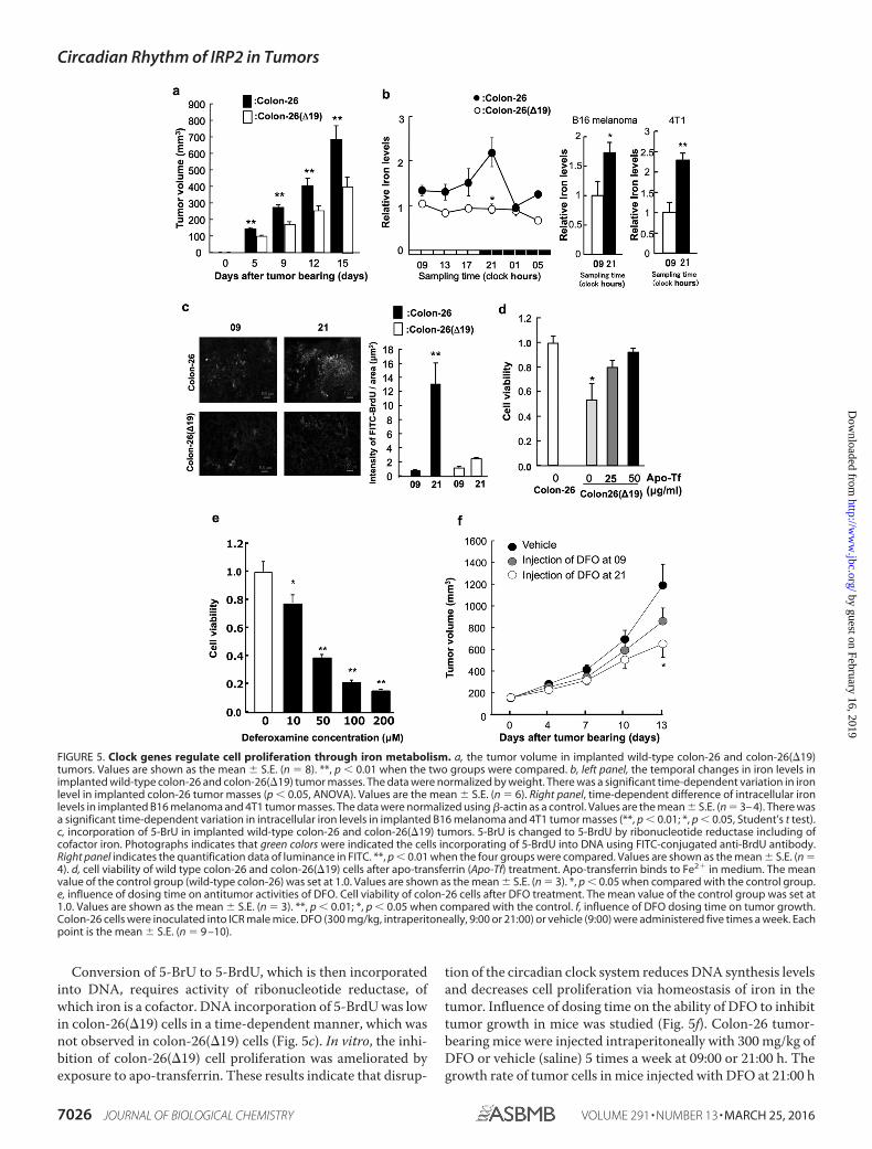
mice implanted with colon-26 or colon-26(�19) cells (Fig.5a). The tumor volume in colon-26(�19)-implanted micewas significantly lower than that in colon-26-implantedmice.
We analyzed temporal changes in iron concentration in thecolon-26 tumors. There was a significant 24-h cycle in the levels
of iron in implanted colon-26 tumors, with higher levels occur-ring during the early dark phase (Fig. 5b). By contrast, the 24-hrhythm of iron levels were not present in implanted colon-26(�19) tumors (Fig. 5b).
Iron is a cofactor of ribonucleotide reductase. 5-BrU is con-verted to 5-BrdU by ribonucleotide reductase, which is then
FIGURE 3. BMAL1/CLOCK regulates the transcription of the IRP2 gene in a time-dependent manner. a, temporal expression profiles of CLOCK (left panel)and BMAL1 (right panel) protein in implanted colon-26 tumor masses. The photographs are shown as representative data. The quantitative data werenormalized using �-ACTIN as a control. Values are the mean � S.E. (n � 3– 4). There were significant time-dependent variations in CLOCK and BMAL1 proteinlevels in implanted colon-26 tumor masses (p 0.05, ANOVA). b, quantification of temporal changes in the binding of BMAL1/CLOCK to E-box in the 5�-flankingregion of IRP2 gene in colon-26 cells implanted in mice. Bindings of Bmal1/CLOCK proteins to each E-box, E-box1 (�7916 bp) and E-box2 (�7576 bp), aredetected by ChIP assay. The photographs are shown as representative data of ChIP assay, which detects bindings of Bmal1/CLOCK proteins on each E-box,E-box1 (�7916 bp) and E-box2 (�7576 bp). Data were normalized to the input control, which consisted of PCR from cross-linked chromatin before immuno-precipitation (IP). The quantitative data were calculated as the ratio to input DNA. The mean value of each assay at 9:00 a.m. was set at 1.0. Values are shown asthe mean � S.E. (n � 3). *, p 0.05 when the two groups were compared. c, the confirming of the expression of �clock mRNA. Photograph was PCR bands ofwild clock mRNA (761 bp) and �clock mRNA (608 bp) in wild-type colon-26 and colon-26(�19) tumor mass. The expression of clock mutant mRNA wasdemonstrated in colon-26(�19). Primer sequences are shown in Tables 1–3. d, 24-h rhythm of Per2 (left panel) and Irp2 (right panel) mRNA in implantedwild-type colon-26 and colon-26(�19) tumor masses. The data were normalized using �-actin as a control. There were significant time-dependent variationsin Per2 and IRP2 mRNA levels in implanted colon-26 tumor masses (p 0.05, ANOVA). Values are shown as the mean � S.E. (n � 3). *, p 0.05; **, p 0.01 whencompared with wild-type colon-26 group at each time.
Circadian Rhythm of IRP2 in Tumors
MARCH 25, 2016 • VOLUME 291 • NUMBER 13 JOURNAL OF BIOLOGICAL CHEMISTRY 7023
by guest on February 16, 2019http://w
ww
.jbc.org/D
ownloaded from
incorporated into DNA. Tumor masses were collected frommice transplanted with colon-26 or colon-26(�19) cells at 09:00and 21:00 h. These tumors were treated with medium contain-ing 5-BrU. In wild-type colon-26 cells, 5-BrdU incorporationinto DNA at 21:00 was higher compared with that at 09:00 h(Fig. 5c). This time-dependent difference of 5-BrdU incorpora-tion was not observed in colon-26(�19) cells.
To investigate the importance of iron in cell proliferation, wemeasured the influence of iron on viability of colon-26(�19)cells using apo-transferrin that binds to iron in culture media
(Fig. 5d). Cell viability of colon-26(�19) was lower than that ofcolon-26 cells. On the other hand, the inhibition of cell viabilityby overexpression of CLOCK�19 was ameliorated by exposureto apo-transferrin (Fig. 5d). Then, influence of dosing time onthe ability of DFO to inhibit tumor growth in vitro and in vivowas studied. The dose-dependent inhibition in administrationof DFO was shown (Fig. 5e). Therefore, colon-26 tumor-bear-ing mice were injected intraperitoneally with 300 mg/kg ofDFO or vehicle (saline) 5 times a week at 09:00 or 21:00 h (Fig.5f). The growth rate of tumor cells in mice injected with DFO at
Circadian Rhythm of IRP2 in Tumors
7024 JOURNAL OF BIOLOGICAL CHEMISTRY VOLUME 291 • NUMBER 13 • MARCH 25, 2016
by guest on February 16, 2019http://w
ww
.jbc.org/D
ownloaded from
21:00 h was significantly lower than that in mice injected withsaline.
Discussion
Iron is essential for biosynthesis and dysregulation of ironmetabolism can cause disease (11–13). Iron is fundamental formany biological processes, including DNA synthesis, electrontransport, and cell cycle regulation (6 – 8). However, excess ironis toxic (11–13) and therefore, cellular iron metabolism must betightly regulated. Iron metabolism is enhanced in varioustumor cells to support increased rates of DNA synthesis andcell proliferation. Thus, iron metabolism is important fortumor growth (22–25). Recent studies have shown that DNAsynthesis and cell proliferation exhibit circadian rhythms intumor cells and that these rhythms are essential for tumor cellproliferation (6 –9). Several factors that contribute to thisrhythm have been reported, but the relevance of iron is unclear.In this study, we hypothesized that iron levels in tumor cellsdisplay circadian rhythms and that these rhythms affect theproliferation of tumor cells.
Because cellular iron levels are regulated by cellular ironmetabolism, we hypothesized that the iron rhythm in tumorcells was generated by the regulation of IRPs, which are keyfactors in iron metabolism (15–17). Therefore, we analyzed theexpression of IRPs. Although no marked rhythmic oscillation ofIrp1 mRNA levels was observed in implanted colon-26 tumorcells, Irp2 mRNA and protein levels in colon-26 tumor cellsimplanted in mice showed a clear 24-h oscillation (Fig. 1a).
Moreover, Irp2 mRNA levels in murine breast cancer 4T1tumor cells or murine B16 melanoma implanted in miceshowed a clear 24-h oscillation (Fig. 1d). These results maysuggest that Irp2 show rhythmic expression in various tumorsthat have the higher proliferating ability than normal cell andtissue (15–17). IRP has two isoforms, IRP1 and IRP2. Both iso-forms stabilize TfR1 and Dmt1 mRNA by binding to 3�-UTR.Our results established that IRP2 may be more important thanIRP1 in inducing circadian expression in implanted tumor cells.
To identify the relationship between circadian clock genesand IRP2, we analyzed whether clock gene products affectedthe expression of Irp2. The luciferase reporter assay and ChIPassay revealed that Irp2 expression was regulated by BMAL1and CLOCK (Figs. 2 and 3).
Furthermore, to clear the relationship between circadianclock gene activity and Irp2 expression in vivo, we established a
mutant colon-26 cell line, colon-26(�19), in which CLOCK�19
(CLOCK protein lacking transcriptional activity) was overex-pressed. clock-mutant mice are a powerful tool for the analysisof molecular clocks (3). These mice have a point mutation inexon 19 of the clock gene and exhibit low-amplitude rhythms inthe expression of clock-controlled genes. Thus, the colon-26(�19) cell line may clarify the relationship between the bio-logical clock and iron metabolism within tumors. In implantedcolon-26(�19) tumor tissue, oscillations in Irp2 mRNA were nolonger apparent, and Irp2 mRNA levels were decreased (Fig.3c). These results suggest that circadian clock regulation oftranscription is important for Irp2 rhythmic expression intumor cells.
Because cellular iron levels are regulated by cellular ironmetabolism, we hypothesized that the iron rhythm was con-trolled by regulation of IRPs, TfR1, and DMT1, which are keyfactors in iron metabolism (15–17). A previous study showedthat the circadian rhythm in Tfr1 mRNA expression is regu-lated by c-Myc (20). However, it was unclear if the stability ofTfR1 was related to IRP2 activity. In this study, temporal anal-ysis of mRNA levels in implanted colon-26 cells suggested thatIRP2 was involved in the circadian regulation of TfR1 and Dmt1mRNA stability. Degradation of TfR1 mRNA at 21:00 h waslower than that at 09:00 h in vivo (Fig. 4d). Moreover, therhythm of IRP2 protein abundance in colon-26 cells correlatedwith the time dependence of its binding to the 3�-UTR of TfR1mRNA (Fig. 4e). These results suggest that oscillation in IRP2protein levels control the 24-h rhythm of TfR1 mRNA stability.Thus, these mechanisms may affect the stability of Dmt1mRNA in vivo. Therefore, the amplitude in the 24-h rhythm ofTfR1 mRNA in Irp2 knockdown colon-26 tumor mass wasdecreased (Fig. 4f).
Iron is an essential element for various biological functions(26). Finally, the results of this study suggest that the circadianclock system controls tumor cell proliferation by regulatingiron metabolism as one of various function of iron. In vivo,colon-26(�19) cell proliferation was lower than that ofcolon-26 cells (Fig. 5a). Iron levels exhibited a 24-h rhythmin colon-26 tumors (Fig. 5b), with higher levels observed duringthe early dark phase. Moreover, the time-dependent differenceof iron levels in mice implanted with murine breast cancer 4T1tumor cells or murine B16 melanoma were revealed (Fig. 5b).On the other hand, oscillations in iron were not apparent inimplanted colon-26(�19) tumor tissues.
FIGURE 4. Stabilization of TfR1 mRNA by DFO in cultured colon-26 cells. a, analysis of IRP2 mRNA and protein levels in colon-26 cells grown for 24 h with orwithout DFO (0, 10, 100, and 200 �M). Upper panel shows the IRP2 protein levels or �-actin protein levels as a control protein. The IRP2 mRNA levels are indicatedunder the graph. The data were normalized using �-actin as a control. The mean value of the control group was set at 1.0. Values are shown as the mean � S.E.(n � 3). *, p 0.01 when compared with the control. b, colon-26 cells were treated with DFO (200 �M) for 24 h, and the stability of TfR1 mRNA extracted fromactinomycin D-treated cells was assessed. The data were normalized using �-actin as a control. The mean value of the control group was set at 1.0. Values areshown as the mean � S.E. (n � 3). *, p 0.01 when compared with the control. c, 24-h rhythm of Irp2 (left panel) and TfR1 (right panel) mRNA in implantedwild-type colon-26 and colon-26 (Irp2 knockdown) tumor masses. The data were normalized using �-actin as a control. There were significant time-dependentvariations in Irp2 and TfR1 mRNA levels in implanted wild-type colon-26 and colon-26 (Irp2 knockdown) tumor (p 0.05, ANOVA). Values are the mean � S.E.(n � 3). *, p 0.05; **, p 0.01 when compared with wild-type colon-26 group at each time. d, quantification of temporal changes in the stability of TfR1 mRNAextracted from actinomycin D-treated implanted colon-26 tumor masses. The data were normalized using �-actin as a control. The value at start time in eachassay was set at 1.0. Values are shown as the mean � S.E. (n � 3). **, p 0.01 and *, p 0.05 when the two groups were compared. e, quantification of temporalchanges in the binding of IRP2 to the IRE located in the 3�-UTR of TfR1 in implanted colon-26 tumors. Data were normalized to the input control, which consistedof PCR from cross-linked total RNA before immunoprecipitation. The mean value at 9:00 a.m. was set at 1.0. Values are shown as the mean � S.E. (n � 3). *, p 0.05 when the two groups were compared. f, 24-h rhythm of TfR1 (left panel) and Dmt1 (right panel) mRNA in implanted wild-type colon-26 and colon-26(�19)tumor masses. The data were normalized using �-actin as a control. There were significant time-dependent variations in TfR1 and Dmt1 mRNA levels inimplanted wild-type colon-26 tumor (p 0.05, ANOVA). Values are the mean � S.E. (n � 3). *, p 0.05; **, p 0.01 when compared with wild-type colon-26group at each time.
Circadian Rhythm of IRP2 in Tumors
MARCH 25, 2016 • VOLUME 291 • NUMBER 13 JOURNAL OF BIOLOGICAL CHEMISTRY 7025
by guest on February 16, 2019http://w
ww
.jbc.org/D
ownloaded from
Conversion of 5-BrU to 5-BrdU, which is then incorporatedinto DNA, requires activity of ribonucleotide reductase, ofwhich iron is a cofactor. DNA incorporation of 5-BrdU was lowin colon-26(�19) cells in a time-dependent manner, which wasnot observed in colon-26(�19) cells (Fig. 5c). In vitro, the inhi-bition of colon-26(�19) cell proliferation was ameliorated byexposure to apo-transferrin. These results indicate that disrup-
tion of the circadian clock system reduces DNA synthesis levelsand decreases cell proliferation via homeostasis of iron in thetumor. Influence of dosing time on the ability of DFO to inhibittumor growth in mice was studied (Fig. 5f). Colon-26 tumor-bearing mice were injected intraperitoneally with 300 mg/kg ofDFO or vehicle (saline) 5 times a week at 09:00 or 21:00 h. Thegrowth rate of tumor cells in mice injected with DFO at 21:00 h
FIGURE 5. Clock genes regulate cell proliferation through iron metabolism. a, the tumor volume in implanted wild-type colon-26 and colon-26(�19)tumors. Values are shown as the mean � S.E. (n � 8). **, p 0.01 when the two groups were compared. b, left panel, the temporal changes in iron levels inimplanted wild-type colon-26 and colon-26(�19) tumor masses. The data were normalized by weight. There was a significant time-dependent variation in ironlevel in implanted colon-26 tumor masses (p 0.05, ANOVA). Values are the mean � S.E. (n � 6). Right panel, time-dependent difference of intracellular ironlevels in implanted B16 melanoma and 4T1 tumor masses. The data were normalized using �-actin as a control. Values are the mean � S.E. (n � 3– 4). There wasa significant time-dependent variation in intracellular iron levels in implanted B16 melanoma and 4T1 tumor masses (**, p 0.01; *, p 0.05, Student’s t test).c, incorporation of 5-BrU in implanted wild-type colon-26 and colon-26(�19) tumors. 5-BrU is changed to 5-BrdU by ribonucleotide reductase including ofcofactor iron. Photographs indicates that green colors were indicated the cells incorporating of 5-BrdU into DNA using FITC-conjugated anti-BrdU antibody.Right panel indicates the quantification data of luminance in FITC. **, p 0.01 when the four groups were compared. Values are shown as the mean � S.E. (n �4). d, cell viability of wild type colon-26 and colon-26(�19) cells after apo-transferrin (Apo-Tf) treatment. Apo-transferrin binds to Fe2� in medium. The meanvalue of the control group (wild-type colon-26) was set at 1.0. Values are shown as the mean � S.E. (n � 3). *, p 0.05 when compared with the control group.e, influence of dosing time on antitumor activities of DFO. Cell viability of colon-26 cells after DFO treatment. The mean value of the control group was set at1.0. Values are shown as the mean � S.E. (n � 3). **, p 0.01; *, p 0.05 when compared with the control. f, influence of DFO dosing time on tumor growth.Colon-26 cells were inoculated into ICR male mice. DFO (300 mg/kg, intraperitoneally, 9:00 or 21:00) or vehicle (9:00) were administered five times a week. Eachpoint is the mean � S.E. (n � 9 –10).
Circadian Rhythm of IRP2 in Tumors
7026 JOURNAL OF BIOLOGICAL CHEMISTRY VOLUME 291 • NUMBER 13 • MARCH 25, 2016
by guest on February 16, 2019http://w
ww
.jbc.org/D
ownloaded from
was significantly lower than in mice injected with saline. Thisresult concurs with a previous study, where DNA synthesis wasenhanced in the dark phase (9). Because of the iron concentra-tion and tumor growth, it was suggested that the circadianrhythm in iron concentration influences proliferation in thetumor.
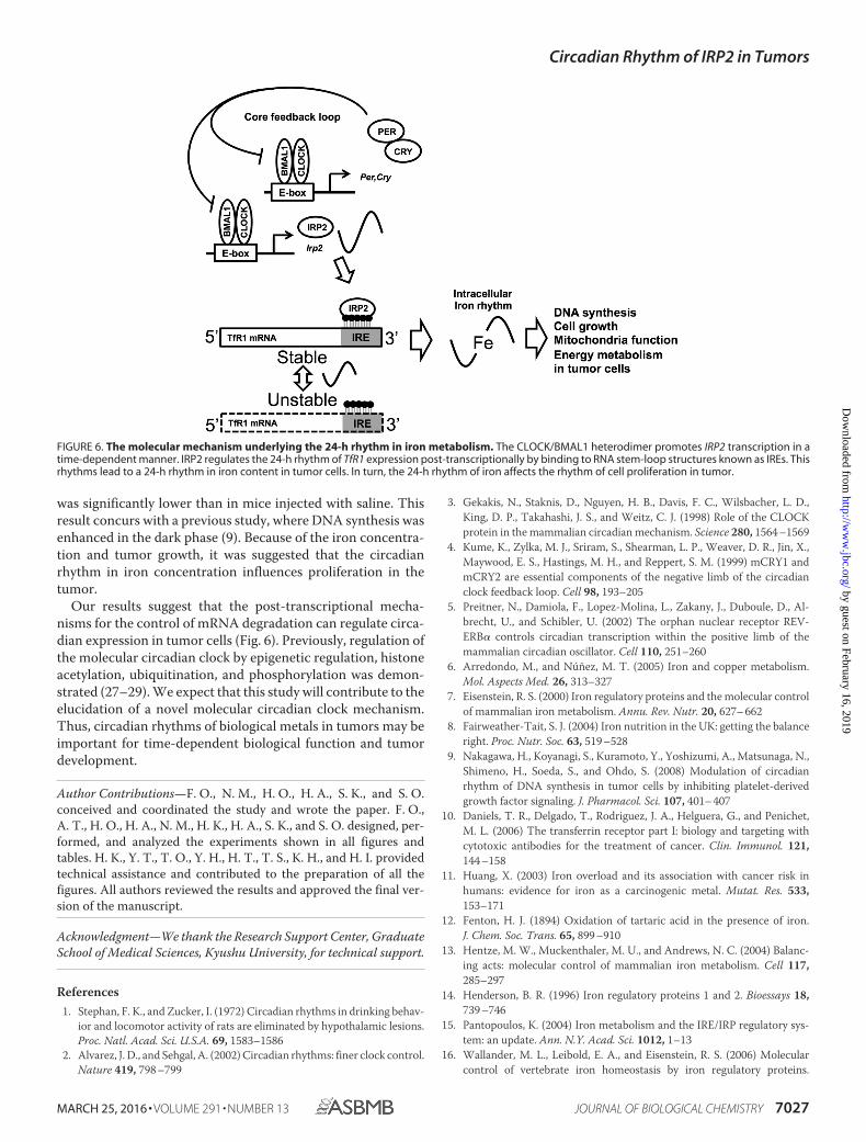
Our results suggest that the post-transcriptional mecha-nisms for the control of mRNA degradation can regulate circa-dian expression in tumor cells (Fig. 6). Previously, regulation ofthe molecular circadian clock by epigenetic regulation, histoneacetylation, ubiquitination, and phosphorylation was demon-strated (27–29). We expect that this study will contribute to theelucidation of a novel molecular circadian clock mechanism.Thus, circadian rhythms of biological metals in tumors may beimportant for time-dependent biological function and tumordevelopment.
Author Contributions—F. O., N. M., H. O., H. A., S. K., and S. O.conceived and coordinated the study and wrote the paper. F. O.,A. T., H. O., H. A., N. M., H. K., H. A., S. K., and S. O. designed, per-formed, and analyzed the experiments shown in all figures andtables. H. K., Y. T., T. O., Y. H., H. T., T. S., K. H., and H. I. providedtechnical assistance and contributed to the preparation of all thefigures. All authors reviewed the results and approved the final ver-sion of the manuscript.
Acknowledgment—We thank the Research Support Center, GraduateSchool of Medical Sciences, Kyushu University, for technical support.
References1. Stephan, F. K., and Zucker, I. (1972) Circadian rhythms in drinking behav-
ior and locomotor activity of rats are eliminated by hypothalamic lesions.Proc. Natl. Acad. Sci. U.S.A. 69, 1583–1586
2. Alvarez, J. D., and Sehgal, A. (2002) Circadian rhythms: finer clock control.Nature 419, 798 –799
3. Gekakis, N., Staknis, D., Nguyen, H. B., Davis, F. C., Wilsbacher, L. D.,King, D. P., Takahashi, J. S., and Weitz, C. J. (1998) Role of the CLOCKprotein in the mammalian circadian mechanism. Science 280, 1564 –1569
4. Kume, K., Zylka, M. J., Sriram, S., Shearman, L. P., Weaver, D. R., Jin, X.,Maywood, E. S., Hastings, M. H., and Reppert, S. M. (1999) mCRY1 andmCRY2 are essential components of the negative limb of the circadianclock feedback loop. Cell 98, 193–205
5. Preitner, N., Damiola, F., Lopez-Molina, L., Zakany, J., Duboule, D., Al-brecht, U., and Schibler, U. (2002) The orphan nuclear receptor REV-ERB� controls circadian transcription within the positive limb of themammalian circadian oscillator. Cell 110, 251–260
6. Arredondo, M., and Núñez, M. T. (2005) Iron and copper metabolism.Mol. Aspects Med. 26, 313–327
7. Eisenstein, R. S. (2000) Iron regulatory proteins and the molecular controlof mammalian iron metabolism. Annu. Rev. Nutr. 20, 627– 662
8. Fairweather-Tait, S. J. (2004) Iron nutrition in the UK: getting the balanceright. Proc. Nutr. Soc. 63, 519 –528
9. Nakagawa, H., Koyanagi, S., Kuramoto, Y., Yoshizumi, A., Matsunaga, N.,Shimeno, H., Soeda, S., and Ohdo, S. (2008) Modulation of circadianrhythm of DNA synthesis in tumor cells by inhibiting platelet-derivedgrowth factor signaling. J. Pharmacol. Sci. 107, 401– 407
10. Daniels, T. R., Delgado, T., Rodriguez, J. A., Helguera, G., and Penichet,M. L. (2006) The transferrin receptor part I: biology and targeting withcytotoxic antibodies for the treatment of cancer. Clin. Immunol. 121,144 –158
11. Huang, X. (2003) Iron overload and its association with cancer risk inhumans: evidence for iron as a carcinogenic metal. Mutat. Res. 533,153–171
12. Fenton, H. J. (1894) Oxidation of tartaric acid in the presence of iron.J. Chem. Soc. Trans. 65, 899 –910
13. Hentze, M. W., Muckenthaler, M. U., and Andrews, N. C. (2004) Balanc-ing acts: molecular control of mammalian iron metabolism. Cell 117,285–297
14. Henderson, B. R. (1996) Iron regulatory proteins 1 and 2. Bioessays 18,739 –746
15. Pantopoulos, K. (2004) Iron metabolism and the IRE/IRP regulatory sys-tem: an update. Ann. N.Y. Acad. Sci. 1012, 1–13
16. Wallander, M. L., Leibold, E. A., and Eisenstein, R. S. (2006) Molecularcontrol of vertebrate iron homeostasis by iron regulatory proteins.
FIGURE 6. The molecular mechanism underlying the 24-h rhythm in iron metabolism. The CLOCK/BMAL1 heterodimer promotes IRP2 transcription in atime-dependent manner. IRP2 regulates the 24-h rhythm of TfR1 expression post-transcriptionally by binding to RNA stem-loop structures known as IREs. Thisrhythms lead to a 24-h rhythm in iron content in tumor cells. In turn, the 24-h rhythm of iron affects the rhythm of cell proliferation in tumor.
Circadian Rhythm of IRP2 in Tumors
MARCH 25, 2016 • VOLUME 291 • NUMBER 13 JOURNAL OF BIOLOGICAL CHEMISTRY 7027
by guest on February 16, 2019http://w
ww
.jbc.org/D
ownloaded from
Biochim. Biophys. Acta 1763, 668 – 68917. Niitsu, Y., Kohgo, Y., Nishisato, T., Kondo, H., Kato, J., Urushizaki, Y., and
Urushizaki, I. (1987) Transferrin receptors in human cancerous tissues.Tohoku J. Exp. Med. 153, 239 –243
18. Smaaland, R., Laerum, O. D., Lote, K., Sletvold, O., Sothern, R. B., andBjerknes, R. (1991) DNA synthesis in human bone marrow is circadianstage dependent. Blood 77, 2603–2611
19. Wood, P. A., Du-Quiton, J., You, S., and Hrushesky, W. J. (2006) Circadianclock coordinates cancer cell cycle progression, thymidylate synthase, and5-fluorouracil therapeutic index. Mol. Cancer Ther. 5, 2023–2033
20. Okazaki, F., Matsunaga, N., Okazaki, H., Utoguchi, N., Suzuki, R., Maruy-ama, K., Koyanagi, S., and Ohdo, S. (2010) Circadian rhythm of transferrinreceptor 1 gene expression controlled by c-Myc in colon cancer-bearingmice. Cancer Res. 70, 6238 – 6246
21. Koyanagi, S., Kuramoto, Y., Nakagawa, H., Aramaki, H., Ohdo, S., Soeda,S., and Shimeno, H. (2003) A molecular mechanism regulating circadianexpression of vascular endothelial growth factor in tumor cells. CancerRes. 63, 7277–7283
22. Trowbridge, I. S., and Lopez, F. (1982) Monoclonal antibody to transferrinreceptor blocks transferrin binding and inhibits human tumor cell growthin vitro. Proc. Natl. Acad. Sci. U.S.A. 79, 1175–1179
23. Levy, J. E., Jin, O., Fujiwara, Y., Kuo, F., and Andrews, N. C. (1999) Trans-ferrin receptor is necessary for development of erythrocytes and the nerv-ous system. Nat. Genet. 21, 396 –399
24. Hanson, E. S., Foot, L. M., Leibold, E. A. (1999) Hypoxia post-translation-ally activates iron-regulatory protein 2. J. Biol. Chem. 274, 5047–5052
25. Richardson, D. R., and Baker, E. (1990) The uptake of iron and transferrinby the human malignant melanoma cell. Biochim. Biophys. Acta 1053,1–12
26. Abbaspour, N., Hurrell, R., Kelishadi, R. (2014) Review on iron and itsimportance for human health. J. Res. Med. Sci. 19, 164 –174
27. Harada, Y., Sakai, M., Kurabayashi, N., Hirota, T., and Fukada, Y. (2005)Ser-557-phosphorylated mCRY2 is degraded upon synergistic phosphor-ylation by glycogen synthase kinase-3�. J. Biol. Chem. 280, 31714 –31721
28. Hirano, A., Yumimoto, K., Tsunematsu, R., Matsumoto, M., Oyama, M.,Kozuka-Hata H, Nakagawa, T., Lanjakornsiripan, D., Nakayama, K. I., andFukada, Y. (2013) FBXL21 regulates oscillation of the circadian clockthrough ubiquitination and stabilization of cryptochromes. Cell 152,1106 –1118
29. Feng, D., Liu, T., Sun, Z., Bugge, A., Mullican, S. E., Alenghat, T., Liu, X. S.,and Lazar, M. A. (2011) A circadian rhythm orchestrated by histonedeacetylase 3 controls hepatic lipid metabolism. Science 331, 1315–1319
Circadian Rhythm of IRP2 in Tumors
7028 JOURNAL OF BIOLOGICAL CHEMISTRY VOLUME 291 • NUMBER 13 • MARCH 25, 2016
by guest on February 16, 2019http://w
ww
.jbc.org/D
ownloaded from
Satoru Koyanagi and Shigehiro OhdoSuzuki, Kenji Hyodo, Hiroshi Ishihara, Hiroshi Kikuchi, Hideto To, Hironori Aramaki,Hamamura, Akito Tsuruta, Yuya Tsurudome, Takashi Ogino, Yukinori Hara, Takuya
Fumiyasu Okazaki, Naoya Matsunaga, Hiroyuki Okazaki, Hiroki Azuma, KengoPromote Tumor Progression
Circadian Clock in a Mouse Colon Tumor Regulates Intracellular Iron Levels to
doi: 10.1074/jbc.M115.713412 originally published online January 21, 20162016, 291:7017-7028.J. Biol. Chem.
10.1074/jbc.M115.713412Access the most updated version of this article at doi:
Alerts:
When a correction for this article is posted•
When this article is cited•
to choose from all of JBC's e-mail alertsClick here
http://www.jbc.org/content/291/13/7017.full.html#ref-list-1
This article cites 29 references, 10 of which can be accessed free at
by guest on February 16, 2019http://w
ww
.jbc.org/D
ownloaded from