Chikungunya Virus Exploits miR-146a to Regulate NF-κB Pathway ...
12
Chikungunya Virus Exploits miR-146a to Regulate NF-kB Pathway in Human Synovial Fibroblasts Sakthi Priya Selvamani . , Ritu Mishra . , Sunit K. Singh ¤ * Laboratory of Neurovirology and Inflammation Biology, CSIR-Centre for Cellular and Molecular Biology (CCMB), Hyderabad, India Abstract Objectives: Chikungunya virus causes chronic infection with manifestations of joint pain. Human synovial fibroblasts get infected with CHIKV and could lead to pro-inflammatory responses. MicroRNAs have potentials to regulate the gene expression of various anti-viral and pro-inflammatory genes. The study aims to investigate the role of miR-146a in modulation of inflammatory responses of human synovial fibroblasts by Chikungunya virus. Methods: To study the role of miR-146a in CHIKV pathogenesis in human synovial cells and underlying inflammatory manifestations, we performed CHIKV infection in primary human synovial fibroblasts. Western blotting, real-time PCR, luciferase reporter assay, overexpression and knockdown of cellular miR-146a strategies have been employed to validate the role of miR-146a in regulation of pro-inflammatory NF-kB pathway. Results: CHIKV infection induced the expression of cellular miR-146a, which resulted into down-regulation of TRAF6, IRAK1, IRAK2 and increased replication of CHIKV in human synovial fibroblasts. Exogenous expression of miR-146a in human synovial fibroblasts led to decreased expression of TRAF6, IRAK1, IRAK2 and decreased replication of CHIKV. Inhibition of cellular miR-146a by anti-miR-146a restored the expression levels of TRAF6, IRAK1 and IRAK2. Downregulation of TRAF6, IRAK1 and IRAK2 led to downstream decreased NF-kB activation through negative feedback loop. Conclusion: This study demonstrated the mechanism of exploitation of cellular miR-146a by CHIKV in modulating the host antiviral immune response in primary human synovial fibroblasts. Citation: Selvamani SP, Mishra R, Singh SK (2014) Chikungunya Virus Exploits miR-146a to Regulate NF-kB Pathway in Human Synovial Fibroblasts. PLoS ONE 9(8): e103624. doi:10.1371/journal.pone.0103624 Editor: Lisa F.P. Ng, Singapore Immunology Network, Agency for Science, Technology and Research (A*STAR), Singapore Received April 10, 2014; Accepted July 3, 2014; Published August 1, 2014 Copyright: ß 2014 Selvamani et al. This is an open-access article distributed under the terms of the Creative Commons Attribution License, which permits unrestricted use, distribution, and reproduction in any medium, provided the original author and source are credited. Data Availability: The authors confirm that all data underlying the findings are fully available without restriction. All relevant data are within the paper and its Supporting Information files. Funding: Authors acknowledge the financial support through Indo-Korean Grant (INT/Korea/P-08) funded by Department of Science and Technology, Govt. of India, New Delhi. The funders had no role in study design, data collection and analysis, decision to publish, or preparation of the manuscript. Competing Interests: The authors have declared that no competing interests exist. * Email: [email protected] . These authors contributed equally to this work. ¤ Current address: Laboratory of Human Molecular Virology & Immunology, Molecular Biology Unit, Faculty of Medicine, Institute of Medical Sciences (IMS), Banaras Hindu University (BHU), Varanasi, India Introduction Chikungunya disease is an arboviral disease caused by CHIKV. CHIKV is transmitted by Aedes species of mosquitoes, and it has emerged as a major public health problem in many parts of the world [1,2]. CHIKV is known as an arthritogenic virus belonging to Togaviridae family of genus Alphavirus [3,4]. CHIKV has a single stranded RNA genome of positive polarity and of approximately 12 kb in size. CHIKV was first reported in East Africa in 1952 [5,6]. During acute infection, CHIKV is known to infect the fibroblast cells of muscle, joint synovium and skin [7,8], affecting wrists, fingers, elbows, toes, ankles and knees causing severe pain collectively known as polyarthralgia and polyarthritis [2,9]. Biopsy from CHIKV infected patients showed high viremia in isolated fibroblasts [10]. Persistence of CHIKV in tissues and organs has been reported in various animal models [8,11,12]. Chikungunya fever is mostly characterised by headache, nausea, polyarthralgia, fever, myalgia and rashes [13,14]. CHIKV has been reported to trigger apoptosis through intrinsic and extrinsic pathways in primary human synovial fibroblasts [15]. CHIKV infected fibroblasts exhibit perturbation in type I interferons production in vitro and in vivo studies [16]. The expression of type I interferons as well as other pro-inflammatory cytokines in CHIKV infected fibroblasts have been demonstrated through MDA5 and RIG-I pathway [7]. In previous studies, CHIKV has been reported to modulate the interferon response in fibroblast cell lines by inhibiting the nuclear translocation of phospho STAT1 [17]. However; microRNA (miRNA/miR) mediated regulation of antiviral response in primary human synovial fibroblasts upon CHIKV infection has not been investigated so far. miRNAs are small non-coding RNAs, 19–22 nucleotides in length, leads to post transcriptional gene regulation by binding to complementary sites in 39UTR of target gene via their seed region [18,19]. MicroRNAs PLOS ONE | www.plosone.org 1 August 2014 | Volume 9 | Issue 8 | e103624
-
Upload
dangnguyet -
Category
Documents
-
view
222 -
download
1
Transcript of Chikungunya Virus Exploits miR-146a to Regulate NF-κB Pathway ...

Chikungunya Virus Exploits miR-146a to Regulate NF-kBPathway in Human Synovial FibroblastsSakthi Priya Selvamani., Ritu Mishra., Sunit K. Singh¤*
Laboratory of Neurovirology and Inflammation Biology, CSIR-Centre for Cellular and Molecular Biology (CCMB), Hyderabad, India
Abstract
Objectives: Chikungunya virus causes chronic infection with manifestations of joint pain. Human synovial fibroblasts getinfected with CHIKV and could lead to pro-inflammatory responses. MicroRNAs have potentials to regulate the geneexpression of various anti-viral and pro-inflammatory genes. The study aims to investigate the role of miR-146a inmodulation of inflammatory responses of human synovial fibroblasts by Chikungunya virus.
Methods: To study the role of miR-146a in CHIKV pathogenesis in human synovial cells and underlying inflammatorymanifestations, we performed CHIKV infection in primary human synovial fibroblasts. Western blotting, real-time PCR,luciferase reporter assay, overexpression and knockdown of cellular miR-146a strategies have been employed to validatethe role of miR-146a in regulation of pro-inflammatory NF-kB pathway.
Results: CHIKV infection induced the expression of cellular miR-146a, which resulted into down-regulation of TRAF6, IRAK1,IRAK2 and increased replication of CHIKV in human synovial fibroblasts. Exogenous expression of miR-146a in humansynovial fibroblasts led to decreased expression of TRAF6, IRAK1, IRAK2 and decreased replication of CHIKV. Inhibition ofcellular miR-146a by anti-miR-146a restored the expression levels of TRAF6, IRAK1 and IRAK2. Downregulation of TRAF6,IRAK1 and IRAK2 led to downstream decreased NF-kB activation through negative feedback loop.
Conclusion: This study demonstrated the mechanism of exploitation of cellular miR-146a by CHIKV in modulating the hostantiviral immune response in primary human synovial fibroblasts.
Citation: Selvamani SP, Mishra R, Singh SK (2014) Chikungunya Virus Exploits miR-146a to Regulate NF-kB Pathway in Human Synovial Fibroblasts. PLoS ONE 9(8):e103624. doi:10.1371/journal.pone.0103624
Editor: Lisa F.P. Ng, Singapore Immunology Network, Agency for Science, Technology and Research (A*STAR), Singapore
Received April 10, 2014; Accepted July 3, 2014; Published August 1, 2014
Copyright: � 2014 Selvamani et al. This is an open-access article distributed under the terms of the Creative Commons Attribution License, which permitsunrestricted use, distribution, and reproduction in any medium, provided the original author and source are credited.
Data Availability: The authors confirm that all data underlying the findings are fully available without restriction. All relevant data are within the paper and itsSupporting Information files.
Funding: Authors acknowledge the financial support through Indo-Korean Grant (INT/Korea/P-08) funded by Department of Science and Technology, Govt. ofIndia, New Delhi. The funders had no role in study design, data collection and analysis, decision to publish, or preparation of the manuscript.
Competing Interests: The authors have declared that no competing interests exist.
* Email: [email protected]
. These authors contributed equally to this work.
¤ Current address: Laboratory of Human Molecular Virology & Immunology, Molecular Biology Unit, Faculty of Medicine, Institute of Medical Sciences (IMS),Banaras Hindu University (BHU), Varanasi, India
Introduction
Chikungunya disease is an arboviral disease caused by CHIKV.
CHIKV is transmitted by Aedes species of mosquitoes, and it has
emerged as a major public health problem in many parts of the
world [1,2]. CHIKV is known as an arthritogenic virus belonging
to Togaviridae family of genus Alphavirus [3,4]. CHIKV has a
single stranded RNA genome of positive polarity and of
approximately 12 kb in size.
CHIKV was first reported in East Africa in 1952 [5,6]. During
acute infection, CHIKV is known to infect the fibroblast cells of
muscle, joint synovium and skin [7,8], affecting wrists, fingers,
elbows, toes, ankles and knees causing severe pain collectively
known as polyarthralgia and polyarthritis [2,9]. Biopsy from
CHIKV infected patients showed high viremia in isolated
fibroblasts [10]. Persistence of CHIKV in tissues and organs has
been reported in various animal models [8,11,12]. Chikungunya
fever is mostly characterised by headache, nausea, polyarthralgia,
fever, myalgia and rashes [13,14]. CHIKV has been reported to
trigger apoptosis through intrinsic and extrinsic pathways in
primary human synovial fibroblasts [15]. CHIKV infected
fibroblasts exhibit perturbation in type I interferons production
in vitro and in vivo studies [16]. The expression of type I
interferons as well as other pro-inflammatory cytokines in CHIKV
infected fibroblasts have been demonstrated through MDA5 and
RIG-I pathway [7]. In previous studies, CHIKV has been
reported to modulate the interferon response in fibroblast cell
lines by inhibiting the nuclear translocation of phospho STAT1
[17].
However; microRNA (miRNA/miR) mediated regulation of
antiviral response in primary human synovial fibroblasts upon
CHIKV infection has not been investigated so far. miRNAs are
small non-coding RNAs, 19–22 nucleotides in length, leads to post
transcriptional gene regulation by binding to complementary sites
in 39UTR of target gene via their seed region [18,19]. MicroRNAs
PLOS ONE | www.plosone.org 1 August 2014 | Volume 9 | Issue 8 | e103624

play important roles in regulation of various biological processes
such as inflammation, infection, immune response and tumori-
genesis etc [20,21]. Viruses are known to modulate the expression
pattern of cellular miRNAs in host cells [22]. The involvement of
miR-146 has been reported in cellular host immune responses
during microbial infections [23,24,25]. The induction of miR-
146a is NF-kB dependent and directly down regulates the signal
transducers like TNF receptor-associated factor 6 (TRAF6) and
IL-1 receptor associated kinase 1 (IRAK1). Therefore miR-146a
suppresses the NF-kB signalling and suppresses the inflammatory
response via negative feedback loop [25,26]. The alterations in the
expression levels of miR-146a have been reported in various
inflammatory conditions [27,28,29]. Taganov et al., 2006;
reported elevated levels of miR-146a expression in THP-1 cells
by LPS, IL-1b and TNFa stimulation [25]. Promoter region of
miR-146a has several NF-kB binding sites, which suggests the NF-
kB dependent induction of miR-146a expression [25,30]. TRAF6
and IRAK1 are key adaptor molecules in TIR signalling pathway,
which have been shown to be suppressed during induction of miR-
146a leading to the suppression of IL-6, IL-8, IL-1b and TNFadue to impaired NF-kB activity [31,32,33,34].
Increased expression levels of miR-146a have been previously
reported in synovial tissues of Rheumatoid Arthritis (RA) patients
[35,36,37]. CHIKV is known to be arthritogenic and their
pathological manifestations have been intricately related to
overactivation of host inflammatory responses. Therefore, we
further investigated the role of miR-146a in CHIKV infected
human synovial fibroblasts. miR-146a is known to play important
role during inflammatory responses. The gene expression studies
in RA and CHIKV arthritis patients show a significant overlap in
the expression profiling of inflammatory genes [38].
The role of miRNA in Alphavirus infection has not been
understood so far. This study is focused to understand the changes
in the expression levels of miR-146a, mechanism of regulation of
the NF-kB pathway and pro-inflammatory responses in primary
human synovial fibroblasts upon CHIKV infection.
Materials and Methods
Cell culturePrimary human synovial fibroblasts isolated from normal knee
synovium of 32 years old Caucasian male were purchased from
Asterand (Asterand, Michigan, USA). Synovial fibroblasts were
cultured in Dulbecco’s Modified Eagle’s Medium (DMEM)
(Invitrogen) supplemented with 20% fetal bovine serum, 100 U/
ml of penicillin and 100 mg/ml streptomycin (Invitrogen). For
reporter assay HEK 293T cells were grown in DMEM (Invitro-
gen) supplemented with 10% fetal bovine serum, 100 U/ml of
penicillin and 100 mg/ml streptomycin (Invitrogen). Vero cells
were cultured in DMEM media supplemented with 10% fetal
bovine serum, 100 U/ml of penicillin and 100 mg/ml streptomy-
cin. All the cultures were maintained at 37uC incubator with a
constant supply of 5% CO2.
Virus propagation and InfectionCHIKV (Ross strain, E1:A226) was obtained from Prof. Duane
Gubler (Emerging Infectious Disease Program, Duke-NUS Med-
ical School, Singapore) as a kind gift. The virus was propagated by
a single passage on Vero cells. After 72 hours of inoculation,
culture supernatants were collected and centrifuged at 35006g for
30 min to remove cell debris and stored at 280uC.Human synovial fibroblasts were infected at a Multiplicity of
Infection (MOI) 2 in serum free DMEM medium for 3 hour at
37uC CO2 incubator. After 3 hours, cells were washed with 1X
PBS and supplemented with complete media containing 20%
serum and harvested at 32 hours post infection for RNA isolation
and protein analysis.
Viral plaque assayTo determine viral titer, viral plaque assay was performed. Vero
cells were grown to 90% confluency in 6-well culture plates and
serially diluted virus (1023 to 10210) in serum free DMEM were
added to the cells. Followed by 1–2 hour of virus absorption at
37uC, the media was removed, washed with 1X PBS (Phosphate
Buffered Saline). Cells were then covered by an overlay of low
melting agarose (2%) (Invitrogen) and incubated at 37uChumidified incubator for 3 days. The cells were fixed by addition
of 10% formaldehyde at room temperature for 4 hours followed by
removal of agarose overlay and staining of plaques by 0.1% crystal
violet stain. The virus titre was determined as plaque forming units
(PFU) per millilitre of the supernatant.
RNA isolation and miRNA assayTotal RNA including the microRNA fractions was isolated by
using miRNeasy kit (Qiagen; Germany). cDNA synthesis of
miRNA has been performed by TaqMan reverse transcription kit
(Applied Biosystems) using primers specific to miRNA as per
manufacture’s instruction. Briefly, thermal incubation for cDNA
synthesis were as follows: 16uC for 30 min, 42uC for 30 min, and
85uC for 5 min. Quantitative Real Time PCR (qRT-PCR) for
miRNA assay was performed using miRNA-specific TaqMan
probes and universal PCR master mix (Applied Biosystems).
Thermal incubation for real time PCR was as follows: 95uC for
10 min, followed by 40 cycles of 95uC for 15 seconds and 60uC for
60 seconds. All the quantitative real time PCR experiments have
been run on thermal cycler ABI 7900 and Roche Light Cycler
480. To normalize miR-146 expression, RNU24 expression levels
were checked in all the experiments and fold changes in miR-146
were calculated by using standard DD ct method.
Reverse transcription and PCRcDNA synthesis of isolated RNA has been performed by reverse
transcription kit, Superscript II (Invitrogen) as per the manufac-
turer’s protocol. Thermal incubations for cDNA synthesis were as
follows: 65uC for 5 min, 25uC for 10 min, 42uC for 50 min, 70uCfor 10 min. Finally the products were treated with RNase H for
20 min at 37uC for removal of residual RNA. CHIKV infection in
synovial fibroblasts was confirmed by PCR with primers specific
Table 1. MicroRNA sequences.
Name of RNA Oligos Sequence
miR-146a UGAGAACUGAAUUCCAUGGGUU
Scramble miR-146a GGAUGUAUGCUGCUGCUAAUAA
doi:10.1371/journal.pone.0103624.t001
Exploitation of Cellular MicroRNA by Chikungunya Virus
PLOS ONE | www.plosone.org 2 August 2014 | Volume 9 | Issue 8 | e103624
for viral 39UTR region in infected samples. Primers used were as
follows: b-actin Forward 59TCATGAAGTGTGACGTGGAC3’,
Reverse 59CAGGAGGAGCAATGATCTTGAT3’, CHIKV For-
ward 59GGAAGCTGAGATAGAAGTTGAAGG3’ and CHIKV
Reverse 59CATCTCCTACGTCCCTGTGGGTT3’). Thermal
cycles were as follows: 98uC for 30 s, followed by 40 cycles of 98uCfor 10 s, 55uC for 30 s, 72uC for 50 s and a final extension of 72uCfor 10 min.
Cell lysate and Western BlottingCell lysates were prepared with RIPA buffer (150 mM NaCl,
1% NP-40, 50 mM Tris-HCl, pH 7.5, 0.1% SDS and 0.5%
sodium deoxycholate supplemented with 1X protease inhibitor
cocktail) for Western blotting. Protein concentrations were
determined by BCA protein assay kit (Novagen). Protein samples
were run on 12% SDS gel and transfer was performed at 100 V
for 2 hour onto PVDF membrane (Millipore). Membranes were
blocked in 5% skimmed milk. Membranes were incubated with
primary antibody at 4uC for overnight. Membranes were washed
with three washes of 15 min each with TBST buffer. HRP
conjugated secondary antibody was incubated onto membrane for
1 hour and followed by three washes for 15 min each with TBST
buffer. The membranes were developed by using Super-Signal
developing reagent (Pierce). Antibodies, anti-TRAF6, anti-
IRAK1, anti-IRAK2, anti-phospho NF-kB p65, anti-NF-kB p65
(Cell Signaling Technology) and anti-b-tubulin (Abcam) have been
used in the study. Western blots band intensities were quantified
by using ImageJ software and normalized by b-tubulin image
density.
MicroRNA overexpressionSynovial fibroblasts were seeded in T-25 flasks and over
expression of miRNA-146a mimics were performed at 70%
confluent monolayer. For over expression of miR-146a, RNA
oligos were used in the study (miR-146a, Table-1). As a negative
control, scrambled oligo sequence of miR-146a was used
(scrambled miR-146a, Table-1). The sequence of mature miR-
146a was retrieved from miRBase and commercially synthesized
by local supplier (Bioserve Ltd, Hyderabad, India). Transfection
mix was prepared in commercial low serum media Opti-MEM
(Invitrogen) and transfection was done in antibiotic-free media
with 100 picomoles of miR-146a mimic with Lipofectamine 2000
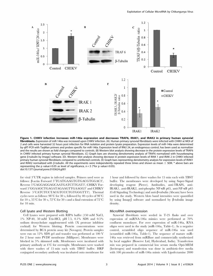
Figure 1. CHIKV infection increases miR-146a expression and decreases TRAF6, IRAK1, and IRAK2 in primary human synovialfibroblasts. Expression of miR-146a was increased upon CHIKV infection. (A). Human primary synovial fibroblasts were infected with CHIKV at MOI of2 and cells were harvested 32 hours post infection for RNA isolation and protein lysate preparation. Expression levels of miR-146a were determinedby qRT-PCR with TaqMan primers and probes specific for miR-146a. Expression level of RNU 24, an endogenous control, has been used as normalizerand the results are shown as fold changes compared to controls. (B) Western blot analysis showing decrease in the protein expression levels of TRAF6in CHIKV infected primary human synovial fibroblasts. (C) Graph bars are showing densitometry analysis of TRAF6 normalized with housekeepinggene b-tubulin by ImageJ software. (D). Western blot analysis showing decrease in protein expression levels of IRAK 1 and IRAK 2 in CHIKV infectedprimary human synovial fibroblasts compared to uninfected controls. (E) Graph bars representing densitometry analysis for expression levels of IRAK1and IRAK2 normalized with b-tubulin. All the experiments were independently repeated three times and shown as mean 6 SEM. * above bars arerepresenting the p value#0.05 as level of significance, n = 3 (*for p value#0.05).doi:10.1371/journal.pone.0103624.g001
Exploitation of Cellular MicroRNA by Chikungunya Virus
PLOS ONE | www.plosone.org 3 August 2014 | Volume 9 | Issue 8 | e103624
(Invitrogen) according to the manufacturer’s protocol. 100
picomoles of scrambled miR-146a mimic were transfected into
synovial fibroblasts as negative control. Transfection efficiency was
monitored by visualization of Green Fluorescent Reporter (GFP)
which was used as positive control for transfection procedures.
Cells were harvested for RNA isolation and protein lysate
preparation 48 hours post transfection. miR-146a over expression
was confirmed by qPCR using TaqMan assay specific to miR-
146a. Expression levels of the target proteins in transfected cells
were analyzed by Western blotting by using the corresponding
antibodies.
Anti-miR (miRNA inhibitor) transfectionSynovial fibroblasts were transfected with 100 picomoles of anti-
miR-146a (Ambion) and Cy3-labeled control anti-miR (Ambion)
with Lipofectamine 2000 as per the manufacturer’s protocols.
Efficiency of transfection was monitored by visualizing the
fluorescence of Cy3-labeled control anti-miR. Cells were harvested
for RNA isolation and protein lysate preparation 48 hours post
transfection. Knockdown of miR-146a in anti-miR transfected
cells were confirmed by quantitative real time PCR using TaqMan
assay specific to miR-146a. Protein expression levels of miR-146a
targets were analysed in the transfected cells by Western blotting
with the corresponding antibodies.
NF-kB Luciferase reporter assayFor reporter assay, HEK 293T cells were seeded in 6-well
culture dishes until ,70% confluency and transfected with 2 mg ofNF-kB -FLuc plasmid (a kind gift from Dr. Adolfo Garcia Sastre,
Mount Sinai School of Medicine, New York, USA) using
Lipofectamine 2000 as per the manufacturer’s protocol. For
infection experiments, 6–8 hours post transfection, cells were
infected with CHIKV at MOI 2 and harvested 32 hours post
infection to measure luciferase activity. For microRNA over
expression experiments, cells were co transfected with reporter
clone of NF-kB and miR-146a/scramble miR-146a mimic and
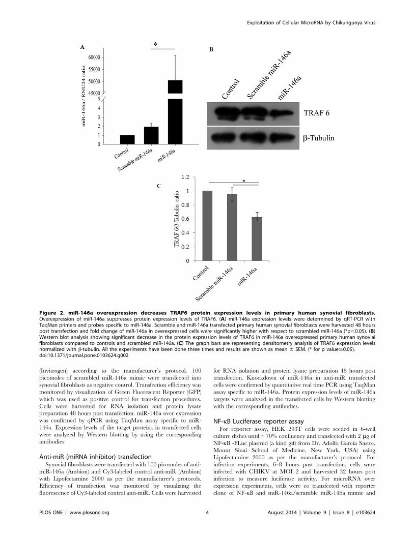
Figure 2. miR-146a overexpression decreases TRAF6 protein expression levels in primary human synovial fibroblasts.Overexpression of miR-146a suppresses protein expression levels of TRAF6. (A) miR-146a expression levels were determined by qRT-PCR withTaqMan primers and probes specific to miR-146a. Scramble and miR-146a transfected primary human synovial fibroblasts were harvested 48 hourspost transfection and fold change of miR-146a in overexpressed cells were significantly higher with respect to scrambled miR-146a (*p,0.05). (B)Western blot analysis showing significant decrease in the protein expression levels of TRAF6 in miR-146a overexpressed primary human synovialfibroblasts compared to controls and scrambled miR-146a. (C) The graph bars are representing densitometry analysis of TRAF6 expression levelsnormalized with b-tubulin. All the experiments have been done three times and results are shown as mean 6 SEM. (* for p value#0.05).doi:10.1371/journal.pone.0103624.g002
Exploitation of Cellular MicroRNA by Chikungunya Virus
PLOS ONE | www.plosone.org 4 August 2014 | Volume 9 | Issue 8 | e103624
harvested 48 hours post transfection to measure the luciferase
activity. In anti-miR experiment, 24 hour post transfection of
reporter plasmids along with anti-miR-146a, cells were infected
with CHIKV and lysates were prepared for luciferase activity.
Luciferase activity was determined as relative light unit by using
luciferase assay kit (Promega) according to the manufacturer’s
protocol and normalized with b-galactosidase expression by b-galactosidase assay kit (Promega).
Statistical analysisResults are expressed as mean 6 SEM from three independent
biologically repeated experiments. Level of significance (p values)
was determined by Student’s t test between treated group versus
untreated (control) group; considering p#0.05 as significant in
two-tailed student’s t-test by applying paired/equal/unequal
variance. The fold changes of miR-146a in treated groups have
been compared with untreated controls by using DD ct methods.
All the comparisons and statistical analysis have been done in
Microsoft excel.
Results
CHIKV infection increases the expression levels of miR-146a in primary human synovial fibroblastsThe roles of microRNAs in the modulation of immune
responses have been reported in various viral infections
[23,39,40]. miR-146a have been reported to play important roles
in the regulation of pro-inflammatory responses [36]. We
investigated the potential perturbation of miR-146a in primary
human synovial fibroblasts after CHIKV infection. Synovial
fibroblasts were infected with CHIKV at MOI of 2 for 32 hours.
Changes in target protein expression levels were initially measured
after 6, 12, 24 and 32 hours of CHIKV infection. However,
significant alterations in the expression pattern of specific target
genes (TRAF6, IRAK1 and IRAK2) were observed only after 32
hours of infection. Therefore, further studies were carried out at
32 hours post-infection. The cellular expression levels of miR-146a
were determined by real time PCR using TaqMan primers and
probes specific for miR-146a. The expression levels of miR-146a
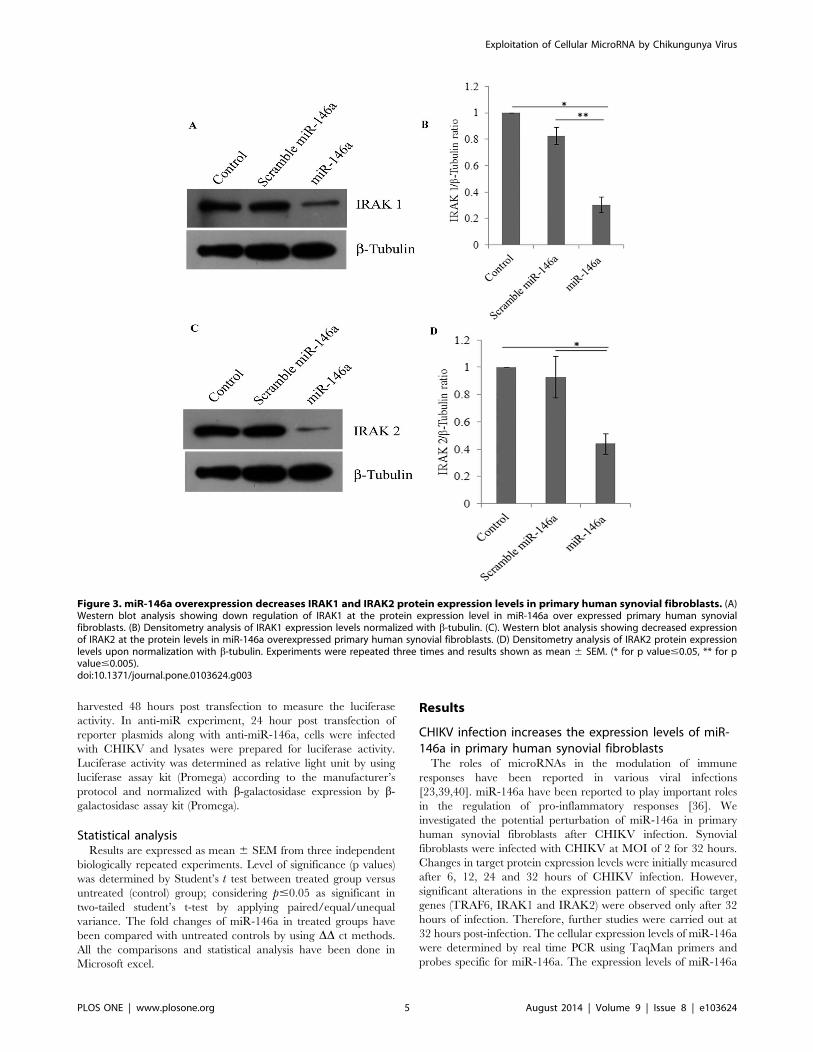
Figure 3. miR-146a overexpression decreases IRAK1 and IRAK2 protein expression levels in primary human synovial fibroblasts. (A)Western blot analysis showing down regulation of IRAK1 at the protein expression level in miR-146a over expressed primary human synovialfibroblasts. (B) Densitometry analysis of IRAK1 expression levels normalized with b-tubulin. (C). Western blot analysis showing decreased expressionof IRAK2 at the protein levels in miR-146a overexpressed primary human synovial fibroblasts. (D) Densitometry analysis of IRAK2 protein expressionlevels upon normalization with b-tubulin. Experiments were repeated three times and results shown as mean 6 SEM. (* for p value#0.05, ** for pvalue#0.005).doi:10.1371/journal.pone.0103624.g003
Exploitation of Cellular MicroRNA by Chikungunya Virus
PLOS ONE | www.plosone.org 5 August 2014 | Volume 9 | Issue 8 | e103624
in CHIKV infected primary human synovial fibroblasts were 1.77
fold higher, compared to uninfected controls (Figure-1A).
CHIKV infection down regulates TRAF6, IRAK1 and IRAK2expression level in primary human synovial fibroblastsTRAF6, IRAK1 and IRAK2 play major roles in regulation of
immune responses during viral infections. In CHIKV infected
primary human synovial fibroblasts, the expression levels of target
genes of miR-146a such as TRAF6, IRAK1 and IRAK2 were
analyzed after 32 hours of CHIKV infection. Significant
downregulation, (69% decrease in TRAF6, 65% decrease in
IRAK1 and 49% decrease in IRAK2) (p#0.05) at the protein
expression levels of TRAF6, IRAK1 and IRAK2 was observed in
CHIKV infected human synovial fibroblasts (Figure-1B, C, D, E).
miR-146a overexpression decreases TRAF6, IRAK1 andIRAK2 protein expression levelsIn primary human synovial fibroblasts, the mimics of miR-146a
were transfected exogenously to analyse the effect on the protein
expression levels of target genes. miR-146a mimics were trans-
fected at the concentration of 100 picomoles and scrambled miR-
146a were transfected as a negative control with same concentra-
tion of 100 picomoles. Scrambled miR-146a were transfected in
primary human synovial fibroblasts to demonstrate the specificity
of miR-146a in regulating the expression levels of target genes
through the binding of the complementary sequences of the seed
region of miRNA with the 39UTR of target mRNA. The
expression levels of miR-146a were significantly higher upto
50,000 fold in overexpressed cells (Figure-2A) (p#0.05); compared
to cells transfected with scrambled miR-146a (p#0.05). Significant
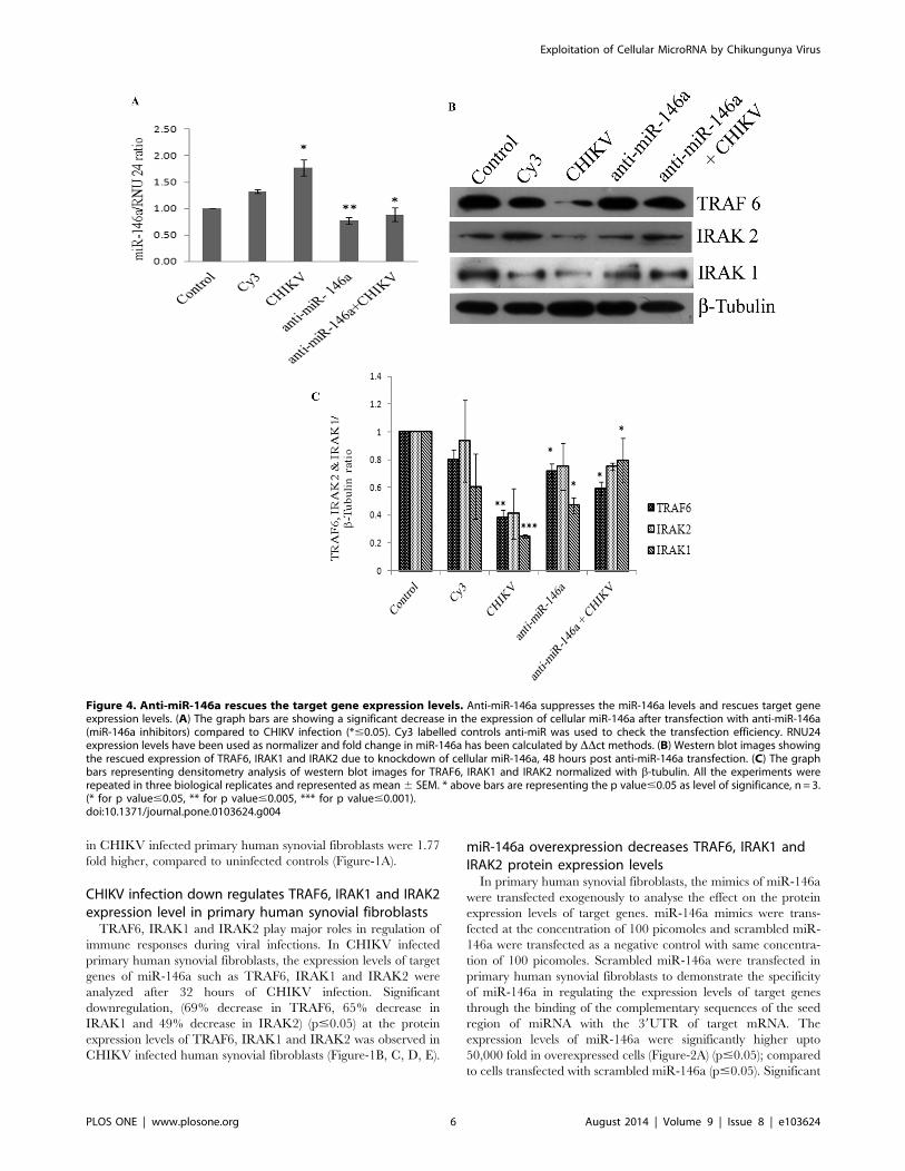
Figure 4. Anti-miR-146a rescues the target gene expression levels. Anti-miR-146a suppresses the miR-146a levels and rescues target geneexpression levels. (A) The graph bars are showing a significant decrease in the expression of cellular miR-146a after transfection with anti-miR-146a(miR-146a inhibitors) compared to CHIKV infection (*#0.05). Cy3 labelled controls anti-miR was used to check the transfection efficiency. RNU24expression levels have been used as normalizer and fold change in miR-146a has been calculated by DDct methods. (B) Western blot images showingthe rescued expression of TRAF6, IRAK1 and IRAK2 due to knockdown of cellular miR-146a, 48 hours post anti-miR-146a transfection. (C) The graphbars representing densitometry analysis of western blot images for TRAF6, IRAK1 and IRAK2 normalized with b-tubulin. All the experiments wererepeated in three biological replicates and represented as mean6 SEM. * above bars are representing the p value#0.05 as level of significance, n = 3.(* for p value#0.05, ** for p value#0.005, *** for p value#0.001).doi:10.1371/journal.pone.0103624.g004
Exploitation of Cellular MicroRNA by Chikungunya Virus
PLOS ONE | www.plosone.org 6 August 2014 | Volume 9 | Issue 8 | e103624
down regulation (38% decrease in TRAF6, 70% decrease in
IRAK1 and 56% decrease in IRAK2) was observed in protein
expression levels of TRAF6, IRAK1, and IRAK2 (Figure-2B,
Figure-3) in miR-146a overexpressed synovial fibroblast cells.
Transfection with scramble miR-146a did not show any significant
change in the protein expression levels of TRAF6, IRAK1, and
IRAK2.
Anti-miR-146a transfection rescues the expression levelsof TRAF6, IRAK1, and IRAK2 in CHIKV infected humansynovial fibroblastsThe knockdown studies of miR-146a were performed to
determine the direct regulatory role of miR-146a on the
expression levels of TRAF6, IRAK1 and IRAK2 and further to
demonstrate the potential of anti-miR-146a in rescuing the
expression levels of the target genes. Cy3-labelled control anti-
miR was used as a negative control as well as to visualize the
transfection efficiency. These are scrambled RNA oligo sequences
which are not possessing any complementary binding region
towards any of hosts mRNA unlike miRNA-146a inhibitor which
has a potential complementary region for seed sequences of
cellular miR-146a. Synovial fibroblasts transfected with anti-miR-
146a showed a significant suppression of cellular miR-146a
(Figure-4A). Primary human synovial fibroblasts having reduced
levels of miR-146a by anti-miR application were infected with
CHIKV and expression levels of TRAF6, IRAK1 and IRAK2
were analysed. Expression levels of TRAF6, IRAK1 and IRAK2
(Figure-4B, C) were rescued significantly in human synovial
fibroblasts transfected with anti-miR-146a. Synovial fibroblasts
transfected with anti-miR-146a followed by CHIKV infection
showed the expression patterns of miR-146a close to controls
(Figure-4A). The expression levels of TRAF6 and IRAK1 proteins
were rescued significantly and their sustained expression levels (p#
0.05) were observed even after CHIKV infection in primary
human synovial fibroblasts (Figure-4B, C). As compared to
CHIKV mediated downregulation of TRAF6 and IRAK1, anti-
miR-146a transfected cells show significant rescue (p#0.05) in
their expression level. This observation demonstrated that
CHIKV modulates the expression levels of miR-146a, which
ultimately suppresses the expression of target genes in primary
human synovial fibroblasts and such effect can be effectively
counter-balanced by suppressing the cellular levels of miR-146a
through microRNA inhibitors.
CHIKV suppresses NF-kB activation in primary humansynovial fibroblastsRegulation of miR-146a is NF-kB dependent and induced by
NF-kB activation; which can further down regulate TRAF6,
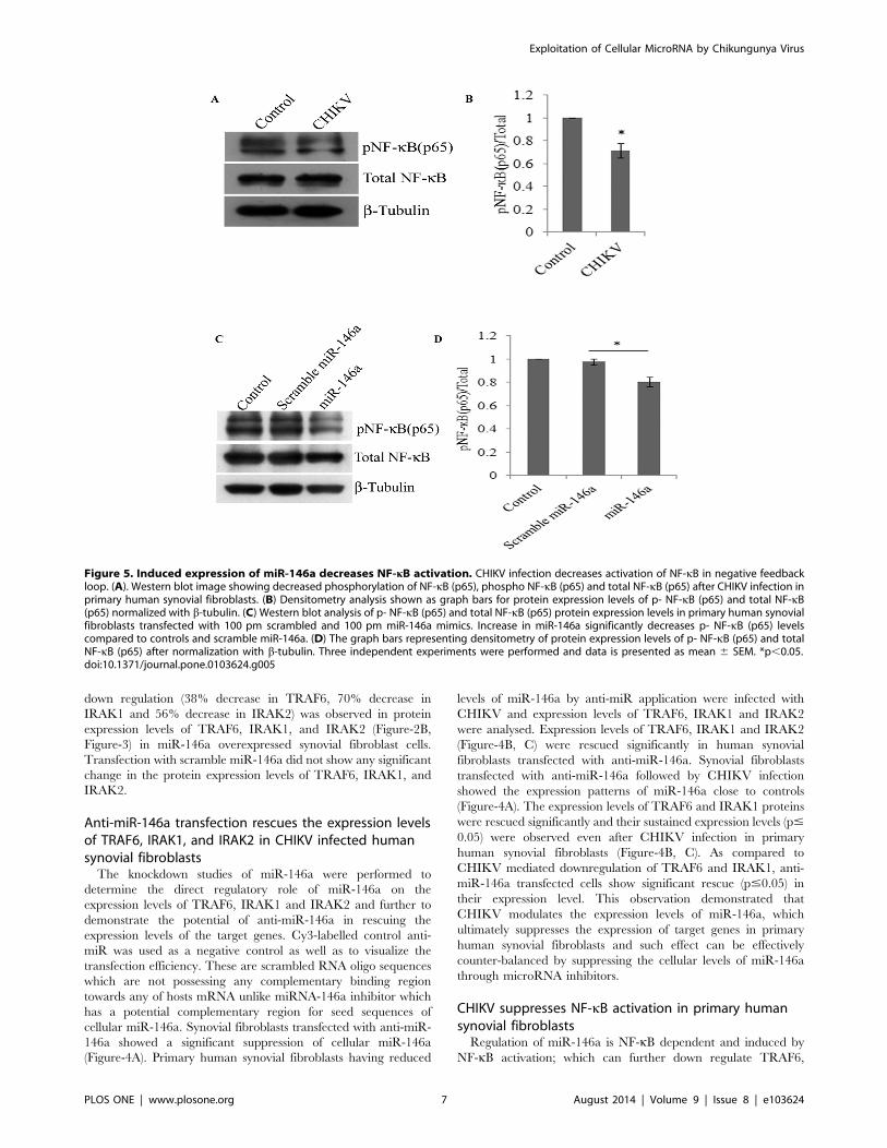
Figure 5. Induced expression of miR-146a decreases NF-kB activation. CHIKV infection decreases activation of NF-kB in negative feedbackloop. (A). Western blot image showing decreased phosphorylation of NF-kB (p65), phospho NF-kB (p65) and total NF-kB (p65) after CHIKV infection inprimary human synovial fibroblasts. (B) Densitometry analysis shown as graph bars for protein expression levels of p- NF-kB (p65) and total NF-kB(p65) normalized with b-tubulin. (C) Western blot analysis of p- NF-kB (p65) and total NF-kB (p65) protein expression levels in primary human synovialfibroblasts transfected with 100 pm scrambled and 100 pm miR-146a mimics. Increase in miR-146a significantly decreases p- NF-kB (p65) levelscompared to controls and scramble miR-146a. (D) The graph bars representing densitometry of protein expression levels of p- NF-kB (p65) and totalNF-kB (p65) after normalization with b-tubulin. Three independent experiments were performed and data is presented as mean 6 SEM. *p,0.05.doi:10.1371/journal.pone.0103624.g005
Exploitation of Cellular MicroRNA by Chikungunya Virus
PLOS ONE | www.plosone.org 7 August 2014 | Volume 9 | Issue 8 | e103624
IRAK1 and IRAK2. In this negative feed-back loop, NF-kB is a
downstream nuclear executor; which regulates the transcription of
various pro-inflammatory cytokines. We checked whether the
elevated levels of cellular miR-146a upon CHIKV infection can
suppress the expression and phosphorylation of NF-kB protein
subunits (p65) in primary human synovial fibroblasts. 30%
decrease in phosphorylation of NF-kB (p65) was observed (p#
0.05) in CHIKV infected synovial fibroblasts; compared to
controls (Figure-5A).
miR-146a overexpression decreases NF-kB activationPerturbation in TRAF6, IRAK1 and IRAK2 expression levels
and their downstream effects on NF-kB activation was investigated
in primary human synovial fibroblasts through overexpression of
miR-146a exogenously. The primary human synovial fibroblasts
were transfected with scrambled and miR-146a mimics. Phos-
phorylation of NF-kB (p65) was significantly decreased after miR-
146a overexpression as compared to the cells transfected with
scramble miR-146a (Figure-5C).
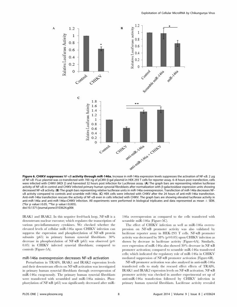
The effect of CHIKV infection as well as miR-146a overex-
pression on NF-kB promoter activity was also validated by
luciferase reporter assay in HEK-293 T cells. NF-kB promoter
activity was decreased by 38% (p#0.05) upon CHIKV infection as
shown by decrease in luciferase activity (Figure-6A). Similarly,
over expression of miR-146a also showed 30% decrease in NF-kBpromoter activation; compared to scramble miR-146a transfected
cells; which indicated the regulatory role of miR-146a in CHIKV
mediated suppression of NF-kB promoter activation (Figure-6B).
NF-kB promoter activation was also analysed in anti-miR-146a
transfected cells to study the rescued effect effects of TRAF6,
IRAK1 and IRAK2 expression levels on NF-kB activation. NF-kBpromoter activity was checked in another experimental set up of
anti-miR-146a transfection followed by CHIKV infection in
primary human synovial fibroblasts. Luciferase activity revealed
Figure 6. CHIKV suppresses NF-kB activity through miR-146a. Increase in miR-146a expression levels suppresses the activation of NF-kB. 2 mgof NF-kB -FLuc plasmid was co-transfected with 700 ng of pCMV-b-gal plasmid in HEK 293 T cells for reporter assay. 6–8 hours post transfection, cellswere infected with CHIKV (MOI 2) and harvested 32 hours post infection for Luciferase assay. (A) The graph bars are representing relative luciferaseactivity of NF-kB in control and CHIKV infected primary human synovial fibroblasts after normalization with b-galactosidase expression units showingdecreased NF-kB activity. (B) The graph bars representing relative luciferase units in miR-146a overexpression. Transfection of miR-146a decreases NF-kB activity compared to controls and scramble miR-146a. (C) HEK cells were infected with CHIKV after the 24 hours of anti-miR-146a transfection.Anti-miR-146a transfection rescues the activity of NF-kB even in cells infected with CHIKV. The graph bars are showing elevated luciferase activity inanti-miR-146a and anti-miR-146a+CHIKV infection. All experiments were performed in biological triplicates and data represented as mean 6 SEM.(*for p value#0.05, **for p value#0.005).doi:10.1371/journal.pone.0103624.g006
Exploitation of Cellular MicroRNA by Chikungunya Virus
PLOS ONE | www.plosone.org 8 August 2014 | Volume 9 | Issue 8 | e103624
significantly sustained activity of NF-kB (71% increase in anti-
miR-146a transfected cells and 79% increase in anti-miR-146a+CHIKV transfected cells) (p#0.005), 24 hours post transfection of
anti-miR-146a followed by CHIKV infection (Figure-6C). These
observations supported the negative regulatory role of miR-146a
upon NF-kB activity.
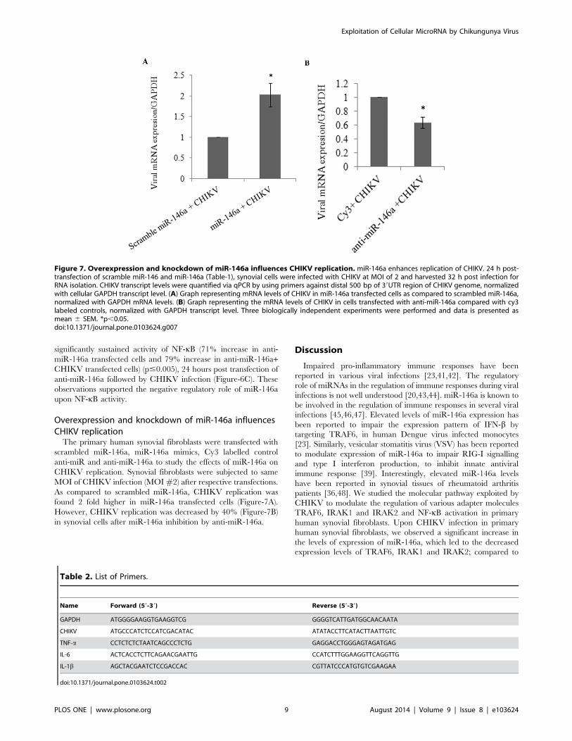
Overexpression and knockdown of miR-146a influencesCHIKV replicationThe primary human synovial fibroblasts were transfected with
scrambled miR-146a, miR-146a mimics, Cy3 labelled control
anti-miR and anti-miR-146a to study the effects of miR-146a on
CHIKV replication. Synovial fibroblasts were subjected to same
MOI of CHIKV infection (MOI#2) after respective transfections.
As compared to scrambled miR-146a, CHIKV replication was
found 2 fold higher in miR-146a transfected cells (Figure-7A).
However, CHIKV replication was decreased by 40% (Figure-7B)
in synovial cells after miR-146a inhibition by anti-miR-146a.
Discussion
Impaired pro-inflammatory immune responses have been
reported in various viral infections [23,41,42]. The regulatory
role of miRNAs in the regulation of immune responses during viral
infections is not well understood [20,43,44]. miR-146a is known to
be involved in the regulation of immune responses in several viral
infections [45,46,47]. Elevated levels of miR-146a expression has
been reported to impair the expression pattern of IFN-b by
targeting TRAF6, in human Dengue virus infected monocytes
[23]. Similarly, vesicular stomatitis virus (VSV) has been reported
to modulate expression of miR-146a to impair RIG-I signalling
and type I interferon production, to inhibit innate antiviral
immune response [39]. Interestingly, elevated miR-146a levels
have been reported in synovial tissues of rheumatoid arthritis
patients [36,48]. We studied the molecular pathway exploited by
CHIKV to modulate the regulation of various adapter molecules
TRAF6, IRAK1 and IRAK2 and NF-kB activation in primary
human synovial fibroblasts. Upon CHIKV infection in primary
human synovial fibroblasts, we observed a significant increase in
the levels of expression of miR-146a, which led to the decreased
expression levels of TRAF6, IRAK1 and IRAK2; compared to
Figure 7. Overexpression and knockdown of miR-146a influences CHIKV replication. miR-146a enhances replication of CHIKV. 24 h post-transfection of scramble miR-146 and miR-146a (Table-1), synovial cells were infected with CHIKV at MOI of 2 and harvested 32 h post infection forRNA isolation. CHIKV transcript levels were quantified via qPCR by using primers against distal 500 bp of 39UTR region of CHIKV genome, normalizedwith cellular GAPDH transcript level. (A) Graph representing mRNA levels of CHIKV in miR-146a transfected cells as compared to scrambled miR-146a,normalized with GAPDH mRNA levels. (B) Graph representing the mRNA levels of CHIKV in cells transfected with anti-miR-146a compared with cy3labeled controls, normalized with GAPDH transcript level. Three biologically independent experiments were performed and data is presented asmean 6 SEM. *p,0.05.doi:10.1371/journal.pone.0103624.g007
Table 2. List of Primers.
Name Forward (59-39) Reverse (59-39)
GAPDH ATGGGGAAGGTGAAGGTCG GGGGTCATTGATGGCAACAATA
CHIKV ATGCCCATCTCCATCGACATAC ATATACCTTCATACTTAATTGTC
TNF-a CCTCTCTCTAATCAGCCCTCTG GAGGACCTGGGAGTAGATGAG
IL-6 ACTCACCTCTTCAGAACGAATTG CCATCTTTGGAAGGTTCAGGTTG
IL-1b AGCTACGAATCTCCGACCAC CGTTATCCCATGTGTCGAAGAA
doi:10.1371/journal.pone.0103624.t002
Exploitation of Cellular MicroRNA by Chikungunya Virus
PLOS ONE | www.plosone.org 9 August 2014 | Volume 9 | Issue 8 | e103624

controls (Figure-1A–E). Exogenous over expression of miR-146a
in primary human synovial fibroblasts resulted into significant
increase in miR-146a (Figure-2A) levels and decreased protein
expression levels of TRAF6 (Figure-2B), IRAK1 and IRAK2
(Figure-3). Transfection with scrambled miR-146a did not exhibit
any significant change in the levels of expression of miR-146a as
well as target genes TRAF6, IRAK1 and IRAK2 (Figure-3). These
observations were in accordance with previous reports, on the
direct targeting of TRAF6, IRAK1 and IRAK2 by complemen-
tary binding between 39UTR of genes and seed region of miR-
146a. Anand Iyer, et.al 2012 reported that miR-146a mimics can
significantly reduce the mRNA expression levels of TRAF6,
IRAK1 and IRAK2 protein levels in IL-1b stimulated human
astrocytes and glioblastoma cell line [49]. The induced expression
of miR-146a by LMP1 mediated activation of NF-kB during EBV
infection in blood cells inhibits NF-kB by down regulation of
TRAF6 [25,40]. Recently, Jin Ho Paik, et.al 2011, also reported
that miR-146a may function as a tumor-suppressor in NK/T cell
lymphoma by down regulating the NF-kB through targeting
TRAF6 [50].
To further delineate the specific role of miR-146a in regulation
of TRAF6, IRAK1 and IRAK2, inhibitor of miR-146a, referred
here as anti-miR-146a was transfected in primary human synovial
fibroblasts. Anti-miR-146a has the complementary sequences of
miR-146a, which resulted into the sequestration of the mature
cellular miR-146a into the synovial fibroblast cells. We observed
that the suppression of cellular miR-146a levels (Figure-4A) led to
the regain in expression levels of TRAF6, IRAK1 and IRAK2
protein expression levels (Figure-4B, C). The ability of anti-miR-
146a in rescuing the expression levels of these proteins were
further elucidated by using anti-miR-146a transfected synovial
fibroblast cells followed by infection with CHIKV. The expression
levels of TRAF6, IRAK1 and IRAK2 were maintained even in
presence of CHIKV (Figure-4B, C). These findings clearly
demonstrated that the CHIKV mediated suppression of TRAF6,
IRAK1 and IRAK2 occurs through upregulation of miR-146a in
primary human synovial fibroblasts.
Cytokine signalling through TRAF6, IRAK1 and IRAK2 leads
to activation of NF-kB pathway. This triggers the translocation of
NF-kB into nucleus and transcriptional activation of set of genes
critical for immune response and inflammatory events [45]. To
emphasize the specificity and exploitation of miR-146a as negative
feedback tool for suppression of NF-kB activity by CHIKV, we
studied the activation of NF-kB in CHIKV infected primary
human synovial fibroblasts. Reduced phosphorylation levels of
NF-kB (p65) and decreased NF-kB activity were observed in
CHIKV infected synovial fibroblasts respectively (Figure-5A,
Figure-6A).
Similarly, miR-146a over expression led to the decreased
phosphorylated form of NF-kB (p65); compared to scramble miR-
146a transfected cells (Figure-5C, D). Decreased luciferase activity
was observed in miR-146a overexpressed cells in NF-kB promoter
Figure 8. CHIKV mediated regulation of NF-kB by miR-146a modulation in primary human synovial fibroblasts. CHIKV infectioninduces the expression of cellular miR-146a in primary human synovial fibroblasts, which in turn downregulates the expression of TRAF6, IRAK1 andIRAK2. Decreased expression of these immune modulators results into reduced NF-kB phosphorylation and activation in primary human synovialfibroblasts.doi:10.1371/journal.pone.0103624.g008
Exploitation of Cellular MicroRNA by Chikungunya Virus
PLOS ONE | www.plosone.org 10 August 2014 | Volume 9 | Issue 8 | e103624

assay (Figure-6B). These findings confirmed that the regulation of
NF-kB activation is mediated via miR-146a in CHIKV infected
human synovial fibroblasts. NF-kB promoter assay showed
restored levels of NF-kB activity in the synovial fibroblasts
transfected with anti-miR-146a (Figure-6C). Significantly an
enhanced level of activation of NF-kB was observed even in
CHIKV infected cells at same MOI (Figure-6C). These results
were compared with down regulation of NF-kB promoter activity
in CHIKV infection to highlight the role of cellular miR-146a in
maintaining the NF-kB activity. We demonstrated that CHIKV
exploits the negative regulatory loop, in which early NF-kBactivation induces miR-146a expression; which results into down-
regulation of TRAF6, IRAK1 and IRAK2 to further restrain the
activity of NF-kB [45]. Negative regulatory function of miR-146a
has been reported in estrogen treated splenic lymphocytes; where
decrease in miR-146a led to increase in LPS induced IFN-c and
iNOS production through TLR signalling [51]. VSV infections
have also been reported to utilize negative feedback pathway of
miR-146a to regulate type I interferon production [39]. A general
induction of the nuclear transcription factor kB (NF-kB) plays anessential role in stimulating the expression of inflammatory genes;
which are particularly involved in the progression of inflammatory
diseases like arthritis [52]. The general repression of NF-kBactivity as well as defective type I interferon response through the
targeting of common adapter molecules TRAF6, IRAK1, IRAK2
by CHIKV explains the susceptibility of neonates and elderly (low
immune strength) for CHIKV infection [4,10]. Our results showed
an enhanced rate of CHIKV replication in presence of higher level
of miR-146a (miR-146a overexpression). In contrast, when cellular
miR-146a levels were inhibited by application of anti-miR-146a,
CHIKV replication decreased (Figure-7), which explained the
exploitation of miR146a mediated repression of NF-kB activity in
favour of increasing viral replication and aggravating pathological
manifestations during CHIKV infections. The expression of
inflammatory cytokines (TNF-a, IL6 and IL-1b) (Table-2)
decreased in miR-146a overexpressing cells. In contrast the
expression of inflammatory cytokines was induced in anti-miR-
146a transfected cells (Figure S1). This indicated a general
suppression of NF-kB mediated cytokine generation in CHIKV
infected synovial fibroblasts, which might be a strategy utilized by
CHIKV to facilitate their replication.
In summary, our study confirms that, CHIKV induces the
expression levels of miR-146a in primary human synovial
fibroblasts; which in turn suppresses TRAF6, IRAK1, IRAK2
expression levels and downstream NF-kB activity through negative
feedback loop (Figure-8). CHIKV utilizes the miR-146a, as a
negative regulator of general antiviral response. This study
provides an understanding about the immune response of human
synovial fibroblasts in CHIKV infection and insights into immune
evasion strategies adapted by CHIKV.
Supporting Information
Figure S1 Overexpression and knockdown of miR-146a affects
TNF-a, IL-6 and IL-1b transcript level. miR-146a suppress the
transcript levels of TNF-a, IL-6 and IL-1b upon CHIKV infection
(Table-2). (A) Graph bar representing the mRNA levels of TNF-ain synovial fibroblast cells upon overexpression and knockdown of
miR-146a. (B) Graph bar showing changes in transcript level of
IL-6 in synovial fibroblast cells upon overexpression and
knockdown of miR-146a. (C) Bar diagram indicating the changes
in transcript level of IL-1b upon overexpression and knockdown of
miR-146a in primary synovial fibroblast cells. Cytokine transcript
level detection was done by qPCR normalized with GAPDH
transcript level (Table-2). All the experiments were repeated three
times and results shown as mean 6 SEM.
(TIF)
Acknowledgments
Authors are thankful to Prof. Adolfo Garcia-Sastre (AGS), Department of
Medicine and Microbiology, Mount Sinai School of Medicine, New York,
USA; for providing the NF-kB -FLuc plasmid as a kind gift. We are
thankful to Prof. Duane Gubler (DG) (Emerging Infectious Disease
Program, Duke-NUS Medical School, Singapore) for providing the
CHIKV (Ross strain, E1:A226) as a kind gift. We are thankful to Ms.
Vaishnavi Jadhav for her support in preparation of this manuscript.
Authors are also thankful to the director, Centre for Cellular and
Molecular Biology (CCMB), Hyderabad for his support.
Author Contributions
Conceived and designed the experiments: SPS. Performed the experi-
ments: SPS RM. Analyzed the data: SPS RM. Contributed to the writing
of the manuscript: SKS.
References
1. Kam YW, Ong EK, Renia L, Tong JC, Ng LF (2009) Immuno-biology of
Chikungunya and implications for disease intervention. Microbes Infect 11:
1186–1196.
2. Singh SK, Unni SK (2011) Chikungunya virus: host pathogen interaction. Rev
Med Virol 21: 78–88.
3. Calisher CH, Shope RE, Brandt W, Casals J, Karabatsos N, et al. (1980)
Proposed antigenic classification of registered arboviruses I. Togaviridae,
Alphavirus. Intervirology 14: 229–232.
4. Abraham R, Mudaliar P, Padmanabhan A, Sreekumar E (2013) Induction of
cytopathogenicity in human glioblastoma cells by chikungunya virus. PLoS One
8: e75854.
5. Robinson MC (1955) An epidemic of virus disease in Southern Province,
Tanganyika Territory, in 1952–53. I. Clinical features. Trans R Soc Trop Med
Hyg 49: 28–32.
6. Lumsden WH (1955) An epidemic of virus disease in Southern Province,
Tanganyika Territory, in 1952–53. II. General description and epidemiology.
Trans R Soc Trop Med Hyg 49: 33–57.
7. Schwartz O, Albert ML (2010) Biology and pathogenesis of chikungunya virus.
Nat Rev Microbiol 8: 491–500.
8. Labadie K, Larcher T, Joubert C, Mannioui A, Delache B, et al. (2010)
Chikungunya disease in nonhuman primates involves long-term viral persistence
in macrophages. J Clin Invest 120: 894–906.
9. Tesh RB (1982) Arthritides caused by mosquito-borne viruses. Annu Rev Med
33: 31–40.
10. Couderc T, Chretien F, Schilte C, Disson O, Brigitte M, et al. (2008) A mouse
model for Chikungunya: young age and inefficient type-I interferon signaling are
risk factors for severe disease. PLoS Pathog 4: e29.
11. Gardner J, Anraku I, Le TT, Larcher T, Major L, et al. (2010) Chikungunya
virus arthritis in adult wild-type mice. J Virol 84: 8021–8032.
12. Teo TH, Lum FM, Lee WW, Ng LF (2012) Mouse models for Chikungunya
virus: deciphering immune mechanisms responsible for disease and pathology.
Immunol Res 53: 136–147.
13. Borgherini G, Poubeau P, Staikowsky F, Lory M, Le Moullec N, et al. (2007)
Outbreak of chikungunya on Reunion Island: early clinical and laboratory
features in 157 adult patients. Clin Infect Dis 44: 1401–1407.
14. Lakshmi V, Neeraja M, Subbalaxmi MV, Parida MM, Dash PK, et al. (2008)
Clinical features and molecular diagnosis of Chikungunya fever from South
India. Clin Infect Dis 46: 1436–1442.
15. Krejbich-Trotot P, Denizot M, Hoarau JJ, Jaffar-Bandjee MC, Das T, et al.
(2011) Chikungunya virus mobilizes the apoptotic machinery to invade host cell
defenses. FASEB J 25: 314–325.
16. Schilte C, Couderc T, Chretien F, Sourisseau M, Gangneux N, et al. (2010)
Type I IFN controls chikungunya virus via its action on nonhematopoietic cells.
J Exp Med 207: 429–442.
17. Thon-Hon VG, Denizot M, Li-Pat-Yuen G, Giry C, Jaffar-Bandjee MC, et al.
(2012) Deciphering the differential response of two human fibroblast cell lines
following Chikungunya virus infection. Virol J 9: 213.
18. Ambros V (2004) The functions of animal microRNAs. Nature 431: 350–355.
19. Singh SK, Pal Bhadra M, Girschick HJ, Bhadra U (2008) MicroRNAs–micro in
size but macro in function. FEBS J 275: 4929–4944.
Exploitation of Cellular MicroRNA by Chikungunya Virus
PLOS ONE | www.plosone.org 11 August 2014 | Volume 9 | Issue 8 | e103624
20. Bartel DP (2004) MicroRNAs: genomics, biogenesis, mechanism, and function.
Cell 116: 281–297.21. Bushati N, Cohen SM (2007) microRNA functions. Annu Rev Cell Dev Biol 23:
175–205.
22. Gottwein E, Cullen BR (2008) Viral and cellular microRNAs as determinants ofviral pathogenesis and immunity. Cell Host Microbe 3: 375–387.
23. Wu S, He L, Li Y, Wang T, Feng L, et al. (2013) miR-146a facilitates replicationof dengue virus by dampening interferon induction by targeting TRAF6. J Infect
67: 329–341.
24. Rusca N, Monticelli S (2011) MiR-146a in Immunity and Disease. Mol Biol Int2011: 437301.
25. Taganov KD, Boldin MP, Chang KJ, Baltimore D (2006) NF-kappaB-dependent induction of microRNA miR-146, an inhibitor targeted to signaling
proteins of innate immune responses. Proc Natl Acad Sci U S A 103: 12481–12486.
26. Tsitsiou E, Lindsay MA (2009) microRNAs and the immune response. Curr
Opin Pharmacol 9: 514–520.27. Perry MM, Moschos SA, Williams AE, Shepherd NJ, Larner-Svensson HM, et
al. (2008) Rapid changes in microRNA-146a expression negatively regulate theIL-1beta-induced inflammatory response in human lung alveolar epithelial cells.
J Immunol 180: 5689–5698.
28. Lukiw WJ, Zhao Y, Cui JG (2008) An NF-kappaB-sensitive micro RNA-146a-mediated inflammatory circuit in Alzheimer disease and in stressed human brain
cells. J Biol Chem 283: 31315–31322.29. Williams AE, Perry MM, Moschos SA, Larner-Svensson HM, Lindsay MA
(2008) Role of miRNA-146a in the regulation of the innate immune responseand cancer. Biochem Soc Trans 36: 1211–1215.
30. Nahid MA, Pauley KM, Satoh M, Chan EK (2009) miR-146a is critical for
endotoxin-induced tolerance: IMPLICATION IN INNATE IMMUNITY.J Biol Chem 284: 34590–34599.
31. Jensen LE, Muzio M, Mantovani A, Whitehead AS (2000) IL-1 signalingcascade in liver cells and the involvement of a soluble form of the IL-1 receptor
accessory protein. J Immunol 164: 5277–5286.
32. Ye H, Arron JR, Lamothe B, Cirilli M, Kobayashi T, et al. (2002) Distinctmolecular mechanism for initiating TRAF6 signalling. Nature 418: 443–447.
33. Bhaumik D, Scott GK, Schokrpur S, Patil CK, Campisi J, et al. (2008)Expression of microRNA-146 suppresses NF-kappaB activity with reduction of
metastatic potential in breast cancer cells. Oncogene 27: 5643–5647.34. Pauley KM, Satoh M, Chan AL, Bubb MR, Reeves WH, et al. (2008)
Upregulated miR-146a expression in peripheral blood mononuclear cells from
rheumatoid arthritis patients. Arthritis Res Ther 10: R101.35. Xu WD, Lu MM, Pan HF, Ye DQ (2012) Association of MicroRNA-146a with
autoimmune diseases. Inflammation 35: 1525–1529.36. Nakasa T, Miyaki S, Okubo A, Hashimoto M, Nishida K, et al. (2008)
Expression of microRNA-146 in rheumatoid arthritis synovial tissue. Arthritis
Rheum 58: 1284–1292.37. Stanczyk J, Pedrioli DM, Brentano F, Sanchez-Pernaute O, Kolling C, et al.
(2008) Altered expression of MicroRNA in synovial fibroblasts and synovialtissue in rheumatoid arthritis. Arthritis Rheum 58: 1001–1009.
38. Nakaya HI, Gardner J, Poo YS, Major L, Pulendran B, et al. (2012) Gene
profiling of Chikungunya virus arthritis in a mouse model reveals significant
overlap with rheumatoid arthritis. Arthritis Rheum 64: 3553–3563.
39. Hou J, Wang P, Lin L, Liu X, Ma F, et al. (2009) MicroRNA-146a feedback
inhibits RIG-I-dependent Type I IFN production in macrophages by targeting
TRAF6, IRAK1, and IRAK2. J Immunol 183: 2150–2158.
40. Cameron JE, Yin Q, Fewell C, Lacey M, McBride J, et al. (2008) Epstein-Barr
virus latent membrane protein 1 induces cellular MicroRNA miR-146a, a
modulator of lymphocyte signaling pathways. J Virol 82: 1946–1958.
41. Kandasamy S, Chattha KS, Vlasova AN, Saif LJ (2013) Prenatal vitamin A
deficiency impairs adaptive immune responses to pentavalent rotavirus vaccine
(RotaTeq) in a neonatal gnotobiotic pig model. Vaccine 23: 816–824.
42. Shi X, Zhou W, Huang H, Zhu H, Zhou P, et al. (2013) Inhibition of the
inflammatory cytokine tumor necrosis factor-alpha with etanercept provides
protection against lethal H1N1 influenza infection in mice. Crit Care 17: R301.
43. Zhuo Y, Gao G, Shi JA, Zhou X, Wang X (2013) miRNAs: biogenesis, origin
and evolution, functions on virus-host interaction. Cell Physiol Biochem 32:
499–510.
44. Sarma NJ, Tiriveedhi V, Crippin JS, Chapman WC, Mohanakumar T (2014)
Hepatitis C Virus Induced Changes in miRNA-107 and miRNA-449a Modulate
CCL2 by Targeting IL6 Receptor Complex in Hepatitis. J Virol 88: 3733–3743.
45. Ma X, Becker Buscaglia LE, Barker JR, Li Y (2011) MicroRNAs in NF-kappaB
signaling. J Mol Cell Biol 3: 159–166.
46. Koval’chuk LV, Gankovskaia LV, Akimova EA (2009) [Role of microRNA in
regulation of innate immunity mechanisms]. Zh Mikrobiol Epidemiol Im-
munobiol: 100–104.
47. Labbaye C, Testa U (2012) The emerging role of MIR-146A in the control of
hematopoiesis, immune function and cancer. J Hematol Oncol 5: 13.
48. Nakasa T, Shibuya H, Nagata Y, Niimoto T, Ochi M (2011) The inhibitory
effect of microRNA-146a expression on bone destruction in collagen-induced
arthritis. Arthritis Rheum 63: 1582–1590.
49. Iyer A, Zurolo E, Prabowo A, Fluiter K, Spliet WG, et al. (2012) MicroRNA-
146a: a key regulator of astrocyte-mediated inflammatory response. PLoS One
7: e44789.
50. Paik JH, Jang JY, Jeon YK, Kim WY, Kim TM, et al. (2011) MicroRNA-146a
downregulates NFkappaB activity via targeting TRAF6 and functions as a tumor
suppressor having strong prognostic implications in NK/T cell lymphoma. Clin
Cancer Res 17: 4761–4771.
51. Dai R, Phillips RA, Zhang Y, Khan D, Crasta O, et al. (2008) Suppression of
LPS-induced Interferon-gamma and nitric oxide in splenic lymphocytes by select
estrogen-regulated microRNAs: a novel mechanism of immune modulation.
Blood 112: 4591–4597.
52. Sehnert B, Burkhardt H, Wessels JT, Schroder A, May MJ, et al. (2013) NF-
kappaB inhibitor targeted to activated endothelium demonstrates a critical role
of endothelial NF-kappaB in immune-mediated diseases. Proc Natl Acad
Sci U S A 110: 16556–16561.
Exploitation of Cellular MicroRNA by Chikungunya Virus
PLOS ONE | www.plosone.org 12 August 2014 | Volume 9 | Issue 8 | e103624