Characteristics of MR Imaging for Staging and Survival ...
9
1 Magnetic Resonance in Medical Sciences Characteristics of MR Imaging for Staging and Survival Analysis of Neuroendocrine Carcinoma of the Endometrium: A Multicenter Study in Japan Kazuhiro Kitajima 1,* , Takako Kihara 2 , Yusuke Kawanaka 1 , Junko Takahama 3 , Yoshiko Ueno 4 , Takamichi Murakami 4 , Kotaro Yoshida 5 , Fumi Kato 6 , Akiko Takahata 7 , Yoshihiko Fukukura 8 , Jiro Munechika 9 , Yasunari Fujinaga 10 , Takeru Fukunaga 11 , Masahiro Tanabe 12 , Yuichiro Kanie 13 , Ayumu Kido 14 , Tsutomu Tamada 14 , Rika Yoshida 15 , Yuki Kamishima 16 , and Koichiro Yamakado 1 Purpose: This study aimed to examine MRI features and staging of neuroendocrine carcinoma (NEC) of the endometrium and evaluate survival. Materials and Methods: Clinical data, pathological, and preoperative pelvic MRI findings in 22 patients with histologically surgery-proven endometrial NEC were retrospectively reviewed. Tumors were pure NEC (n = 10) or mixed histotype (n = 12), with 13 large and nine small cell type. Results: International Federation of Gynecology and Obstetrics (FIGO) staging was I, II, III, and IV in 6, 2, 12, and 2 patients, respectively. In 13 (76.4%) of 17 patients with pathological deep myometrial invasion, MRI showed abnormal diffusely infiltrative high T 2 signal intensity throughout the myometrium with loss of normal uterine architecture. All tumors had restricted diffusion (apparent diffusion coefficient map low signal intensity, diffusion weighted imaging high signal intensity). Accuracy of T staging by MRI for all cases was 81.8%, with reference to pathology staging, while patient-based sensitivity, specificity, and accuracy for detecting metastatic pelvic lymph nodes was 60.0%, 100%, and 77.8%, respectively. Two intrapelvic peritoneal dissemination cases were detected by MRI. During follow-up (mean 30.4, range 3.3–138.4 months), 16 patients (72.7%) experienced recurrence and 12 (54.5%) died of disease. Two-year disease-free and overall survival rates for FIGO I, II, III, and IV were 66.7% and 83.3%, 50% and 100%, 10% and 33.3%, and 0% and 0%, respectively. Conclusion: Abnormal diffusely infiltrative high T 2 signal intensity throughout the myometrium with normal uterine architecture loss and obvious restricted diffusion throughout the tumor are suggestive features of endometrial NEC. Pelvic MRI is reliable for intrapelvic staging of affected patients. Keywords: endometrial cancer, magnetic resonance imaging, neuroendocrine carcinoma, survival Published Online: July 22, 2020 Magn Reson Med Sci 2020; XX; XXX–XXX doi:10.2463/mrms.mp.2020-0056 MAJOR PAPER 1 Department of Radiology, Hyogo College of Medicine, Hyogo, Japan 2 Department of Surgical Pathology, Hyogo College of Medicine, Hyogo, Japan 3 Department of Radiology, Nara Medical University, Nara, Japan 4 Department of Radiology, Kobe University Graduate School of Medicine, Hyogo, Japan 5 Department of Radiology, Kanazawa University, Graduate School of Medicine Science, Ishikawa, Japan Received: March 28, 2020 | Accepted: May 13, 2020 6 Department of Diagnostic and Interventional Radiology, Hokkaido University Hospital, Hokkaido, Japan 7 Department of Radiology, Kyoto Prefectural University of Medicine, Kyoto, Japan 8 Department of Radiology, Graduate School of Medical and Dental Sciences, Kagoshima University, Kagoshima, Japan 9 Department of Radiology, Showa University School of Medicine, Tokyo, Japan 10 Department of Radiology, Shinshu University School of Medicine, Nagano, Japan 11 Department of Pathophysiological and Therapeutic Sciences, Tottori University, Tottori, Japan 12 Department of Radiology, Yamaguchi University Graduate School of Medicine, Yamaguchi, Japan 13 Department of Radiology, Japanese Red Cross Society Himeji Hospital, Hyogo, Japan 14 Department of Radiology, Kawasaki Medical School, Okayama, Japan 15 Department of Radiology, Faculty of Medicine, Shimane University, Shimane, Japan 16 Department of Radiology, Nagoya City West Medical Center, Aichi, Japan * Corresponding author: Department of Radiology, Hyogo College of Medicine, 1-1, Mukogawa-cho, Nishinomiya, Hyogo 663-8501, Japan. Phone: +81-798-45- 6362, Fax: +81-798-45-6361, E-mail: [email protected] ©2020 Japanese Society for Magnetic Resonance in Medicine This work is licensed under a Creative Commons Attribution-NonCommercial- NoDerivatives International License.
Transcript of Characteristics of MR Imaging for Staging and Survival ...
1Magnetic Resonance in Medical Sciences
Characteristics of MR Imaging for Staging and Survival Analysis of Neuroendocrine Carcinoma of the Endometrium:
A Multicenter Study in Japan
Kazuhiro Kitajima1,*, Takako Kihara2, Yusuke Kawanaka1, Junko Takahama3, Yoshiko Ueno4, Takamichi Murakami4, Kotaro Yoshida5, Fumi Kato6,
Akiko Takahata7, Yoshihiko Fukukura8, Jiro Munechika9, Yasunari Fujinaga10, Takeru Fukunaga11, Masahiro Tanabe12, Yuichiro Kanie13, Ayumu Kido14,
Tsutomu Tamada14, Rika Yoshida15, Yuki Kamishima16, and Koichiro Yamakado1
Purpose: This study aimed to examine MRI features and staging of neuroendocrine carcinoma (NEC) of the endometrium and evaluate survival.Materials and Methods: Clinical data, pathological, and preoperative pelvic MRI findings in 22 patients with histologically surgery-proven endometrial NEC were retrospectively reviewed. Tumors were pure NEC (n = 10) or mixed histotype (n = 12), with 13 large and nine small cell type.Results: International Federation of Gynecology and Obstetrics (FIGO) staging was I, II, III, and IV in 6, 2, 12, and 2 patients, respectively. In 13 (76.4%) of 17 patients with pathological deep myometrial invasion, MRI showed abnormal diffusely infiltrative high T2 signal intensity throughout the myometrium with loss of normal uterine architecture. All tumors had restricted diffusion (apparent diffusion coefficient map low signal intensity, diffusion weighted imaging high signal intensity). Accuracy of T staging by MRI for all cases was 81.8%, with reference to pathology staging, while patient-based sensitivity, specificity, and accuracy for detecting metastatic pelvic lymph nodes was 60.0%, 100%, and 77.8%, respectively. Two intrapelvic peritoneal dissemination cases were detected by MRI. During follow-up (mean 30.4, range 3.3–138.4 months), 16 patients (72.7%) experienced recurrence and 12 (54.5%) died of disease. Two-year disease-free and overall survival rates for FIGO I, II, III, and IV were 66.7% and 83.3%, 50% and 100%, 10% and 33.3%, and 0% and 0%, respectively.Conclusion: Abnormal diffusely infiltrative high T2 signal intensity throughout the myometrium with normal uterine architecture loss and obvious restricted diffusion throughout the tumor are suggestive features of endometrial NEC. Pelvic MRI is reliable for intrapelvic staging of affected patients.Keywords: endometrial cancer, magnetic resonance imaging, neuroendocrine carcinoma, survival
Published Online: July 22, 2020Magn Reson Med Sci 2020; XX; XXX–XXXdoi:10.2463/mrms.mp.2020-0056
MAJOR PAPER
1Department of Radiology, Hyogo College of Medicine, Hyogo, Japan2Department of Surgical Pathology, Hyogo College of Medicine, Hyogo, Japan3Department of Radiology, Nara Medical University, Nara, Japan4Department of Radiology, Kobe University Graduate School of Medicine, Hyogo, Japan5Department of Radiology, Kanazawa University, Graduate School of Medicine Science, Ishikawa, Japan
Received: March 28, 2020 | Accepted: May 13, 2020
6Department of Diagnostic and Interventional Radiology, Hokkaido University Hospital, Hokkaido, Japan7Department of Radiology, Kyoto Prefectural University of Medicine, Kyoto, Japan8Department of Radiology, Graduate School of Medical and Dental Sciences, Kagoshima University, Kagoshima, Japan9Department of Radiology, Showa University School of Medicine, Tokyo, Japan10Department of Radiology, Shinshu University School of Medicine, Nagano, Japan11Department of Pathophysiological and Therapeutic Sciences, Tottori University, Tottori, Japan12Department of Radiology, Yamaguchi University Graduate School of Medicine, Yamaguchi, Japan13Department of Radiology, Japanese Red Cross Society Himeji Hospital, Hyogo, Japan14Department of Radiology, Kawasaki Medical School, Okayama, Japan15Department of Radiology, Faculty of Medicine, Shimane University, Shimane, Japan16Department of Radiology, Nagoya City West Medical Center, Aichi, Japan
*Corresponding author: Department of Radiology, Hyogo College of Medicine, 1-1, Mukogawa-cho, Nishinomiya, Hyogo 663-8501, Japan. Phone: +81-798-45-6362, Fax: +81-798-45-6361, E-mail: [email protected]
©2020 Japanese Society for Magnetic Resonance in MedicineThis work is licensed under a Creative Commons Attribution-NonCommercial- NoDerivatives International License.

2 Magnetic Resonance in Medical Sciences
K. Kitajima et al.
IntroductionA neuroendocrine carcinoma (NEC) of the female genital tract is an aggressive uncommon tumor usually involving the uterine cervix and ovaries, while endometrium occur-rence is very rare.1 An NEC of the uterine endometrium, an uncommon histologic subtype of endometrial carcinoma, is extremely rare and found in <1% of all primary endometrial carcinoma cases. The current World Health Organization (WHO) defines small cell NEC (SCNEC) as an undifferen-tiated carcinoma with cellular and nuclear features that include small-sized cells, scant cytoplasm, and hyperchro-matic features, including inconspicuous finely granular and molded nuclei.2 In contrast, a large cell NEC (LCNEC) is characterized by undifferentiated large cells lacking cyto-logic and architectural features of a small cell carcinoma, with glandular or squamous differentiation, and defined as a malignant tumor composed of large cells showing neuroen-docrine differentiation.
An endometrial NEC is a highly malignant tumor and affected patients have an extremely poor prognosis.3–6 Methods used for definitive diagnosis of a biopsy specimen from the endometrium are insensitive and inconclusive, because of tumor heterogeneity and non-specific patholog-ical findings.7 For diagnosis and staging of endometrial tumors, MRI is currently well established. If MRI can be shown to correctly diagnose endometrial NEC and provide accurate staging prior to treatment, it would be utilized as a helpful clinical tool. To the best of our knowledge, only four case reports discussing the MRI characteristics of endome-trial LCNEC have been presented,8–11 while none are known to include discussion of MRI findings of endometrial SCNEC, thus the MRI features of endometrial NEC are not well understood. The aim of this study was to identify dis-tinct features as well as staging accuracy of endometrial NEC shown by MRI, including SCNEC and LCNEC. Further-more, we evaluated survival of patients affected by this disease.
Materials and MethodsPatientsTen institutions participated in this retrospective multi-center study of MRI findings of endometrial NEC in which 50% or more of component were NEC. The appropriate review board at each approved the study protocol and waived any informed patient consent requirement. Twenty-two patients (average age at diagnosis 63.4 years, range 39–82 years) with endometrial NEC underwent pre-opera-tive pelvic MRI examinations between August 2004 and February 2018 at one of the participating institutions. For whole-body staging, 20 patients also underwent chest/abdomen/pelvis CT and two 18F-fluorodeoxyglucose- positron emission tomography/CT examinations. Additionally, four
patients under went an abdominal hysterectomy and bilateral salpingo-oophorectomy with or without an omentectomy, three an abdominal hysterectomy, bilateral salpingo- oophorectomy, and pelvic lymphadenectomy with or without an omentectomy, and 15 an abdominal hysterec-tomy, bilateral salpingo-oophorectomy, pelvic lymphad-enectomy, and para-aortic lymphadenectomy with or without an omentectomy. Resected tumors and biopsy sam-ples were processed for conventional hematoxylin–eosin and immunochemistry staining for various markers, including synaptophysin, chromogranin A, CD56, neuron-specific enolase (NSE), p53, SSRT2a, and Ki-67. Tumor staging was determined according to the 2008 International Federation of Gynecology and Obstetrics (FIGO) staging system for carcinomas of the endometrium.12
Pelvic MRIPelvic MRI was performed using a 1.5T system (n = 17) (1.5T Gyroscan Intera NT or Intera Achieva 1.5T nova dual; Philips Medical Systems, Best, the Netherlands; SIGNA HDxt 1.5T or SIGNA EXCITE; GE Healthcare, Waukesha, WI, USA. MAGNETON Vision, MAGNETON symphony, or MAG-NETOM Avanto; Siemens Healthcare, Erlangen, Germany. NT MRT-200 SP5; Canon Medical Systems, Tochigi, Japan) or a 3.0T system (n = 5) (Intera Achieva Quasar dual; Philips Medical Systems. DISCOVERY MR750w or SIGNA EXCITE 3T-HD, GE Healthcare. MAGNETON Trio; Siemens Healthcare) using a body coil for excitation and pelvic phased-array coil for signal reception. At all institutions, butyl sco-polamine (Buscopan; Boehringer Ingelheim, Tokyo, Japan) was given intramuscularly immediately before the examina-tion to reduce artifacts from bowel peristalsis (unless con-traindicated). The MRI parameters varied among the institutions. Unenhanced axial and sagittal fast-spin-echo T2-weighted images (T2WIs) were acquired with a TR/TE of 3400–4800/90–120 ms and a 4–5-mm slice thickness. Unen-hanced spin-echo T1-weighted images (T1WIs) using a TR/TE of 532–699/7–11 ms and a 4–5-mm slice thickness were acquired in the axial and sagittal planes (n = 12) and axial or sagittal plane (n = 10). Axial diffusion weighted imaging (DWI) was performed in three orthogonal directions using spin-echo-type single-shot echo planar imaging with a 4–5-mm slice thickness and b-values of 0 and 1000 s/mm2 (n = 15), 0 and 800 s/mm2 (n = 3), 50 and 800 s/mm2 (n = 2), 0 and 1500 s/mm2 (n = 1) or 0, 500, and 1000 s/mm2 (n = 1). Apparent diffusion coefficient (ADC) maps were made using all b-values. Twenty patients underwent contrast-enhanced scanning. In 15, after administration of 0.1 mmol/kg gado-linium diethylenetriaminepentaacetic acid (Gd-DTPA) at a rate of 2.0–3.0 mL/s, followed by a saline flush (15 mL at 2.0–3.0 mL/s), multiphase dynamic images with four phases (n = 7), three phases (n = 4), or five phases (n = 4) were scanned with fast-gradient-echo, fat-suppressed T1-weighted, axial or sagittal sequences (2–3 mm slice thickness). Finally,

3Epub ahead for print
Endometrial Neuroendocrine Carcinoma MRI
delayed (4–6 min after Gd-DTPA administration) T1-weighted fat-suppressed axial and sagittal sequences were obtained sequentially, with parameters similar to those used before injection of Gd-DTPA. In the other 5 patients, after a single injection of Gd-DTPA at a dose of 0.1 mmol/kg body weight, T1-weighted fat-suppressed axial and sagittal sequences in the late phase were obtained sequentially using parameters sim-ilar to those used prior to injection of Gd-DTPA.
Imaging analysisAll pelvic MRI were reviewed by 2 experienced radiologists (K.K. and Y.K. with 18 and 10 years of experience with gynecological MRI) who were blinded to patient informa-tion. MR images were presented on a Digital Imaging and Communications in Medicine viewer (Osirix; Pixmeo, Geneva, Switzerland). Tumor location, size, margin (well- or ill-defined), shape (mass-forming or endometrial thick-ness type), signal intensity (compared with uterine myometrium), lesion texture (homogeneous or heteroge-neous), contrast enhancement patterns, and degrees were assessed. The greatest diameter in either the transverse or sagittal plane was used for tumor size determination. Dynamic contrast enhancement patterns were categorized as washout/plateau or gradual. Time-intensity curves were cat-egorized into progressive, plateau, washout, and indetermi-nate types. Progressive enhancement was defined as a gradual increase in signal throughout all phases of enhance-ment. Plateau enhancement was defined as an initial increase in signal followed by a plateau in which the MRI signal units remained unchanged within a range of a 5% difference. A washout curve was defined as a steep initial increase and peak in signal followed by a decrease of at least 5–10%.13 The degree of tumor enhancement in the late phase was compared with enhancement of normal myometrium and classified as mild, moderate, or strong. Decisions were reached by consensus.
The same 2 experienced readers also evaluated tumor extension into the myometrium, uterine serosa, cervical stroma, adnexa, parametrium, vagina, and urinary bladder or rectum mucosa, and pelvic lymph nodes (LNs), as well as distant metastasis to the bone and peritoneum using methods previously reported.14,15 Lymphadenopathy was considered to be present based on size criteria (greater than 1 cm in short diameter) and/or signal intensity (with central necrosis).
Statistical analysisValues are shown as the mean ± standard deviation (SD) and number (%). Analysis of disease-free survival (DFS) was done using Kaplan–Meier plots and a log-rank test, and defined as time from surgery to tumor recurrence or progres-sion, as determined by histologic or follow-up findings including imaging. Overall survival (OS) was defined as the time interval from surgery to the date of death from this disease. Statistical analysis was performed using the
SAS software package, version 9.3 (SAS Institute, Cary, NC, USA), with P-values < 0.05 considered to indicate significance.
ResultsPatient characteristicsThe clinicopathologic features of the 22 patients in this study are summarized in Table 1. Patient age ranged from 39 to 82 years (median 67.0, mean 63.4 years). Presenting symptoms included vaginal bleeding (n = 16 patients), abnormal dis-charge (n = 3), uterine swelling (n = 2), and menstruation irregularity (n = 1). Abnormal levels of tumor markers included NSE in 4 (44.4%) of 9, proGRP in 1 (33.3%) of 3, CA125 in 6 (37.5%) of 16, CEA in 4 (22.2%) of 18, CA19-9 in 2 (11.8%) of 17, SCC in 1 (9.1%) of 11, AFP in 0 (0%) of 5,
Table 1 Patient and tumor characteristics
Character nPercentage
(%)
Age at diagnosis (years)
Median (range) 67.0 (39–82)
NEC
pure type 10 45.5
LCNEC 6 27.3
SCNEC 4 18.2
mixed with other histotype 12 54.5
LCNEC with other histotype 7 31.8
SCNEC with other histotype 5 22.7
Positivity for synaptophysin 19/20 95.0
Positivity for chromogranin A 15/19 78.9
Positivity for CD56 17/19 89.5
FIGO stage
IA 2 9.1
IB 4 18.2
II 2 9.1
IIIA 1 4.5
IIIB 2 9.1
IIIC1 4 18.2
IIIC2 5 22.7
IVB 2 9.1
Treatment
Surgery 2 9.1
Surgery + chemotherapy 15 78.9
Surgery + radiotherapy 2 9.1
Surgery + chemotherapy + radiotherapy
3 13.6
FIGO, Federation of Gynecology and Obstetrics; LCNEC, large cell neuroendocrine carcinoma; NEC, neuroendocrine carcinoma; SCNEC, small cell neuroendocrine carcinoma.
4 Magnetic Resonance in Medical Sciences
K. Kitajima et al.
Table 2 MRI findings of endometrial neuroendocrine carcinoma
Character nPercentage
(%)
Margin
Well-defined 16 72.7
Ill-defined 6 27.3
Shape
Mass-forming type 17 77.3
Endometrial thickness type 5 22.7
Internal appearance
Homogeneous 12 54.5
Heterogeneous 10 45.5
T1WI
Slightly low 6 27.3
Iso 13 59.1
Slightly high 3 13.6
T2WI
Slightly high 15 68.2
High 7 31.8
Diffusion weighted imaging
Intense high 22 100
Apparent diffusion coefficient map
Intense low 22 100
Dynamic contrast enhancement pattern
Gradual pattern 12 80.0
Washout/plateau pattern 3 20.0
The enhancement degree on the late phase
Mild 16 80.0
Moderate 4 20.0
T1WI, T1-weighted image; T2WI, T2-weighted image.
and CYFRA in 0 (0%) of 2 patients. Endometrial biopsy results revealed NEC in 10 (45.5%) of the present patients, whereas the preoperative pathological diagnoses were endo-metrioid adenocarcinoma in 8 patients (G1 in 4, G2 in 2, G3 in 2), undifferentiated carcinoma in 2 patients, and carcino-sarcoma in 2 patients.
Pathological findings by the surgery showed pure NEC in 10 patients (45.5%), including pure LCNEC in 6 and SCNEC in 4. The remaining 12 (54.5%) had LCNEC com-bined with other pathologies, such as endometrioid adeno-carcinoma or serous carcinoma (n = 7), or SCNEC combined with other pathologies, such as endometrioid adenocarci-noma or squamous cell carcinoma (n = 5).
All tumors were positive for one or more neuroendocrine markers (synaptophysin, chromogranin A, CD56). Immuno-histochemical examinations of the specimens from patients examined for neuroendocrine markers yielded positive reac-tions for synaptophysin in 19 (95.0%) of 20, chromogranin A in 15 (78.9%) of 19, CD56 in 17 (89.5%) of 19, NSE in two (66.6%) of 3, p53 in 3 (60%) of 5, and SSRT2a in 1 (100%) of 1 patient. In addition, Ki-67 labelling index was identified in 5 patients and ranged from 7% to 90%, with a median of 46%. The mitotic index was high for 7 patients, ranging from 15 to 63 mitoses per 10 high power field (median 25.0). Lym-phovascular invasion was observed in all of the present patients.
Eight patients were presented with early FIGO stage dis-ease (stage IA, 2; stage IB, 4; stage II, 2) and 14 with advanced FIGO stage disease (stage IIIA, 1; stage IIIB, 2; stage IIIC1, 4; stage IIIC2, 5; stage IVB, 2). Two patients had peritoneal dissemination.
MRI findingsMagnetic resonance imaging findings are summarized in Table 2. The greatest tumor diameter was 62.0 ± 27.1 mm [mean ± SD] (range 25–123 mm). Sixteen (72.7%) tumors showed well-defined margins, while the remaining 6 (27.3%) had ill-defined margins. A mass-forming pattern was noted in 17 patients (77.3%) and the other 5 (22.7%) had an endometrial thickness pattern. In T1WI results, 6 (27.3%) tumors showed slightly low signal intensity, 13 (59.1%) iso signal intensity, and 3 (13.6%) slightly high signal intensity. T2WI results showed 15 (68.2%) tumors with slightly high signal intensity and 7 (31.8%) with high signal intensity. Images of 13 (76.4%) of 17 patients with pathologically deep myometrial invasion showed abnormal diffusely infiltrative high T2 signal intensity throughout the myometrium along with loss of uterine normal architecture. All 22 tumors showed intense low signal intensity on ADC maps as well as intense high signal intensity on DWI throughout the mass. The internal tumor appearance was homogeneous in 12 (54.5%) cases and heterogeneous in 10 (45.5%). Dynamic contrast enhancement scanning results of 15 patients showed a gradual pattern in 12 (80.0%) and a washout/plateau pattern in 3 (20.0%). The degree of tumor
enhancement in the late phase was mild for 16 tumors (80.0%) and moderate for four (20.0%). Two representative cases are presented in Figs. 1 and 2.
MRI and pathological stagingFindings of MRI diagnostic accuracy of tumor extension in neighboring organs are summarized in Table 3. All 22 patients showed myometrial invasion proven pathologically and MRI was able to detect that in 18 (81.8%). Furthermore, deep myometrial (≥1/2) invasion was noted in 17 (77.3%) and correctly detected by MRI in 14 (Fig. 1). On the other hand, 2 of 5 patients without deep myometrial invasion were incorrectly over-diagnosed by MRI (Fig. 2). Cervical stroma invasion was pathologically clarified in 10 (45.5%) of 22 patients and correctly detected in 7 patients by MRI, while uterine serosa invasion was pathologically clarified in 7 (31.8%) and correctly detected in 5, adnexa invasion was pathologically clarified in 6 (27.3%) and correctly detected

5Epub ahead for print
Endometrial Neuroendocrine Carcinoma MRI
Fig. 1 A 69-year-old woman with pT1bN0M0 and FIGO stage IB endometrial small cell neuroendocrine carcinoma. (a) Sagittal T2WI showing abnormal diffuse infiltrative heterogeneous and slightly high signal intensity throughout the myometrium, along with loss of normal uterine architecture (arrow). (b) Sagittal T1WI showing mild enlargement of uterine body (arrow). Dynamic contrast- enhanced axial fat-suppressed T1WI, including (c) pre-contrast, and (d) 30, (e) 60, (f) 120, and (g) 180 s following administration of Gd-DTPA, demonstrating a weak and gradual enhancement pattern in the tumor (arrows). (h) Sagittal late-phase enhanced fat- suppressed T1WI (240 s after Gd-DTPA administration) showing weak enhancement of tumor invading deep into myometrium (arrow). (i) Axial T2WI showing abnormal diffuse infiltrative heterogeneous and slightly high T2 signal intensity throughout the myo-metrium, along with loss of normal uterine architecture (arrow). (j) Axial T1WI showing mild enlargement of uterine body (arrow). (k) Axial DWI showing uterine mass involving both endometrium and myometrium, with remarkably high signal intensity throughout the tumor (arrow). (l) Axial Apparent diffusion coefficient (ADC) map showing obvious diffusion restriction of uterine body tumor (arrow). (m) Axial late-phase enhanced fat-suppressed T1WI obtained 6 min after Gd-DTPA showing weak enhancement of uterine tumor invading deep into myometrium (arrow). Deep myometrial invasion of the primary tumor was confirmed by surgical pathology findings. The patient showed progression at 42.1 months and died 78.6 months after surgery. ADC, apparent diffusion coefficient; DWI, diffusion weighted imaging; FIGO, Federation of Gynecology and Obstetrics; Gd-DTPA, gadolinium diethylenetriaminepen-taacetic acid; T1WI, T1-weighted image; T2WI, T2-weighted image.
a
e
i
m
b
f
j
c
g
k
d
h
l
6 Magnetic Resonance in Medical Sciences
K. Kitajima et al.
Table 3 MRI staging of endometrial neuroendocrine carcinoma on a per patient basis
Sensitivity Specificity PPV NPV Accuracy
Myometrial invasion 81.8% (18/22) - 100% (18/18) - 81.8% (18/22) 95% CI 65.7–97.9 - 100 - 65.7–97.9Deep myometrial invasion (≥50%) 82.3% (14/17) 60.0% (3/5) 87.5% (14/16) 50.0% (3/6) 77.3% (17/22) 95% CI 64.2–100 57.6–84.5 71.3–100 17.3–82.7 59.8–94.8Invasion of cervical stroma 70.0% (7/10) 91.7% (11/12) 87.5% (7/8) 78.6% (11/14) 81.8% (18/22) 95% CI 41.6–98.4 76.0–100 64.6–100 57.1–100 65.7–97.9Invasion of uterine serosa 71.4% (5/7) 100% (15/15) 100% (5/5) 88.2% (15/17) 90.9% (20/22)
95% CI 38.0–100 100 100 72.9–100 78.9–100
Invasion of adnexa 83.3% (5/6) 100% (16/16) 100% (5/5) 94.1% (16/17) 95.5% (21/22) 95% CI 53.5–100 100 100 82.9–100 86.7–100Invasion of parametrium 50.0% (1/2) 100% (20/20) 100% (1/1) 95.2% (20/21) 95.5% (21/22) 95% CI 0–100 100 100 86.1–100 86.7–100Metastatic pelvic lymph node 60.0% (6/10) 100% (8/8) 100% (6/6) 75.0% (8/12) 77.8% (14/18) 95% CI 29.6–90.4 100 100 45.0–100 65.7–97.9Peritoneal dissemination 100% (2/2) 100% (20/20) 100% (2/2) 100% (20/20) 100% (22/22) 95% CI 100 100 100 100 100
PPV, positive predictive value; NPV, negative predictive value; CI, confidence interval.
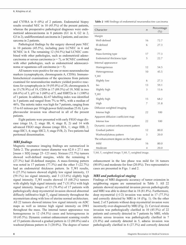
Fig. 2 A 65-year-old woman with pT3aN1M1 (peritoneal dissemination) and FIGO stage IV and endometrial small cell neuroendocrine carcinoma. (a) Axial T2WI showing slightly hyperintense and homogeneous mass invading from endometrium to myometrium (arrow). Deep myometrial invasion (>50%) was suspected. (b) Axial DWI showing remarkably high signal intensity throughout tumor (arrow). (c) Axial late-phase enhanced fat-suppressed T1WI obtained 5 min after Gd-DTPA administration showing weak enhancement of tumor (arrow). (d) Axial T2WI showing swollen lymph node, exceeding 1 cm in the short diameter, in the left obturator area (upper arrow), suggesting nodal metastasis, along with a slightly hyperintense peritoneal mass 1 cm in size (lower arrow), indicating peritoneal dis-semination. (e) Axial DWI showing remarkably high signal intensity in the left obturator node (upper arrow) and peritoneal mass (lower arrow), indicating node metastasis and peritoneal dissemination, respectively. (f) Axial late-phase enhanced fat-suppressed T1WI obtained 5 min after Gd-DTPA administration showing weak enhancement of left obturator node (upper arrow) and peritoneal mass (lower arrow), confirming node metastasis and peritoneal dissemination, respectively. Myometrial invasion of the primary tumor (<50%), invasion of left adnexa left obturator node metastasis, and peritoneal dissemination were confirmed by surgical pathology findings. The patient showed recurrence at 7.5 months and died 12.1 months after surgery. DWI, diffusion weighted imaging; FIGO, Federation of Gynecology and Obstetrics; Gd-DTPA, gadolinium diethylenetriaminepentaacetic acid; T1WI, T1-weighted image; T2WI, T2-weighted image.
a b
e
c
fd

7Epub ahead for print
Endometrial Neuroendocrine Carcinoma MRI
Table 4 Correlation between MRI T staging and pathological staging in patients with endometrial neuroendocrine carcinoma
MRI staging
Pathological staging
T1a T1b T2 T3a T3b T4 All
T1a 3 1 1 0 0 0 5
T1b 0 5 0 1 0 0 6
T2 0 0 2 0 1 0 3
T3a 0 0 0 7 0 0 7
T3b 0 0 0 0 1 0 1
T4 0 0 0 0 0 0 0
All 3 6 3 8 2 0 22
in 5, and parametrium invasion was pathologically clarified in 2 and correctly detected in 1. No vaginal, urinary bladder, or rectum mucosa invasion was observed in any of our cases.
According to the most recent report of the American Joint Committee on Cancer (AJCC) cancer staging,16 T stage was classified as pT1a in 3, pT1b in 6, pT2 in 3, pT3a in 8, and pT3b in 2 patients. Agreements between MRI staging and pathological staging are shown in Table 4. Two patients with pathological myometrial invasion of <50% have also proved to have pathological cervical stroma invasion (pT2) in 1 patient and pathological adnexal invasion (pT3a) in 1 patient. Agreement was noted in 18 patients, with an overall accuracy of MRI for local staging of 81.8%. A discrepancy occurred in 4 patients that resulted from false negative findings for pT1b (n = 1), pT2 (n = 1), pT3a (n = 1), and pT3b (n = 1).
Pelvic lymph nodal metastasis was pathologically clari-fied in 10 (55.5%) of the 18 patients who had undergone lym-phadenectomy and correctly detected in 6 patients by MRI. The values for patient-based sensitivity, specificity, positive predictive value (PPV), negative predictive value (NPV), and accuracy for detecting metastatic pelvic LN were 60.0% (6/10), 100% (8/8), 100% (6/6) 75.0% (8/12), and 77.8% (14/18), respectively (Table 3 and Fig. 2).
Survival analysisAfter surgery, 15 patients received adjuvant chemotherapy, 3 chemoradiation therapy, 2 radiation therapy, and 2 no therapy. During a mean follow-up period of 30.4 months (3.3–138.4 months), 16 (72.7%) experienced recurrence. Of those, 2 (33.3%) of 6 patients classified as FIGO I, 1 (50.0%) of 2 as FIGO II, 11 (91.7%) of 12 as FIGO III, and 2 (100%) of 2 as FIGO IV had recurrence. The sites of recurrence in these 16 patients were LN (n = 4), peritoneum (n = 4), local (n = 2), brain (n = 1), liver and lung (n = 1), liver and perito-neum (n = 1), LN, lung, and liver (n = 1), LN, lung, and bone (n = 1), and LN, lung, adrenal gland, and brain (n = 1).
The median time to recurrence in the 16 patients was 6.75 months (range 1.5–20.2 months). Two-year DFS rates for patients classified as FIGO I, II, III, and IV were
66.7% (4/6), 50.0% (1/2), 8.3% (1/12), and 0% (0/2), respectively, while that was 27.3% (6/22) for all patients. The 14 patients with advanced FIGO stage disease (III and IV) showed significantly shorter PFS as compared with the 8 with early FIGO staged disease (I and II, P = 0.0005; Fig. 3).
During a mean follow-up period of 30.4 months (3.3–138.4 months), 12 (54.5%) of the 22 patients died of disease, including 1 (16.7%) classified as FIGO I, zero (0%) as FIGO II, 9 (75.0%) as FIGO III, and 2 (100%) as FIGO IV. Median OS for all patients was 22.6 months, while that was 26.1 months for FIGO I, 80.9 months for FIGO II, 18.9 months for FIGO III, and 9.8 months for FIGO IV classifications. Additionally, the 2-year OS rate for FIGO I, II, III, and IV was 83.3% (5/6), 100% (2/2), 33.3% (4/12), and 0% (0/2), respectively, while that was 50.0% (11/22) for all patients. The 14 patients with advanced FIGO stage disease (III and IV) showed signifi-cantly worse OS as compared with the 8 with early FIGO stage disease (I and II, P = 0.0028; Fig. 4).
DiscussionThe present analysis of 22 patients with histologically and surgery proven endometrial NEC provides important infor-mation in regard to MRI findings and patient prognosis. An abnormal diffuse infiltrative high T2 signal intensity throughout the myometrium with loss of uterine normal architecture and obvious restricted diffusion throughout the tumor are likely suggestive features of endometrial NEC. Furthermore, pelvic MRI is reliable for intrapelvic staging of endometrial NEC, which has a poor prognosis, especially advanced stage patients.
Fig. 3 Kaplan–Meier progression-free survival curves for 22 patients with endometrial neuroendocrine carcinoma according to FIGO stage. Patients with advanced FIGO stage disease (III and IV, n = 14) showed significantly shorter DFS as compared with those with early FIGO stage disease (I and II; n = 8, P = 0.0005). DWI, diffusion weighted imaging; FIGO, Federation of Gynecology and Obstetrics.

8 Magnetic Resonance in Medical Sciences
K. Kitajima et al.
Fig. 4 Kaplan–Meier overall survival curves for 22 patients with endometrial neuroendocrine carcinoma according to FIGO stage. Patients with advanced FIGO stage disease (III and IV, n = 14) showed significantly worse OS as compared with those with early FIGO stage disease (I and II; n = 8, P = 0.0028). FIGO, Federation of Gynecology and Obstetrics; OS, overall survival.
We compared 4 previous case reports that discussed MRI findings of endometrial LCNEC8–11 and found similar findings in the present patients, as follows: (1) The tumors showed diffuse involvement of both the endometrium and myometrium, and an ill-defined endometrial–myometrial border was seen with loss of normal architecture; (2) the masses showed diffusely infiltrative and heterogeneous high signal intensity on T2WI; (3) DWI showed abnormal high signal intensity throughout the tumors, reflecting high cellu-larity of malignant cells. MRI findings of endometrial NEC mimic those of other types of cancer, including type II endo-metrial carcinoma (poorly differentiated endometrioid carci-noma, papillary serous carcinoma, clear cell carcinoma),17 a malignant tumor of the uterine corpus that invades the endo-metrium, malignant lymphoma, endometrial carcinosar-coma, uterine sarcoma, and metastatic cancer. Definitive diagnosis based on preoperative MRI findings is difficult.
The average age of 42 patients with endometrial SCNEC at the time of diagnosis was reported to be 60 years old,3 while Ogura et al.11 noted ages ranging from 40 to 88 years (mean 62 years) for 19 cases of endometrial LCNEC. In a series of 25 patients with endometrial NEC (LCNEC 15, SCNEC 4, mixture of both 6), patient age ranged from 37 to 87 years (median 57 years).7 A comparison showed that the average age of the present cases (63.4 years) was about 10 years older than that of endometrial adenocarcinoma cases.4
The clinical behavior of NEC of the endometrium is aggressive and highly malignant, with an advanced stage commonly observed at the time of diagnosis. To date, 83 known cases of endometrial SCNEC have been reported in English literature, of which 40.8% (29 of 71) were FIGO stage I or II and 59.2% (42 of 71) stage III or IV among cases with available staging data.3 In a series of 25 patients with endometrial NEC, Pocrnich et al.7 found that most (68%)
were advanced stage (FIGO III or IV). Similar to those reports, our investigation showed an advanced stage for about two-thirds (63.6%) of the present cases.7
Patients with NEC of the endometrium have been reported to have a poor prognosis. In a literature review, Katahira et al.4 found that only 23% (3/13) of endometrial SCNEC patients with stage I disease died of the disease within 5 years, while 73% (17/22) of those with stage II–IV died within 2 years of diagnosis. In addition, Korcum et al.5 reviewed 19 endometrial SCNEC cases with stage I disease, including one under their care and 18 others previously reported. Of those, 13 (68.4%) patients had no evidence of disease at the time of follow-up and there were four (21.1%) long-term survivors with no evidence of disease after 5 years. FIGO stage is the most important prognostic factor. Matsumoto et al.6 published a review of long-term survivors (>1 year) diagnosed with endometrial SCNEC. Of 53 with detailed information available, 20 (37.7%) patients were long-term survivors, including 17 (85.0%) with stage I or II disease, and only 3 (15.0%) with stage III or IV disease. In that study, 4 (66.7%) of 6 patients survived more than 1 year after surgery and 2 of those 4 had advanced stage IIIB or IIIC disease. Similar to these reports, our study also demonstrated a 2-year OS rate of 87.5% for early-stage disease (I or II) as compared with 28.6% for advanced- stage (III or IV).
Neuroendocrine carcinoma of the uterine body is rarely encountered, though affected patients have a poor prognosis and no treatment strategy has been established. Although clear treatment recommendations for endometrial NEC have not been defined, patients should be given multimodality therapy, including surgery, chemotherapy and radio-therapy,3–5 with surgery generally accepted to be the corner-stone. As for chemotherapy, patients are generally administered 6 cycles of cisplatin (60 mg/m2, day 1) and etoposide (60 mg/m2, days 1, 8, and 15).7,8
Our study had several limitations. First, our study include its retrospective design and variations in the MRI scanning protocols among the centers over the 14-year study period, primarily due to the rarity of this entity. Second, the patho-logical diagnosis of 22 endometrial NECs was made by each pathologist at each institution, not by one pathologist (central diagnosis).
ConclusionNeuroendocrine carcinoma of the uterine endometrium is a rare disease and affected patients show poor prognosis, espe-cially those in an advanced stage. Definitive diagnosis based on preoperative MRI results is difficult. However, abnormal diffuse infiltrative high T2 signal intensity throughout the myometrium with loss of uterine normal architecture and obvious restricted diffusion throughout the tumor may sug-gest endometrial NEC. Pelvic MRI is a reliable imaging technique for intrapelvic staging of endometrial NEC.

9Epub ahead for print
Endometrial Neuroendocrine Carcinoma MRI
FundingThis work was supported by Hyogo Science and Technology Association and JSPS KAKENHI grant (number 19K08187).
AcknowledgmentsThe authors wish to thank None Yasuyo Urase, MD (Depart-ment of Radiology, Kobe University Graduate School of Medicine, Hyogo, Japan), Yasunari Mizumoto, MD, PhD (Department of Obstetrics and Gynecology, Kanazawa Uni-versity, Graduate School of Medicine Science, Ichikawa, Japan), Yoshimitstu Ohgiya, MD, PhD (Department of Radi-ology, Showa University School of Medicine, Tokyo, Japan), Ayumi Ohya, MD, PhD (Department of Radiology, Shinshu University School of Medicine, Nagano, Japan), Shinya Fujii, MD, PhD (Division of Radiology, Department of Pathophysiological and Therapeutic Sciences, Tottori Uni-versity, Tottori, Japan), and Katsuyoshi Ito, MD, PhD (Department of Radiology, Yamaguchi University Graduate School of Medicine, Yamaguchi, Japan).
Conflicts of InterestAll authors declare no actual or potential conflicts of interest.
References1. Rouzbahman M, Clarke B. Neuroendocrine tumors of the
gynecologic tract: select topics. Semin Diagn Pathol 2013; 30:224–233.
2. Kurman RJ, Carcanju ML, Herrington CS, Young, RH. World Health Organization classification of tumours of female reproductive organs. 4th ed. Lyon: IARC Press; 2014.
3. Koo YJ, Kim DY, Kim KR, et al. Small cell neuroendocrine carcinoma of the endometrium: a clinicopathologic study of six cases. Taiwan J Obstet Gynecol 2014; 53:355–359.
4. Katahira A, Akahira J, Niikura H, et al. Small cell carcinoma of the endometrium: report of three cases and literature review. Int J Gynecol Cancer 2014; 14:1018–1023.
5. Korcum AF, Aksu G, Ozdogan M, Erdogan G, Taskin O. Stage I small cell carcinoma of the endometrium: survival and management options. Acta Obstet Gynecol Scand 2008; 87:122–126.
6. Matsumoto H, Takai N, Nasu K, Narahara H. Small cell carcinoma of the endometrium: a report of two cases. J Obstet Gynaecol Res 2011; 37:1739–1743.
7. Pocrnich CE, Ramalingam P, Euscher ED, Malpica A. Neuroendocrine carcinoma of the endometrium: a clinicopathologic study of 25 cases. Am J Surg Pathol 2016; 40:577–586.
8. Makihara N, Maeda T, Nishimura M, et al. Large cell neuroendocrine carcinoma originating from the uterine endometrium: a report on magnetic resonance features of 2 cases with very rare and aggressive tumor. Rare Tumors 2012; 4:e37.
9. Nguyen ML, Han L, Minors AM, et al. Rare large cell neuroendocrine tumor of the endometrium: a case report and review of the literature. Int J Surg Case Rep 2013; 4:651–655.
10. Kobayashi A, Yahata T, Nanjo S, et al. Rapidly progressing large-cell neuroendocrine carcinoma arising from the uterine corpus: a case report and review of the literature. Mol Clin Oncol 2017; 6:881–885.
11. Ogura J, Adachi Y, Yasumoto K, et al. Large-cell neuroendocrine carcinoma arising in the endometrium: a case report. Mol Clin Oncol 2018; 8:571–574.
12. Pecorelli S. Revised FIGO staging for carcinoma of the vulva, cervix, and endometrium. Int J Gynaecol Obstet 2009; 105:103–104.
13. Garza A, Elsherif SB, Faria SC, et al. Staging MRI of uterine malignant mixed Müllerian tumors versus endometrial carcinomas with emphasis on dynamic enhancement characteristics. Abdom Radiol (NY) 2020; 45:1141–1154.
14. Freeman SJ, Aly AM, Kataoka MY, Addley HC, Reinhold C, Sala E. The revised FIGO staging system for uterine malignancies: implications for MR imaging. Radiographics 2012; 32:1805–1827.
15. Rauch GM, Kaur H, Choi H, et al. Optimization of MR imaging for pretreatment evaluation of patients with endometrial and cervical cancer. Radiographics 2014; 34:1082–1098.
16. Edge SB, Compton CC. The American joint committee on cancer: the 7th edition of the AJCC cancer staging manual and the future of TNM. Ann Surg Oncol 2010; 17: 1471–1474.
17. Fukunaga T, Fujii S, Inoue C, et al. Accuracy of semiquantitative dynamic contrast-enhanced MRI for differentiating type II from type I endometrial carcinoma. J Magn Reson Imaging 2015; 41:1662–1668.