Cdk5 phosphorylates PLD2 to mediate EGF-dependent insulin secretion
8
Cdk5 phosphorylates PLD2 to mediate EGF-dependent insulin secretion Hye Young Lee a , Hyuntae Jung b , Il Ho Jang a , Pann-Ghill Suh a , Sung Ho Ryu a,b, ⁎ a Division of Molecular and Life Sciences, Pohang University of Science and Technology, Pohang, 790-784, Republic of Korea b School of Interdisciplinary Bioscience and Bioengineering, Pohang University of Science and Technology, Pohang, 790-784, Republic of Korea ABSTRACT ARTICLE INFO Article history: Received 4 March 2008 Received in revised form 11 June 2008 Accepted 12 June 2008 Available online 25 June 2008 Keywords: Phospholipase D2 Cyclin-dependent kinase 5 Insulin secretion Epidermal growth factor Insulin secretion from pancreatic β-cells is an important process that affects the regulation of glucose level in the blood. In our previous study, we suggested that epidermal growth factor (EGF) stimulates insulin secretion by activating phospholipase D2 (PLD2) [H.Y. Lee, K. Yea, J. Kim, B.D. Lee, Y.C. Chae, H.S. Kim, D.W. Lee, S.H. Kim, J.H. Cho, C.J. Jin, D.S. Koh, K.S. Park, P.G. Suh, S.H. Ryu, 2007. Epidermal Growth Factor Increases Insulin Secretion and Lowers Blood Glucose in Diabetic Mice. J. Cell. Mol. Med. 5:5]. However, the specific mechanism by which PLD2 activation leads to insulin secretion was not determined. In this study, we suggest that the phosphorylation and activation of PLD2 by cyclin-dependent kinase 5 (Cdk5) is critical for EGF- dependent insulin secretion. We found that a Cdk5 inhibitor, roscovitine, and dominant-negative Cdk5 inhibited EGF-dependent PLD2 activation and insulin secretion. EGF stimulation activated Cdk5 activity in rat insulinoma RINm5F cells, and PLD2 phosphorylation by Cdk5 was observed in vitro and in cells. The kinetics of PLD2 phosphorylation correlates with the interaction between PLD2 and Cdk5 and its effect on EGF signaling. We determined that the phosphorylation site of PLD2 was located at Ser 134 . PLD2-S134A did not show EGF-dependent phosphorylation and activation by Cdk5. Furthermore, this mutant was unable to mediate EGF-dependent insulin secretion in pancreatic β cell lines, suggesting that the phosphorylation of PLD2 at Ser 134 by Cdk5 is critical for this process. The study results suggest that PLD2 is a new substrate of Cdk5 and that the phosphorylation of PLD2 by Cdk5 is involved in EGF-dependent insulin secretion. © 2008 Elsevier Inc. All rights reserved. 1. Introduction Insulin is stored in secretary granules in pancreatic β-cells and is released by exocytosis upon stimulation with secretagogues [1]. It has been suggested that the molecular machinery that regulates insulin secretion is similar to the machinery that regulates neurotransmitter release from synaptic vesicles [15,35,46,47,49]. Regulated insulin secretion from pancreatic β-cells has to be tightly controlled to ensure glucose homeostasis in the body. Several different signaling pathways determine the capacity of the β-cell to respond to stimulation. The level of β-cell activity is determined by several different stimulators, including glucose, amino acids, fatty acids, neurotransmitters, and hormones [12]. Among them, epidermal growth factor (EGF) is a potent insulin stimulator that regulates plasma glucose levels [23]. Despite the investigation into the mechanism of EGF-induced insulin secretion, the processes that are involved in this stimulus-secretion coupling and those that maintain exquisite control of insulin release are still incompletely understood. Cyclin-dependent kinase 5 (Cdk5) is a member of the cyclin- dependent kinase family, but plays a different role than other Cdks. Cdk5 is widely expressed although its kinase activity has been primarily described in neuronal systems [6]. The function of Cdk5 was thought to be restricted to the nervous system because its kinase activity and the expression of its cofactors, p35 and p39, were initially detected in the nervous system [6,51]. However, several groups have demonstrated the presence of active Cdk5 in many non-neuronal tissues [6,20,34]. Especially, Cdk5 is expressed in insulin-secreting pancreatic β-cells, and the inhibition of Cdk5 by roscovitine altered insulin secretion, suggesting that Cdk5 is a critical regulator of insulin secretion [26,27,44]. Cdk5 in association with its regulatory protein, p39, stimulates Ca 2+ -dependent exocytosis in primary β-cells through the phosphorylation of Munc18-1 [26]. The L-type voltage-dependent Ca 2+ channel (L-VDCC) is also a substrate of Cdk5, and it has been shown to regulate insulin secretion under the condition of high glucose concentration [44]. However, the effect of secretagogue on Cdk5 activity and the detailed mechanism of Cdk5-medited insulin secretion have hardly been studied. Insulin secretion is mainly triggered by the elevation of intracel- lular Ca 2+ , but it can be modulated by several cellular signaling proteins. Of these proteins, phospholipase D (PLD) is a membrane- bound enzyme that hydrolyzes phosphatidylcholine (PC) to generate a multifunctional lipid, phosphatidic acid (PA), in response to a variety of signals, including growth factors [7]. Interestingly, PLD activity is inhibited in diabetic animals [32]. PLD activity is involved in various trafficking processes, particularly in the regulation of insulin Cellular Signalling 20 (2008) 1787–1794 ⁎ Corresponding author. Division of Molecular and Life Sciences, Pohang University of Science and Technology, Pohang, 790-784, Republic of Korea. Tel.: +82 54 279 2292; fax: +82 54 279 0645. E-mail addresses: [email protected], [email protected] (S.H. Ryu). 0898-6568/$ – see front matter © 2008 Elsevier Inc. All rights reserved. doi:10.1016/j.cellsig.2008.06.009 Contents lists available at ScienceDirect Cellular Signalling journal homepage: www.elsevier.com/locate/cellsig
-
Upload
hye-young-lee -
Category
Documents
-
view
214 -
download
2
Transcript of Cdk5 phosphorylates PLD2 to mediate EGF-dependent insulin secretion

Cellular Signalling 20 (2008) 1787–1794
Contents lists available at ScienceDirect
Cellular Signalling
j ourna l homepage: www.e lsev ie r.com/ locate /ce l l s ig
Cdk5 phosphorylates PLD2 to mediate EGF-dependent insulin secretion
Hye Young Lee a, Hyuntae Jung b, Il Ho Jang a, Pann-Ghill Suh a, Sung Ho Ryu a,b,⁎a Division of Molecular and Life Sciences, Pohang University of Science and Technology, Pohang, 790-784, Republic of Koreab School of Interdisciplinary Bioscience and Bioengineering, Pohang University of Science and Technology, Pohang, 790-784, Republic of Korea
⁎ Corresponding author. Division of Molecular and LifeScience and Technology, Pohang, 790-784, Republic offax: +82 54 279 0645.
E-mail addresses: [email protected], lazypink@p
0898-6568/$ – see front matter © 2008 Elsevier Inc. Aldoi:10.1016/j.cellsig.2008.06.009
A B S T R A C T
A R T I C L E I N F OArticle history:
Insulin secretion from pancr Received 4 March 2008Received in revised form 11 June 2008Accepted 12 June 2008Available online 25 June 2008Keywords:Phospholipase D2Cyclin-dependent kinase 5Insulin secretionEpidermal growth factor
eatic β-cells is an important process that affects the regulation of glucose level inthe blood. In our previous study, we suggested that epidermal growth factor (EGF) stimulates insulinsecretion by activating phospholipase D2 (PLD2) [H.Y. Lee, K. Yea, J. Kim, B.D. Lee, Y.C. Chae, H.S. Kim, D.W.Lee, S.H. Kim, J.H. Cho, C.J. Jin, D.S. Koh, K.S. Park, P.G. Suh, S.H. Ryu, 2007. Epidermal Growth Factor IncreasesInsulin Secretion and Lowers Blood Glucose in Diabetic Mice. J. Cell. Mol. Med. 5:5]. However, the specificmechanism by which PLD2 activation leads to insulin secretionwas not determined. In this study, we suggestthat the phosphorylation and activation of PLD2 by cyclin-dependent kinase 5 (Cdk5) is critical for EGF-dependent insulin secretion. We found that a Cdk5 inhibitor, roscovitine, and dominant-negative Cdk5inhibited EGF-dependent PLD2 activation and insulin secretion. EGF stimulation activated Cdk5 activity in ratinsulinoma RINm5F cells, and PLD2 phosphorylation by Cdk5 was observed in vitro and in cells. The kineticsof PLD2 phosphorylation correlates with the interaction between PLD2 and Cdk5 and its effect on EGFsignaling. We determined that the phosphorylation site of PLD2 was located at Ser134. PLD2-S134A did notshow EGF-dependent phosphorylation and activation by Cdk5. Furthermore, this mutant was unable tomediate EGF-dependent insulin secretion in pancreatic β cell lines, suggesting that the phosphorylation ofPLD2 at Ser134 by Cdk5 is critical for this process. The study results suggest that PLD2 is a new substrate ofCdk5 and that the phosphorylation of PLD2 by Cdk5 is involved in EGF-dependent insulin secretion.
© 2008 Elsevier Inc. All rights reserved.
1. Introduction
Insulin is stored in secretary granules in pancreatic β-cells and isreleased by exocytosis upon stimulation with secretagogues [1]. It hasbeen suggested that the molecular machinery that regulates insulinsecretion is similar to the machinery that regulates neurotransmitterrelease from synaptic vesicles [15,35,46,47,49]. Regulated insulinsecretion from pancreatic β-cells has to be tightly controlled toensure glucose homeostasis in the body. Several different signalingpathways determine the capacity of the β-cell to respond tostimulation. The level of β-cell activity is determined by severaldifferent stimulators, including glucose, amino acids, fatty acids,neurotransmitters, and hormones [12]. Among them, epidermalgrowth factor (EGF) is a potent insulin stimulator that regulatesplasma glucose levels [23]. Despite the investigation into themechanism of EGF-induced insulin secretion, the processes that areinvolved in this stimulus-secretion coupling and those that maintainexquisite control of insulin release are still incompletely understood.
Cyclin-dependent kinase 5 (Cdk5) is a member of the cyclin-dependent kinase family, but plays a different role than other Cdks.
Sciences, Pohang University ofKorea. Tel.: +82 54 279 2292;
ostech.ac.kr (S.H. Ryu).
l rights reserved.
Cdk5 is widely expressed although its kinase activity has beenprimarily described in neuronal systems [6]. The function of Cdk5 wasthought to be restricted to the nervous system because its kinaseactivity and the expression of its cofactors, p35 and p39, were initiallydetected in the nervous system [6,51]. However, several groups havedemonstrated the presence of active Cdk5 in many non-neuronaltissues [6,20,34]. Especially, Cdk5 is expressed in insulin-secretingpancreatic β-cells, and the inhibition of Cdk5 by roscovitine alteredinsulin secretion, suggesting that Cdk5 is a critical regulator of insulinsecretion [26,27,44]. Cdk5 in association with its regulatory protein,p39, stimulates Ca2+-dependent exocytosis in primary β-cells throughthe phosphorylation of Munc18-1 [26]. The L-type voltage-dependentCa2+ channel (L-VDCC) is also a substrate of Cdk5, and it has beenshown to regulate insulin secretion under the condition of highglucose concentration [44]. However, the effect of secretagogue onCdk5 activity and the detailed mechanism of Cdk5-medited insulinsecretion have hardly been studied.
Insulin secretion is mainly triggered by the elevation of intracel-lular Ca2+, but it can be modulated by several cellular signalingproteins. Of these proteins, phospholipase D (PLD) is a membrane-bound enzyme that hydrolyzes phosphatidylcholine (PC) to generate amultifunctional lipid, phosphatidic acid (PA), in response to a varietyof signals, including growth factors [7]. Interestingly, PLD activity isinhibited in diabetic animals [32]. PLD activity is involved in varioustrafficking processes, particularly in the regulation of insulin

1788 H.Y. Lee et al. / Cellular Signalling 20 (2008) 1787–1794
exocytosis [16]. To date, two types of mammalian PLD, PLD1 and PLD2,have been cloned, and they show differences in localization andregulatory protein interactions [9]. PLD2 has been shown to be criticalfor vesicle exocytosis at the plasma membrane [5]. In our previousreport, we suggested that EGF stimulates insulin secretion byactivating PLD2 [23], but the mechanism of EGF-dependent PLD2activation in this process remains unknown.
In this study, we revealed that Cdk5 is a key regulator of EGF-dependent insulin secretion in a pancreatic β-cell line. In this process,Cdk5 activation by EGF phosphorylates and activates PLD2. Wedetermined that Cdk5 phosphorylates PLD2 at Ser134 of PLD2 andthat this phosphorylation was suggested to be important for EGF-dependent insulin secretion.
2. Materials and methods
The abbreviations used are: PLD, phospholipase D; Cdk5, cyclin-dependent kinase 5; EGF, epidermal growth factor; PC, phosphati-dylcholine; PA, phosphatidic acid; PBt, phosphatidyl-butanol; RT,room temperature; sf9, spodoptera frugiperda 9; ECL, enhancedchemiluminescence; EGTA, ethylene glycol bis (β-aminoethyl ether)N,N,N′,N′-tetraacetic acid; GST, glutathione S-transferase; His6, hexa-histidine; PAGE, polyacrylamide gel electrophoresis; PCR, polymerasechain reaction; DMEM, Dulbecco's modified Eagle's medium; PBS,phosphate buffered saline.
2.1. Materials
The enhanced chemiluminescence (ECL) kit, chelating-Sepharose,and glutathione-Sepharose 4B were purchased from AmershamPharmacia Biotech (Buckinghamshire, United Kingdom); [3H]myristicacid from Dupont NEN (Boston, MA); Silica Gel 60 thin-layerchromatography plates from MERCK (Darmstadt, Germany); ProteinA-Sepharose from RepliGen (Needham, MA); Dulbecco's modifiedEagle's medium and LipofectAMINE from Invitrogen (Carlsbad, CA);Fetal bovine serum from HyClone (Logan, UT); EGF from the DaewoongPharmaceutical Company (Seoul, Republic of Korea); Cholic acid fromUSB (Cleveland, OH); Horseradish peroxidase-conjugated goat anti-rabbit IgG and anti-mouse IgG, IgM, and IgA from Kirkegaard and PerryLaboratories, Inc. (Gaitherburg, MD); Monoclonal and polyclonal anti-Cdk5 antibody from Santa Cruz biotechnology (Santa Cruz, CA); andPhospho-(Ser) Cdks substrate antibody from Cell Signaling technology(Danvers, MA). Polyclonal antibody (mSTP4) recognizing both PLD1 andPLD2 was produced as described previously [18]. All other chemicalswere purchased from Sigma.
2.2. DNA constructs
Full-length cDNAs of rat PLD1 or human PLD2 were generated aspreviously reported [24]. The full-length cDNA of human PLD2 with aserine to alanine mutation at amino acid 134 was ligated into thepcDNA 3.1 vector for transfection into RINm5F cells. pCMV vectorcontaining a wild-type or a dominant-negative Cdk5 plasmid wasgenerously provided by Dr. Li-Hei Tsai (Massachusetts Institute ofTechnology, Boston, MA). A Cdk5 insert was obtained by PCR andligated into pGEX4T1. The full-length cDNA of human PLD2 weredigested into fragments containing PX domain and ligated into thepGEX4T3 vector. Murine Munc-18-1 plasmid in pGEX vector wasgenerously provided by Dr. Thomas C. Südhof (Howard HughesMedical Institute, The University of Texas Southwestern MedicalSchool, Dallas).
2.3. Protein purification
His6-tagged rat PLD1 and human PLD2 were purified fromdetergent extracts of baculovirus-infected Sf9 cells by chelating-
Sepharose affinity column chromatography, as described previously[17]. E. coli BL21 cells were transformed with individual expressionvectors encoding the GST fusion proteins and purified by standardmethods [21] that involved lysing E. coli with a buffer containing 1%Triton X-100 and 1% cholic acid, centrifuging the lysate at 100,000 ×gfor 30 min at 4 °C, and a final purification with glutathione-Sepharose4B. To obtain the soluble GST fusion Munc-18-1, we eluted the proteintwo times using buffer (50 mM Tris/HCl, pH 7.0, 10 mM glutathione,1 mM DTT) for 1 h at 4 °C.
2.4. Cell culture
RINmF5 cells were maintained in a growth medium composed ofRPMI supplemented with 10% fetal bovine serum at 37 °C in ahumidified CO2-controlled (5%) incubator. The mouse insulin-produ-cing MIN6m9 cells provided by Dr. Susumu Seino (Division of Cellularand Molecular Medicine, Kobe University Graduate School ofMedicine, Kobe, Japan) were used between passages 19 and 25, andwere cultured in DMEM containing 25mMglucose,10mMHEPES,10%(v/v) fetal calf serum, 50 IU/ml penicillin, and 50 µg/ml streptomycinat 37 °C in a humidified CO2-controlled (5%) incubator. HEK293 cellswere maintained in a growth medium composed of DMEM supple-mented with 10% fetal bovine serum at 37 °C in a humidified CO2-controlled (5%) incubator. Cells were transfected using LipofectAMINE,as described previously [18].
2.5. Insulin secretion assay
Cells plated at a density of 1×10⁶ cells/well in 24-well plates werewashed twicewith KRB supplementedwith 0.2% bovine serumalbumin(BSA) and then incubated for 60min at 37 °C in the KRB solution. At theendof incubation, the solutionswere replacedwith freshKRBcontainingtest reagents and incubated for the designated amount of time. Theincubation medium was sampled and centrifuged to remove cells, andthe supernatant was assayed for insulin levels with a rat insulinradioimmunoassay (RIA) kit (Linco, St. Louis, MO, Cat. # RI-13K).
2.6. PLD activity assay in cells
PLD activity was assayed bymeasuring the formation of PBt [18]. Theintensities of PBt spots in the presence of 1-butanol weremeasured, andPLD activity was obtained by subtracting the corresponding intensitiesof spots obtained in the absence of 1-butanol.
2.7. Immunoblot analysis
Proteins were denatured by boiling for 5 min at 95 °C in Laemmlisample buffer [19], separated by SDS-PAGE, and immunoblotted asdescribed previously [18].
2.8. Immunoprecipitation
Immunoprecipitation was performed as described previously [22].Briefly, cells were lysed with PLD assay buffer (50 mM HEPES/NaOH,pH 7.3, 3 mM EGTA, 3 mM CaCl2, 3 mM MgCl2, and 80 mM KC1)containing 1% Triton X-100, 1% cholic acid, and 1 mM phenylmethyl-sulfonyl fluoride. After brief sonication, the cell lysates werecentrifuged at 100,000 ×g for 30 min at 4 °C. The supernatants(1 mg of protein) were then incubated with anti-Cdk5 polyclonal oranti-PLD polyclonal antibody immobilized on protein A-Sepharose for6 h at 4 °C. The resulting immune complexes werewashed three timeswith 1ml of PLD assay buffer containing 1% Triton X-100 and 1% cholicacid and once with a buffer without detergent. Samples were thensubjected to SDS-PAGE and immunoblotting using anti-Cdk5 mono-clonal, anti-PLD polyclonal antibody, phospho-(Ser) Cdks substrateantibody or anti-actin monoclonal antibody.

Fig. 1. Cdk5 mediates EGF-dependent PLD activity and insulin secretion in RINm5F cells. RINm5F cells were plated onto 6-well plates or 24-well plates and grown for 24 h. The cellswere washed twice with KRB supplemented with 0.2% BSA and then incubated for 60 min at 37 °C in KRB solution. (A) To measure PLD activity, the cells werewashed twice with KRBand then incubated for 4 h at 37 °C in the KRB solution in the presence of [3H]myristic acid. At the end of the incubation period, the solutions were replacedwith fresh KRB containingDMSO or roscovitine (10 µM), incubated for 4 h, and 15 nM EGF stimulation was performed for 0 or 5 min. The intensities of PBt spots were measured after 5 min of accumulation inthe presence of 0.4% 1-butanol and EGF. ⁎, Pb0.05 compared with EGF-treated cells. (B) At the end of incubation, the solutions were replaced with fresh KRB containing DMSO orroscovitine (10 µM), incubated for 4 h, and 15 nM EGF stimulationwas performed for 0 or 5min. The incubation mediumwas sampled in order to measure the insulin levels. ⁎, Pb0.05compared with EGF-treated cells. (C–D) RINm5F cells were plated onto 6-well plates or 24-well plates and grown for 24 h. Cells were transfected with wild-type (WT) or dominant-negative (DN) Cdk5 and grown for 24 h. The cells were washed twice with KRB supplemented with 0.2% BSA and then incubated for 60 min at 37 °C in KRB solution. (C) To measurePLD activity, the cells werewashed twice with KRB and then incubated for 4 h at 37 °C in the KRB solution in the presence of [3H]myristic acid. At the end of the incubation, 15 nM EGFstimulationwas performed for 0 or 5min. The intensities of PBt spots weremeasured after 5min of accumulation in the presence of 0.4% 1-butanol and EGF. ⁎, Pb0.05 comparedwithEGF-treated cells. (D) At the end of incubation, 15 nM EGF stimulation was performed for 0 or 5 min. The incubation medium was sampled in order to measure the insulin levels.⁎, Pb0.05 compared with EGF-treated cells. (A–D) Means±S.D. of two independent assays are shown.
Fig. 2. Cdk5 is activated by EGF treatment in RINm5F cells. RINm5F cells were platedonto 100–mm plates and grown for 24 h. After serum starvation for 24 h, EGF (15 nM)stimulationwas performed for 0, 0.5, 1, 2, or 5min. Cells were immunoprecipitated withanti-Cdk5 polyclonal antibody and incubated with histone (1 µg) and (γ-32P) ATP(10 µCi). Samples were subjected to SDS-PAGE followed by autoradiographic analysis, asdescribed in the Materials and methods section. The data shown are representative oftwo independent experiments.
1789H.Y. Lee et al. / Cellular Signalling 20 (2008) 1787–1794
2.9. Cdk5 kinase assay
A cyclin-dependent kinase 5 kinase assay was performed aspreviously described [43]. Immunoprecipitated Cdk5 was washed twotimeswith lysis buffer and twicewith kinase buffer [20mmol/LTris–HCl(pH 7.5), 20 mmol/L MgCl2, 1 mmol/L EDTA, and 1 mmol/L EGTA]containing a phosphatase inhibitor cocktail. The isolated complexeswere then incubated at 30 °C for 20 min with kinase buffer containing1 µg of histone H1 (Sigma) or 200 ng of purified PLDs and 10 µCi of (γ-32P) ATP in a final volume of 40 µl. Kinase activity was determined bySDS-PAGE and autoradiography. For detecting the phosphorylationlevels of PLD in cells, immunoprecipitated PLD was performed by SDS-PAGE and immunoblotted with phospho-(Ser) Cdks substrate antibody.Phosphorylation levelsweremeasuredby the ImageGauge Programandshown in each figures as small numbers.
2.10. Binding analysis in vitro
In vitro binding was performed in 300 µl of PLD assay buffer(50 mM HEPES/NaOH, pH 7.3, 3 mM EGTA, 3 mM CaCl2, 3 mM MgCl2,and 80 mM of KC1) containing 0.1% Triton X-100 and 0.1% cholic acidat 4 °C for 2 h. The resulting immune complexes were washed threetimes with 1 ml of PLD assay buffer containing 0.1% Triton X-100 and
0.1% cholic acid and oncewith a buffer without detergent. The sampleswere then subjected to SDS-PAGE and immunoblotting using anti-Cdk5 or anti-PLD antibody.

1790 H.Y. Lee et al. / Cellular Signalling 20 (2008) 1787–1794
2.11. Immunocytochemical analysis
RINm5F cells were grown on coverslips and transfected. Afterstimulation with 100 nM EGF for 0, 0.5, 1, 2 or 5 min, immunocyto-
Fig. 3. PLD2 is a substrate of Cdk5. (A) HEK293 cells were plated onto 100-mm plates and grwith ice-cold PLD buffer and immunoprecipitated with anti-PLD antibody, as described in thATP (10 µCi) in the presence or absence of p25/Cdk5. Samples were subjected to SDS-PAGindependent experiments. (B–E) The RINm5F cells were plated onto 100-mm plates and grow0.5, 1, 2, or 5 min. Cells were lysed with ice-cold PLD buffer and immunoprecipitated wiImmunoprecipitated Cdk5 was incubated with purified PLD1 or PLD2 (200 ng) and (γ-32P) ATThe data shown are representative of two independent experiments. (C) Cells were transfecte1min. (D) Cells were transfected with PLD2. At the end of serum starvation for 24 h, the cellsperformed for 1min. (E) At the end of serum starvation for 24 h, the cells were pretreatedwith(F) Cells were transfected with wild-type (WT) or dominant-negative (DN) Cdk5. At the endwere lysedwith ice-cold PLD buffer, as described in theMaterials andmethods section. TotalSDS-PAGE, followed by immunoblot analysis. Actin and Cdk5 were blotted as controls. The
chemical analysis was performed as described previously [27]. Cellswere washed with PBS, fixed in 4% (w/v) paraformaldehyde and thenincubated in blocking buffer (1% horse serum in PBS containing 0.2%Triton X-100). Subsequently, the cells were incubated with primary
own for 24 h. Cells were transfected with PLD2 and grown for 24 h. The cells were lysede Materials and methods section. Immunoprecipitated PLD2 was incubated with (γ-32P)E followed by autoradiographic analysis. The data shown are representative of threen for 24 h. (B) After serum starvation for 24 h, cells were treated with EGF (15 nM) for 0,
th anti-Cdk5 polyclonal antibody, as described in the Materials and methods section.P (10 µCi). Samples were subjected to SDS-PAGE followed by autoradiographic analysis.d with PLD1 or PLD2. After serum starvation for 24 h, EGF stimulationwas performed forwere pretreated with DMSO or roscovitine (10 µM) for 4 h, and then EGF stimulationwasDMSO or roscovitine (10 µM) for 4 h, and EGF stimulationwas performed for 0 or 1min.of serum starvation for 24 h, EGF stimulation was performed for 0 or 1 min. (C–F) Cellscell lysates (Total) and immune complexes (IP) with anti-PLD antibodywere subjected todata shown are representative of two independent experiments.

1791H.Y. Lee et al. / Cellular Signalling 20 (2008) 1787–1794
antibodies for 2 h at RT and then with the secondary antibodiesrhodamine-conjugated anti-rabbit antibody (for PLD) or fluoresceinisothiocyanate (FITC)-conjugated anti-mouse antibody (for Cdk5) for1 h at RT. Slides were then mounted and examined under a confocalmicroscope (Carl Zeiss, Germany).
3. Results
3.1. Cdk5 inhibition attenuates EGF-dependent PLD activity and insulinsecretion
To find a modulator of EGF-dependent PLD activation and insulinsecretion, several kinase inhibitors (GF109203X: cPKC inhibitor,Rottlerin: PKC δ and PKC θ inhibitor, Go: PKC α and βI inhibitor,roscovitine: Cdk5 inhibitor) were tested (data not shown). Roscov-itine, a Cdk5 inhibitor, was the only kinase inhibitor to significantlyinhibit both EGF-dependent PLD activity (51% inhibition comparedwith DMSO treatment) and insulin secretion (75% inhibition com-pared with DMSO treatment) in rat insulinoma RINm5F cells (Fig. 1Aand B). Inhibition by roscovitine alone suggests that this event isspecifically dependent on Cdk5 activity. Notably, roscovitine did not
Fig. 4. PLD2 interacts with Cdk5. (A–B) RINm5F cells were plated onto 100-mm plates and gro0, 0.5, 1, 2, or 5 min. The cells were lysed with ice-cold PLD buffer, as described in the Materanti-Cdk5 polyclonal antibody were subjected to SDS-PAGE, followed by immunoblot analyindependent experiments. (B) Total cell lysates (Total) and immune complexes (IP) with anti-Actin and Cdk5 were blotted as controls. The data shown are representative of two independand grown for 24 h. Cells were cotransfected with PLD2 and Cdk5. After serum starvaImmunocytochemical analysis was then performed using polyclonal anti-PLD antibody ormocells as described in the Materials and methods section. The FITC-conjugated anti-mouse antthe merged image (right) were obtained using a confocal microscope. The data shown are r
inhibit basal PLD activity or basal insulin secretion, but only inhibitedEGF-induced PLD activity or insulin secretion. From this result, it isreasonable to question whether or not dominant-negative Cdk5 hasan effect on EGF-dependent PLD activity or insulin secretion. Toaddress this question, dominant-negative Cdk5 was tested andcompared with wild-type Cdk5 on EGF-dependent PLD activity andinsulin secretion (Fig. 1C and D). Dominant-negative Cdk5 signifi-cantly inhibited both EGF-dependent PLD activity (88% inhibitioncompared with wild-type Cdk5) and insulin secretion (88% inhibitioncompared with wild-type Cdk5) in rat insulinoma RINm5F cells(Fig. 1C and D). Therefore, we assumed that Cdk5 activity is critical forEGF-dependent PLD activation and insulin secretion.
3.2. EGF activates Cdk5 in RINm5F cells
To confirm the activation of Cdk5 by EGF, we performed a kinaseassay with immunoprecipitated Cdk5 after EGF treatment in RINm5Fcells. The results showed that Cdk5 was activated by EGF treatment,reached a maximum rate at 1 min (1.81 fold increase; Fig. 2), and thendecreased. These findings showed that Cdk5 is transiently activated byEGF signaling.
wn for 24 h. After serum starvation for 24 h, EGF (15 nM) stimulationwas performed forials and methods section. (A) Total cell lysates (Total) and immune complexes (IP) withsis. Actin and Cdk5 were blotted as controls. The data shown are representative of twoPLD polyclonal antibody were subjected to SDS-PAGE, followed by immunoblot analysis.ent experiments. (C) The RINm5F cells were plated onto 6-well plates with cover glasstion for 24 h, EGF (15 nM) stimulation was performed for 0, 0.5, 1, 2, or 5 min.noclonal anti-Cdk5 antibody to determine the localizations of Cdk5 and PLD2 in RINm5Fibody image (left), the rhodamine-conjugated anti-rabbit antibody image (middle), andepresentative of five independent experiments.

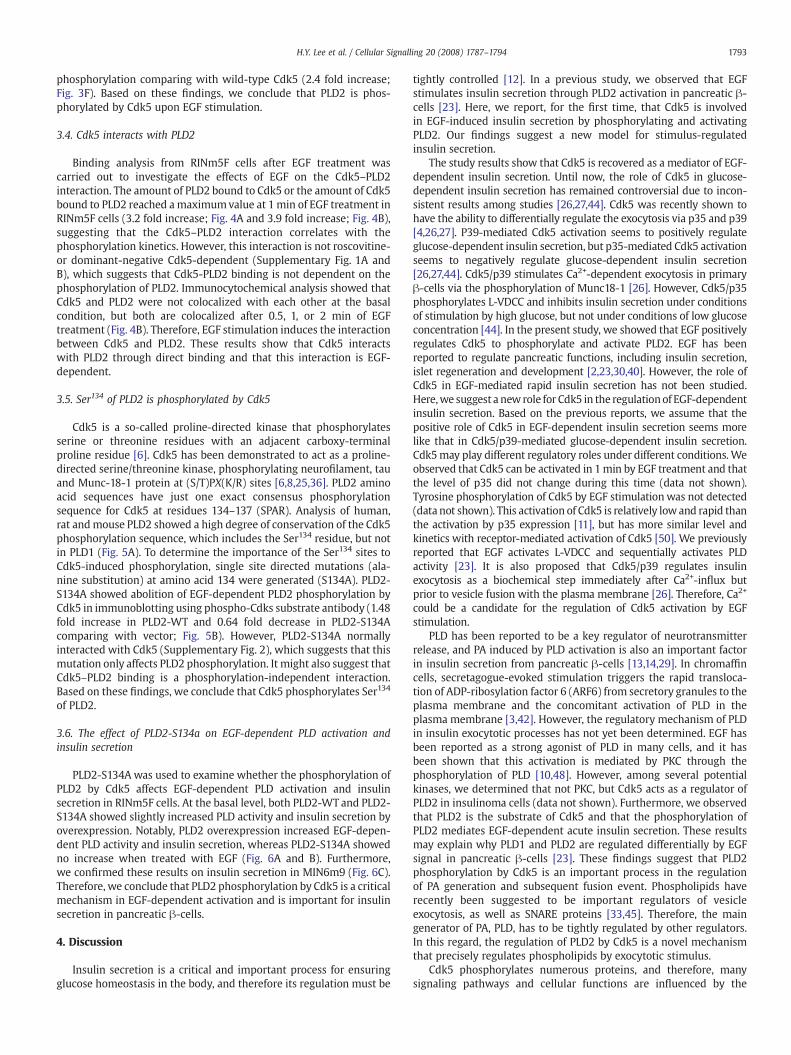
Fig. 5. Cdk5 phosphorylates PLD2 at Ser134. (A) Domain structure of PLD2. Expected sites of major consensus by Cdk5 phosphorylation at PLD1 and PLD2 were determined. Oneexpected site (SPAR) in PLD2 is representatively shown in domain structure of PLD2 (upper panel). Sequences of human (h), rat (r), or mouse (m) PLD1 and PLD2 were aligned, andonly the aligned region of expectation sequences (SPAR in PLD2) is shown (lower panel). The name of each protein is indicated on the left. The GenBank™ accession numbers are asfollows: hPLD1, U69550; rPLD1, NP_112254; mPLD1, NP_032901; hPLD2, NP_002654; rPLD2, NP_150641.2; mPLD2, AAH68317 (B) The RINm5F cells were plated onto 100-mm platesand grown for 24 h. The cells were transfected with vector, PLD2-WT, or PLD2-S134A. After serum starvation for 24 h, EGF stimulation was performed for 1 min. RINm5F cells werelysed with ice-cold PLD buffer as described in the Materials and methods section. Total cell lysates (Total) and immune complexes (IP) with anti-PLD antibody were subjected to SDS-PAGE, followed by immunoblot analysis. Actin and Cdk5 were blotted as controls. The data shown are representative of three independent experiments.
1792 H.Y. Lee et al. / Cellular Signalling 20 (2008) 1787–1794
3.3. PLD2 is phosphorylated by Cdk5
We showed that EGF-dependent PLD2 activity is regulated by aCdk5 inhibitor. Since Cdk5 is a kinase, we hypothesized that PLD2 is anew substrate of Cdk5. Immunoprecipitated PLD2 was incubated withor without purified p25/Cdk5 in the presence of (γ-32P) ATP. PLD2washighly phosphorylated (3.81 fold increase; Fig. 3A), which indicatesthat PLD2 is a new substrate of Cdk5. To determine whether thisphosphorylation is EGF-dependent or PLD2-specific, we performed akinase assay with immunoprecipitated Cdk5 from RINm5F cellstreated with EGF for 0, 0.5, 1, 2 or 5 min using purified PLD1 orPLD2 as a substrate. The results showed that treatment with EGF for1 min significantly increased the levels of PLD2 phosphorylation(2.15 fold increase), while PLD1 does not, which indicates that PLD2phosphorylation by Cdk5 is EGF-dependent and PLD2-specific
Fig. 6. PLD2-S134A leads to a decrease in the EGF-dependent PLD activation and insulin secre24 h. Cells were transfected with vector, PLD2-WT, or PLD2-S134A, and grown for 24 h. The60 min at 37 °C in the KRB solution. (A) To measure PLD activity, the cells were washed twicemyristic acid. At the end of incubation, 15 nM EGF stimulationwas performed for 0 or 5 min. T0.4% 1-butanol and EGF. ⁎, Pb0.05 compared with EGF-treated cells. (B) At the end of incubatincubated for 5 min. The incubation mediumwas sampled in order to measure the insulin levwell plates and grown for 24 h. Cells were transfected with vector, PLD2-WT, or PLD2-S134A,and then incubated for 60min at 37 °C in the KRB solution. At the end of incubation, the solut5min. The incubationmediumwas sampled in order tomeasure the insulin levels. ⁎, Pb0.05 co
(Fig. 3B). This phosphorylation of PLD2 was confirmed using anti-phospho-Cdks substrate antibody in RINm5F cells. To determinewhether PLD2 phosphorylation is detectable with anti-phospho-Cdkssubstrate antibody in intact cells, we performed an immunoblot withimmunoprecipitated PLDs after 1 min of EGF treatment. The resultsshowed that PLD phosphorylation is detectable with anti-phospho-Cdks substrate antibody only at a PLD2 size and that this PLD2phosphorylation is increased by PLD2 overexpression (1.67 fold in-crease; Fig. 3C). Moreover, roscovitine pretreatment significantlydecreased this PLD2 phosphorylation by EGF stimulation, indicatingthat it is Cdk5-dependent (0.44 fold decrease; Fig. 3D). Furthermore,endogenous PLD2 was detected with anti-phospho-Cdks substrateantibody (3.9 fold increase), and this phosphorylation was not ob-served by roscovitine treatment (no increase; Fig. 3E). And dominant-negative Cdk5 has an inhibitory effect (no increase) on EGF-induced
tion. (A–B) RINm5F cells were plated onto 6-well plates or 24-well plates and grown forcells were washed twice with KRB supplemented with 0.2% BSA and then incubated forwith KRB and then incubated for 4 h at 37 °C in the KRB solution in the presence of [3H]he intensities of PBt spots were measured after 5 min of accumulation in the presence ofion, the solutions were replaced with fresh KRB containing none (NT) or 15 nM EGF andels. ⁎, Pb0.05 compared with EGF-treated cells. (C) MIN6 m9 cells were plated onto 24-and grown for 24 h. The cells were washed twice with KRB supplemented with 0.2% BSAions were replaced with fresh KRB containing none (NT) or 15 nM EGF and incubated formparedwith EGF-treated cells. (A–C)Means±S.D. of two independent assays are shown.
1793H.Y. Lee et al. / Cellular Signalling 20 (2008) 1787–1794
phosphorylation comparing with wild-type Cdk5 (2.4 fold increase;Fig. 3F). Based on these findings, we conclude that PLD2 is phos-phorylated by Cdk5 upon EGF stimulation.
3.4. Cdk5 interacts with PLD2
Binding analysis from RINm5F cells after EGF treatment wascarried out to investigate the effects of EGF on the Cdk5–PLD2interaction. The amount of PLD2 bound to Cdk5 or the amount of Cdk5bound to PLD2 reached amaximumvalue at 1min of EGF treatment inRINm5F cells (3.2 fold increase; Fig. 4A and 3.9 fold increase; Fig. 4B),suggesting that the Cdk5–PLD2 interaction correlates with thephosphorylation kinetics. However, this interaction is not roscovitine-or dominant-negative Cdk5-dependent (Supplementary Fig. 1A andB), which suggests that Cdk5-PLD2 binding is not dependent on thephosphorylation of PLD2. Immunocytochemical analysis showed thatCdk5 and PLD2 were not colocalized with each other at the basalcondition, but both are colocalized after 0.5, 1, or 2 min of EGFtreatment (Fig. 4B). Therefore, EGF stimulation induces the interactionbetween Cdk5 and PLD2. These results show that Cdk5 interactswith PLD2 through direct binding and that this interaction is EGF-dependent.
3.5. Ser134 of PLD2 is phosphorylated by Cdk5
Cdk5 is a so-called proline-directed kinase that phosphorylatesserine or threonine residues with an adjacent carboxy-terminalproline residue [6]. Cdk5 has been demonstrated to act as a proline-directed serine/threonine kinase, phosphorylating neurofilament, tauand Munc-18-1 protein at (S/T)PX(K/R) sites [6,8,25,36]. PLD2 aminoacid sequences have just one exact consensus phosphorylationsequence for Cdk5 at residues 134–137 (SPAR). Analysis of human,rat andmouse PLD2 showed a high degree of conservation of the Cdk5phosphorylation sequence, which includes the Ser134 residue, but notin PLD1 (Fig. 5A). To determine the importance of the Ser134 sites toCdk5-induced phosphorylation, single site directed mutations (ala-nine substitution) at amino acid 134 were generated (S134A). PLD2-S134A showed abolition of EGF-dependent PLD2 phosphorylation byCdk5 in immunoblotting using phospho-Cdks substrate antibody (1.48fold increase in PLD2-WT and 0.64 fold decrease in PLD2-S134Acomparing with vector; Fig. 5B). However, PLD2-S134A normallyinteracted with Cdk5 (Supplementary Fig. 2), which suggests that thismutation only affects PLD2 phosphorylation. It might also suggest thatCdk5–PLD2 binding is a phosphorylation-independent interaction.Based on these findings, we conclude that Cdk5 phosphorylates Ser134
of PLD2.
3.6. The effect of PLD2-S134a on EGF-dependent PLD activation andinsulin secretion
PLD2-S134A was used to examine whether the phosphorylation ofPLD2 by Cdk5 affects EGF-dependent PLD activation and insulinsecretion in RINm5F cells. At the basal level, both PLD2-WT and PLD2-S134A showed slightly increased PLD activity and insulin secretion byoverexpression. Notably, PLD2 overexpression increased EGF-depen-dent PLD activity and insulin secretion, whereas PLD2-S134A showedno increase when treated with EGF (Fig. 6A and B). Furthermore,we confirmed these results on insulin secretion in MIN6m9 (Fig. 6C).Therefore, we conclude that PLD2 phosphorylation by Cdk5 is a criticalmechanism in EGF-dependent activation and is important for insulinsecretion in pancreatic β-cells.
4. Discussion
Insulin secretion is a critical and important process for ensuringglucose homeostasis in the body, and therefore its regulation must be
tightly controlled [12]. In a previous study, we observed that EGFstimulates insulin secretion through PLD2 activation in pancreatic β-cells [23]. Here, we report, for the first time, that Cdk5 is involvedin EGF-induced insulin secretion by phosphorylating and activatingPLD2. Our findings suggest a new model for stimulus-regulatedinsulin secretion.
The study results show that Cdk5 is recovered as a mediator of EGF-dependent insulin secretion. Until now, the role of Cdk5 in glucose-dependent insulin secretion has remained controversial due to incon-sistent results among studies [26,27,44]. Cdk5 was recently shown tohave the ability to differentially regulate the exocytosis via p35 and p39[4,26,27]. P39-mediated Cdk5 activation seems to positively regulateglucose-dependent insulin secretion, but p35-mediated Cdk5 activationseems to negatively regulate glucose-dependent insulin secretion[26,27,44]. Cdk5/p39 stimulates Ca2+-dependent exocytosis in primaryβ-cells via the phosphorylation of Munc18-1 [26]. However, Cdk5/p35phosphorylates L-VDCC and inhibits insulin secretion under conditionsof stimulation by high glucose, but not under conditions of low glucoseconcentration [44]. In the present study, we showed that EGF positivelyregulates Cdk5 to phosphorylate and activate PLD2. EGF has beenreported to regulate pancreatic functions, including insulin secretion,islet regeneration and development [2,23,30,40]. However, the role ofCdk5 in EGF-mediated rapid insulin secretion has not been studied.Here,we suggest a newrole for Cdk5 in the regulationof EGF-dependentinsulin secretion. Based on the previous reports, we assume that thepositive role of Cdk5 in EGF-dependent insulin secretion seems morelike that in Cdk5/p39-mediated glucose-dependent insulin secretion.Cdk5may play different regulatory roles under different conditions. Weobserved that Cdk5 can be activated in 1min by EGF treatment and thatthe level of p35 did not change during this time (data not shown).Tyrosine phosphorylation of Cdk5 by EGF stimulationwas not detected(data not shown). This activation of Cdk5 is relatively lowand rapid thanthe activation by p35 expression [11], but has more similar level andkinetics with receptor-mediated activation of Cdk5 [50]. We previouslyreported that EGF activates L-VDCC and sequentially activates PLDactivity [23]. It is also proposed that Cdk5/p39 regulates insulinexocytosis as a biochemical step immediately after Ca2+-influx butprior to vesicle fusion with the plasma membrane [26]. Therefore, Ca2+
could be a candidate for the regulation of Cdk5 activation by EGFstimulation.
PLD has been reported to be a key regulator of neurotransmitterrelease, and PA induced by PLD activation is also an important factorin insulin secretion from pancreatic β-cells [13,14,29]. In chromaffincells, secretagogue-evoked stimulation triggers the rapid transloca-tion of ADP-ribosylation factor 6 (ARF6) from secretory granules to theplasma membrane and the concomitant activation of PLD in theplasma membrane [3,42]. However, the regulatory mechanism of PLDin insulin exocytotic processes has not yet been determined. EGF hasbeen reported as a strong agonist of PLD in many cells, and it hasbeen shown that this activation is mediated by PKC through thephosphorylation of PLD [10,48]. However, among several potentialkinases, we determined that not PKC, but Cdk5 acts as a regulator ofPLD2 in insulinoma cells (data not shown). Furthermore, we observedthat PLD2 is the substrate of Cdk5 and that the phosphorylation ofPLD2 mediates EGF-dependent acute insulin secretion. These resultsmay explain why PLD1 and PLD2 are regulated differentially by EGFsignal in pancreatic β-cells [23]. These findings suggest that PLD2phosphorylation by Cdk5 is an important process in the regulationof PA generation and subsequent fusion event. Phospholipids haverecently been suggested to be important regulators of vesicleexocytosis, as well as SNARE proteins [33,45]. Therefore, the maingenerator of PA, PLD, has to be tightly regulated by other regulators.In this regard, the regulation of PLD2 by Cdk5 is a novel mechanismthat precisely regulates phospholipids by exocytotic stimulus.
Cdk5 phosphorylates numerous proteins, and therefore, manysignaling pathways and cellular functions are influenced by the

1794 H.Y. Lee et al. / Cellular Signalling 20 (2008) 1787–1794
activity of Cdk5. Several categories of substrates have been studied onthe basis of their functions [6,38,39]. Recent reports have suggestedthat Cdk5 can regulate vesicle exocytosis by phosphorylatingMunc-18and synapsin 1 [8,28,37]. One of the proposed functions of Munc-18-1is to hold syntaxin 1 in a closed conformation, thereby preventingsyntaxin 1 from participating in trans-SNARE formation [31]. Themechanisms for the disassembly of the syntaxin 1/Munc-18-1complex have been proposed, and the phosphorylation of bothMunc-18-1 and syntaxin 1 by different kinases are known tomodulatetheir binding properties. For example, the phosphorylation of Munc-18-1 by Cdk5 in vitro disrupts the complex, and the phosphorylationof syntaxin 1A by death-associated protein kinase (DAP-kinase)significantly reduces its binding affinity for Munc-18-1 [31,41]. Ourprevious report showed that Munc-18 inhibits PLD activity by directinteraction [22]. As with the syntaxin 1/Munc-18-1 complex, PLD2 isnegatively regulated in the resting state by the Munc-18-1 interaction,but EGF stimulation rapidly dissociates Munc-18-1 from PLD2. In thepresent study, the kinetics of the PLD2/Munc-18-1 interactionreversely correlates with the PLD2/Cdk5 interaction and phosphor-ylation by EGF treatment (Figs. 4A and 3B), as well as the activity ofCdk5 (Fig. 2). Furthermore, both syntaxin 1 and PLD2 bind to Munc-18-1 in a pocket-like structure [21,31], which suggests that these twoproteins can be regulated by a similar mechanism. And we observedthat Cdk5 phosphorylation mediates the dissociation of the PLD2/Munc-18-1 complex in vitro (Supplementary Fig. 3). Thus, wespeculate that Cdk5 might regulate SNARE proteins and regulatephospholipids in order to mediate vesicle exocytosis. It might beinteresting to determine the effect of PLD2/Munc-18-1 dissociation onthe exocytosis as future studies.
Acknowledgments
We thank L.-H. Tsai (Massachusetts Institute of Technology, Boston,MA) for providing p35 and Cdk5 cDNA. Andwe thank T. C. Südhof (TheUniversity of Texas Southwestern Medical School, Dallas, TX). Thiswork was supported by the 21st Frontier Functional ProteomicsProgram of the Ministry of Science and Technology, Republic of Korea.
Appendix A. Supplementary data
Supplementary data associated with this article can be found, inthe online version, at doi:10.1016/j.cellsig.2008.06.009.
References
[1] S. Barg, L. Eliasson, E. Renstrom, P. Rorsman, Diabetes 51 (2002) S74.[2] S.J. Brand, S. Tagerud, P. Lambert, S.G. Magil, K. Tatarkiewicz, K. Doiron, Y. Yan,
Pharmacol. Toxicol. 91 (2002) 414.[3] A.S. Caumont,M.C. Galas, N. Vitale, D. Aunis,M.F. Bader, J. Biol. Chem. 273 (1998) 1373.[4] K. Chergui, P. Svenningsson, P. Greengard, Proc. Natl. Acad. Sci. U. S. A. 101 (2004)
2191 (Epub 2004 Feb 2199).[5] W.S. Choi, Y.M. Kim, C. Combs, M.A. Frohman, M.A. Beaven, J. Immunol. 168 (2002)
5682.[6] R. Dhavan, L.H. Tsai, Nat. Rev. Mol. Cell Biol. 2 (2001) 749.[7] J.H. Exton, Biochim. Biophys. Acta 1439 (1999) 121.[8] A.I. Fletcher, R. Shuang, D.R. Giovannucci, L. Zhang, M.A. Bittner, E.L. Stuenkel,
J. Biol. Chem. 274 (1999) 4027.[9] M.A. Frohman, T.C. Sung, A.J. Morris, Biochim. Biophys. Acta 1439 (1999) 175.
[10] J.M. Han, Y. Kim, J.S. Lee, C.S. Lee, B.D. Lee, M. Ohba, T. Kuroki, P.G. Suh, S.H. Ryu,Mol. Biol. Cell 13 (2002) 3976.
[11] T. Harada, T. Morooka, S. Ogawa, E. Nishida, Nat. Cell Biol. 3 (2001) 453.[12] J.C. Henquin, M.A. Ravier, M. Nenquin, J.C. Jonas, P. Gilon, Eur. J. Clin. Investig. 33
(2003) 742.[13] W.E. Hughes, Z. Elgundi, P. Huang,M.A. Frohman, T.J. Biden, J. Biol. Chem. 279 (2004)
27534 (Epub 22004 Apr 27514).[14] Y. Humeau, N. Vitale, S. Chasserot-Golaz, J.L. Dupont, G. Du, M.A. Frohman, M.F.
Bader, B. Poulain, Proc. Natl. Acad. Sci. U. S. A. 98 (2001) 15300.[15] G. Jacobsson, A.J. Bean, R.H. Scheller, L. Juntti-Berggren, J.T. Deeney, P.O. Berggren,
B. Meister, Proc. Natl. Acad. Sci. U. S. A. 91 (1994) 12487.[16] D. Jones, C. Morgan, S. Cockcroft, Biochim. Biophys. Acta 1439 (1999) 229.[17] J.H. Kim, Y. Kim, S.D. Lee, I. Lopez, R.S. Arnold, J.D. Lambeth, P.G. Suh, S.H. Ryu, FEBS
Lett. 454 (1999) 42.[18] J.H. Kim, B.D. Lee, Y. Kim, S.D. Lee, P.G. Suh, S.H. Ryu, J. Immunol. 163 (1999) 5462.[19] U.K. Laemmli, Nature 227 (1970) 680.[20] J.B. Lazaro, M. Kitzmann, M.A. Poul, M. Vandromme, N.J. Lamb, A. Fernandez, J. Cell
Sci. 110 (1997) 1251.[21] C. Lee, S.R. Kim, J.K. Chung, M.A. Frohman, M.W. Kilimann, S.G. Rhee, J. Biol. Chem.
275 (2000) 18751.[22] H.Y. Lee, J.B. Park, I.H. Jang, Y.C. Chae, J.H. Kim, I.S. Kim, P.G. Suh, S.H. Ryu, J. Biol.
Chem. 279 (2004) 16339 (Epub 12004 Jan 16326).[23] H.Y. Lee, K. Yea, J. Kim, B.D. Lee, Y.C. Chae, H.S. Kim, D.W. Lee, S.H. Kim, J.H. Cho, C.J.
Jin, D.S. Koh, K.S. Park, P.G. Suh, S.H. Ryu, J. Cell. Mol. Med. 5 (2007) 5.[24] S. Lee, J.B. Park, J.H. Kim, Y. Kim, J.H. Kim, K.J. Shin, J.S. Lee, S.H. Ha, P.G. Suh, S.H.
Ryu, J. Biol. Chem. 276 (2001) 28252 (Epub 22001 May 28223).[25] J. Lew, R.J. Winkfein, H.K. Paudel, J.H. Wang, J. Biol. Chem. 267 (1992) 25922.[26] L. Lilja, J.U. Johansson, J. Gromada, S.A. Mandic, G. Fried, C. Bark, P.O. Berggren,
J. Biol. Chem. 279 (2004) 29534 (Epub 22004 Apr 29526).[27] L. Lilja, S.N. Yang, D.L. Webb, L. Juntti-Berggren, P.O. Berggren, C. Bark, J. Biol. Chem.
276 (2001) 34199 (Epub 32001 Jul 34196).[28] M.Matsubara,M.Kusubata, K. Ishiguro, T. Uchida, K. Titani, H. Taniguchi, J. Biol. Chem.
271 (1996) 21108.[29] S.A. Metz, M. Dunlop, Biochem. J. 270 (1990) 427.[30] P.J. Miettinen, M. Huotari, T. Koivisto, J. Ustinov, J. Palgi, S. Rasilainen, E. Lehtonen, J.
Keski-Oja, T. Otonkoski, Development 127 (2000) 2617.[31] K.M. Misura, R.H. Scheller, W.I. Weis, Nature 404 (2000) 355.[32] J. Ortmeyer, V. Mohsenin, Life Sci. 59 (1996) 255.[33] S.L. Osborne, P.J. Wen, F.A. Meunier, J. Neurochem. 98 (2006) 336.[34] A. Philpott, E.B. Porro, M.W. Kirschner, L.H. Tsai, Genes Dev. 11 (1997) 1409.[35] K. Sadoul, J. Lang, C. Montecucco, U. Weller, R. Regazzi, S. Catsicas, C.B.Wollheim, P.A.
Halban, J. Cell Biol. 128 (1995) 1019.[36] K.T. Shetty, W.T. Link, H.C. Pant, Proc. Natl. Acad. Sci. U. S. A. 90 (1993) 6844.[37] R. Shuang, L. Zhang, A. Fletcher, G.E. Groblewski, J. Pevsner, E.L. Stuenkel, J. Biol.
Chem. 273 (1998) 4957.[38] D.S. Smith, P.L. Greer, L.H. Tsai, Cell Growth Differ. 12 (2001) 277.[39] D.S. Smith, L.H. Tsai, Trends Cell Biol. 12 (2002) 28.[40] W.L. Suarez-Pinzon, Y. Yan, R. Power, S.J. Brand, A. Rabinovitch, Diabetes 54 (2005)
2596.[41] J.H. Tian, S. Das, Z.H. Sheng, J. Biol. Chem. 278 (2003) 26265 (Epub 22003 May
26262).[42] N. Vitale, S. Chasserot-Golaz, M.F. Bader, Ann. N. Y. Acad. Sci. 971 (2002) 193.[43] C.X. Wang, J.H. Song, D.K. Song, V.W. Yong, A. Shuaib, C. Hao, Cell Death Differ. 13
(2006) 1203 (Epub 2005 Nov 1204).[44] F.Y. Wei, K. Nagashima, T. Ohshima, Y. Saheki, Y.F. Lu, M. Matsushita, Y. Yamada, K.
Mikoshiba, Y. Seino, H. Matsui, K. Tomizawa, Nat. Med. 11 (2005) 1104 (Epub 2005Sep 1111).
[45] M.R. Wenk, P. De Camilli, Proc. Natl. Acad. Sci. U. S. A. 101 (2004) 8262 (Epub 2004May 8214).
[46] M.B. Wheeler, L. Sheu, M. Ghai, A. Bouquillon, G. Grondin, U. Weller, A.R. Beaudoin,M.K. Bennett, W.S. Trimble, H.Y. Gaisano, Endocrinology 137 (1996) 1340.
[47] S.N. Yang, O. Larsson, R. Branstrom, A.M. Bertorello, B. Leibiger, I.B. Leibiger, T.Moede, M. Kohler, B. Meister, P.O. Berggren, Proc. Natl. Acad. Sci. U. S. A. 96 (1999)10164.
[48] E.J. Yeo, J.H. Exton, J. Biol. Chem. 270 (1995) 3980.[49] W. Zhang, A. Efanov, S.N. Yang, G. Fried, S. Kolare, H. Brown, S. Zaitsev, P.O.
Berggren, B. Meister, J. Biol. Chem. 275 (2000) 41521.[50] X. Zhen, S. Goswami, S.A. Abdali, M. Gil, K. Bakshi, E. Friedman, Mol. Pharmacol. 66
(2004) 1500.[51] M. Zheng, C.L. Leung, R.K. Liem, J. Neurobiol. 35 (1998) 141.