Cataract-linked mutation R188H promotes βB2-crystallin aggregation and fibrillization during acid...
6
Cataract-linked mutation R188H promotes bB2-crystallin aggregation and fibrillization during acid denaturation Yi-Bo Xi a,b , Kai Zhang c , An-Bang Dai b , Shang-Rong Ji a , Ke Yao c , Yong-Bin Yan b,⇑ a MOE Key Laboratory of Cell Activities and Stress Adaptations, School of Life Sciences, Lanzhou University, Lanzhou 730000, China b State Key Laboratory of Biomembrane and Membrane Biotechnology, School of Life Sciences, Tsinghua University, Beijing 100084, China c Eye Center of the 2nd Affiliated Hospital, Medical College of Zhejiang University, Hangzhou 310009, China article info Article history: Received 19 March 2014 Available online 1 April 2014 Keywords: bB2-crystallin Inherited mutation Autosomal dominant congenital nuclear cataract Protein aggregation Protein fibrillization Acid denaturation abstract Cataract is characterized by the formation of light-scattering protein aggregates in the lens. b/c-Crystallins are the predominant structural proteins in the cytosol of lens fiber cells, and more than fifty b/c-crystallin mutations have been linked to autosomal dominant congenital cataract. However, the structural role of these mutations in the formation of the core structures of amorphous aggregates or amyloid-like fibrils has not been elucidated yet. In this research, we studied the effects of the V187M and R188H mutations on the aggregation and fibrillization of bB2-crystallin during acid denaturation. The behavior of V187M was the same as the WT protein, suggesting that the residue at position 187 contributed little to the aggregation/fibrillization process. R188H promoted the formation of amorphous aggregates at pH above 3 and accelerated fibrillization at pH 3. The distinct behaviors of the mutants suggested that the residue at position 188 might play a regulatory role in bB2-crystallin aggregation/fibrillization but not reside in the core of the aggregates/fibrils. Ó 2014 Elsevier Inc. All rights reserved. 1. Introduction Protein misfolding and aggregation are prevalent phenomena observed in numerous diseases, which are generally called protein folding diseases or conformational diseases [1]. The occurrence of aggregation under certain conditions can be regarded as an intrin- sic property of proteins due to the marginable stability of proteins [2]. In the cells, incompletely folded proteins, misfolded proteins and aggregates are abolished by the protein quality-control system [3]. Once the aggregated proteins escape the cellular quality-control mechanism, cellular functions may be impaired by the lack of func- tional proteins, the toxic effects of the aggregates or the accumula- tion of large aggregates. Cataract, which is characterized by the opacification of the lens, is the most obvious and well-known disease caused by the appearance of large protein aggregates [4]. Lens is a highly differentiated organ with the function of trans- mitting and focusing light on the retina, and has been used as an excellent model to investigate fundamental biological processes for several decades [5]. In mature vertebrate lens fiber cells, the organelles are fully degraded to reduce the scattering of visible light [6]. Due to the lack of turnover machineries, the proteins in the lens are required to maintain a lifelong stability against various intracellular and environmental stresses. Consistently, crystallins, the most abundant soluble proteins in vertebrate lens, are unique in their high stability and extreme solubility up to 300 mg/ml [7,8]. There are three classes of crystallins in vertebrate lens: a-, b- and c-crystallins. a-Crystallins are small heat shock proteins with chaperone-like function to inhibit protein aggregation [9]. b/c-Crystallins are structural proteins with similar tertiary struc- tures composing four Greek-key motifs divided into two domains [7]. The major difference between b- and c-crystallins is their olig- omeric states: b-crystallins exist as homo- or hetero-oligomers, while c-crystallins are exclusively monomers. The importance of crystallins in lens structure and function has been revealed by the extremely high concentration of crystallins in lens fiber cells and a number of cataract-linked mutations [7,10,11]. Although cataract can be induced by numerous factors, the pre- cipitation of b/c-crystallins is a general feature for both congenital and aging cataract. Thus, understanding crystallin aggregation is one of the key points to elucidate the molecular mechanism of cat- aract and to develop non-surgical methods to prevent or delay cat- aract [12]. In cataract lens, crystallins may deposit in amorphous http://dx.doi.org/10.1016/j.bbrc.2014.03.119 0006-291X/Ó 2014 Elsevier Inc. All rights reserved. Abbreviations: ANS, 1-anilinonaphtalene-8-sulfonate; BSA, bovine serum albu- min; CD, circular dichroism; DTT, dithiothreitol; EM, electron microscope; E max , maximum emission wavelength of intrinsic Trp fluorescence; IPTG, isopropyl-1- thio-b-D-galactopyranoside; SEC, size-exclusion chromatography; SDS, sodium dodecyl sulfate; SDS–PAGE, SDS–polyacrylamide gel electrophoresis; ThT, thioflavin T; WT, wild type. ⇑ Corresponding author. Fax: +86 10 6277 2245. E-mail address: [email protected] (Y.-B. Yan). Biochemical and Biophysical Research Communications 447 (2014) 244–249 Contents lists available at ScienceDirect Biochemical and Biophysical Research Communications journal homepage: www.elsevier.com/locate/ybbrc
Transcript of Cataract-linked mutation R188H promotes βB2-crystallin aggregation and fibrillization during acid...

Biochemical and Biophysical Research Communications 447 (2014) 244–249
Contents lists available at ScienceDirect
Biochemical and Biophysical Research Communications
journal homepage: www.elsevier .com/locate /ybbrc
Cataract-linked mutation R188H promotes bB2-crystallin aggregationand fibrillization during acid denaturation
http://dx.doi.org/10.1016/j.bbrc.2014.03.1190006-291X/� 2014 Elsevier Inc. All rights reserved.
Abbreviations: ANS, 1-anilinonaphtalene-8-sulfonate; BSA, bovine serum albu-min; CD, circular dichroism; DTT, dithiothreitol; EM, electron microscope; Emax,maximum emission wavelength of intrinsic Trp fluorescence; IPTG, isopropyl-1-thio-b-D-galactopyranoside; SEC, size-exclusion chromatography; SDS, sodiumdodecyl sulfate; SDS–PAGE, SDS–polyacrylamide gel electrophoresis; ThT, thioflavinT; WT, wild type.⇑ Corresponding author. Fax: +86 10 6277 2245.
E-mail address: [email protected] (Y.-B. Yan).
Yi-Bo Xi a,b, Kai Zhang c, An-Bang Dai b, Shang-Rong Ji a, Ke Yao c, Yong-Bin Yan b,⇑a MOE Key Laboratory of Cell Activities and Stress Adaptations, School of Life Sciences, Lanzhou University, Lanzhou 730000, Chinab State Key Laboratory of Biomembrane and Membrane Biotechnology, School of Life Sciences, Tsinghua University, Beijing 100084, Chinac Eye Center of the 2nd Affiliated Hospital, Medical College of Zhejiang University, Hangzhou 310009, China
a r t i c l e i n f o
Article history:Received 19 March 2014Available online 1 April 2014
Keywords:bB2-crystallinInherited mutationAutosomal dominant congenital nuclearcataractProtein aggregationProtein fibrillizationAcid denaturation
a b s t r a c t
Cataract is characterized by the formation of light-scattering protein aggregates in the lens. b/c-Crystallinsare the predominant structural proteins in the cytosol of lens fiber cells, and more than fifty b/c-crystallinmutations have been linked to autosomal dominant congenital cataract. However, the structural role ofthese mutations in the formation of the core structures of amorphous aggregates or amyloid-like fibrilshas not been elucidated yet. In this research, we studied the effects of the V187M and R188H mutationson the aggregation and fibrillization of bB2-crystallin during acid denaturation. The behavior of V187Mwas the same as the WT protein, suggesting that the residue at position 187 contributed little to theaggregation/fibrillization process. R188H promoted the formation of amorphous aggregates at pH above3 and accelerated fibrillization at pH 3. The distinct behaviors of the mutants suggested that the residueat position 188 might play a regulatory role in bB2-crystallin aggregation/fibrillization but not reside inthe core of the aggregates/fibrils.
� 2014 Elsevier Inc. All rights reserved.
1. Introduction excellent model to investigate fundamental biological processes
Protein misfolding and aggregation are prevalent phenomenaobserved in numerous diseases, which are generally called proteinfolding diseases or conformational diseases [1]. The occurrence ofaggregation under certain conditions can be regarded as an intrin-sic property of proteins due to the marginable stability of proteins[2]. In the cells, incompletely folded proteins, misfolded proteinsand aggregates are abolished by the protein quality-control system[3]. Once the aggregated proteins escape the cellular quality-controlmechanism, cellular functions may be impaired by the lack of func-tional proteins, the toxic effects of the aggregates or the accumula-tion of large aggregates. Cataract, which is characterized by theopacification of the lens, is the most obvious and well-known diseasecaused by the appearance of large protein aggregates [4].
Lens is a highly differentiated organ with the function of trans-mitting and focusing light on the retina, and has been used as an
for several decades [5]. In mature vertebrate lens fiber cells, theorganelles are fully degraded to reduce the scattering of visiblelight [6]. Due to the lack of turnover machineries, the proteins inthe lens are required to maintain a lifelong stability against variousintracellular and environmental stresses. Consistently, crystallins,the most abundant soluble proteins in vertebrate lens, are uniquein their high stability and extreme solubility up to �300 mg/ml[7,8]. There are three classes of crystallins in vertebrate lens: a-,b- and c-crystallins. a-Crystallins are small heat shock proteinswith chaperone-like function to inhibit protein aggregation [9].b/c-Crystallins are structural proteins with similar tertiary struc-tures composing four Greek-key motifs divided into two domains[7]. The major difference between b- and c-crystallins is their olig-omeric states: b-crystallins exist as homo- or hetero-oligomers,while c-crystallins are exclusively monomers. The importance ofcrystallins in lens structure and function has been revealed bythe extremely high concentration of crystallins in lens fiber cellsand a number of cataract-linked mutations [7,10,11].
Although cataract can be induced by numerous factors, the pre-cipitation of b/c-crystallins is a general feature for both congenitaland aging cataract. Thus, understanding crystallin aggregation isone of the key points to elucidate the molecular mechanism of cat-aract and to develop non-surgical methods to prevent or delay cat-aract [12]. In cataract lens, crystallins may deposit in amorphous

Fig. 1. Changes in the oligomeric states of the WT and mutated bB2-crystallinsduring acid denaturation. (A) Representative SEC profiles of the proteins. The SECanalysis was performed using a protein concentration of 1 mg/ml and carried out at4 �C in buffer A. The positions of the standard proteins used for calibration arelabeled on the top of the panel. (B) pH-dependence of the elution volumes of themain peaks in the SEC profiles. Both the WT protein and V187M eluted as a singlepeak under various pH conditions. The elution profiles of R188H contained a mainpeak from monomers and a minor peak from dimers under neutral conditions, andonly the elution volume of the main peak was measured and presented.
Y.-B. Xi et al. / Biochemical and Biophysical Research Communications 447 (2014) 244–249 245
aggregates or amyloid-like fibrils. Despite of the morphologicaldifference, the formation of both forms of aggregates is generallyinitiated by partially or fully unfolding of crystallins, which reorga-nize to pack via intermolecular b-sheet structures and grow intolarge aggregates thereafter [12,13]. The extensive studies in themonomeric protein cD-crystallin have led to several aggregationmodels depending on the conditions at which the aggregates areformed. It seems that the C-terminal domain of cD-crystallin isresponsible to the formation of the core of amyloid-like fibrils,while both N- and C-terminal domain contribute to thermalaggregation [14–19]. However up to now, little is known aboutthe details regarding the aggregation mechanism of oligomeric b-crystallins. Moreover, it is also unclear whether the cataract-linkedmutations just destabilize crystallins or could play a role in theprogression of aggregation. In this research, we addressed thisproblem by using two cataract-linked mutations (V187M andR188H) located at the last b-strand of Greek-key motif 4 in bB2-crystallin [20–22]. Acid-induced denaturation and aggregationwas selected as a model system to distinguish the effects of muta-tions on protein stability and aggregation. Our results suggestedthat R188H but not V187M significantly promoted bB2-crystallinaggregation and fibrillization during acid-induced denaturation.Based on the results herein and the previous results [17,22], weproposed that His188 in the R188H mutant may promote aggrega-tion by stabilizing intermolecular interactions among regionsflanking the core of the aggregates.
2. Materials and methods
2.1. Materials
Bovine serum albumin (BSA), dithiothreitol (DTT), EDTA, isopro-pyl-1-thio-b-D-galactopyranoside (IPTG), sodium dodecyl sulfate(SDS), 1-anilinonaphtalene-8-sulfonate (ANS) and thioflavin T(ThT) were Sigma products. All the other chemicals were localproducts of analytical grade.
2.2. Protein expression and purification
The expression and purification of the wild type (WT) and mu-tated human bB2-crystallins were the same as those described pre-viously [22,23]. In brief, recombinant proteins were overexpressedin Escherichia coli Rosetta (DE3) cells. After induction by 0.1 mMIPTG, the cells were grown in the Luria–Bertani medium for 4 hat 37 �C for the WT protein and V187M or 15 h at 20 �C forR188H. The recombinant proteins were purified from the solublefractions of cell lysis by a Ni–NTA affinity column, followed by a Hi-load 16/60 Superdex 200 prep-grade column. The purity of the fi-nal products was checked by SDS–PAGE and size-exclusionchromatography (SEC) using a Superdex 200HR 10/30 columnequipped on an ÄKTA purifier. The protein concentration wasdetermined by the Bradford method using BSA as a standard[24]. The purified proteins were dissolved in buffer A containing20 mM sodium phosphate, 150 mM NaCl, 0.5 mM EDTA, 0.5 mMDTT. The pH of the protein solutions was adjusted by adding HClor NaOH.
2.3. Spectroscopy
Details regarding the spectroscopic measurements were thesame as those described previously [22,25]. In brief, the proteinconcentration was 0.2 mg/ml for all spectroscopic experiments.The high-concentration protein solutions used for fibrillizationwere diluted in buffer A before spectroscopic measurements. Thefar-UV circular dichroism (CD) spectra were measured on a JascoJ-715 spectropolarimeter. The fluorescence spectra were recorded
on a Hitachi F-2500 fluorescence spectrophotometer. Theexcitation wavelengths of the intrinsic Trp fluorescence, ANS fluo-rescence and ThT fluorescence were 295 nm, 380 nm and 440 nm.Parameter A, a sensitive parameter to reflect the position andshape of the Trp fluorescence spectra [26], was calculated by divid-ing the fluorescence intensity at 320 nm by that at 365 nm (I320/I365). The resonance Rayleigh light scattering was measured withan excitation wavelength at 295 nm [27]. The turbidity of the pro-tein solutions was monitored by the absorbance at 400 nm (A400)using an Ultraspec 4300 pro UV/Visible spectrophotometer.
2.4. Size-exclusion chromatography
Size-exclusion chromatography (SEC) analysis was conductedusing a Superdex 200HR 10/30 column equipped on an ÄKTA FPLCas described previously [22,23]. In brief, the column was pre-equil-ibrated with buffer A at a given pH, and then 100 ll protein solu-tions were injected into the column and run at a flow rate of0.4 ml/min at 4 �C. The protein concentration used for SEC analysiswas 1 mg/ml. The column was calibrated using standard molecularweight markers as described elsewhere [22].
246 Y.-B. Xi et al. / Biochemical and Biophysical Research Communications 447 (2014) 244–249
2.5. Acid denaturation
The stock protein solutions were prepared in buffer A, pH 7.2.Acid denaturation was conducted by mixing the prepared bufferA at various pH adjusted by HCl or NaOH. The pH of the final solu-tions was checked after mixing. Spectroscopic experiments andSEC analysis were carried out after 1 h, 12 h or 24 h equilibrationat 25 �C. The spectrum of the buffer at each pH was also recorded,and the resultant spectrum was obtained by subtracting the spec-trum of the buffer from that of the protein solutions. Fitting of thedata were performed by non-linear least squares algorithm usingGraphPad Prism 5.01 as described previously [28].
2.6. Protein aggregation and fibrillization
The optimal conditions of the formation of amorphous aggre-gates and amyloid-like fibrils were screened for pH ranging from2.0 to 5.0, temperature ranging from 25 �C to 37 �C and proteinconcentration ranging from 2 mg/ml to 18 mg/ml. After equili-brated for 24 h, turbidity and ThT fluorescence were determinedusing diluted samples with a protein concentration of 0.2 mg/ml.For the screening of the optimal pH and temperature conditions,a solution volume of 10 ll was used. The samples were sealed intubes and were incubated simultaneously in a water bath. Forthe protein concentration-dependent fibrillization analysis, theproteins were incubated at pH 3.0 with a solution volume of50 ll to minimize the experimental errors. The samples for
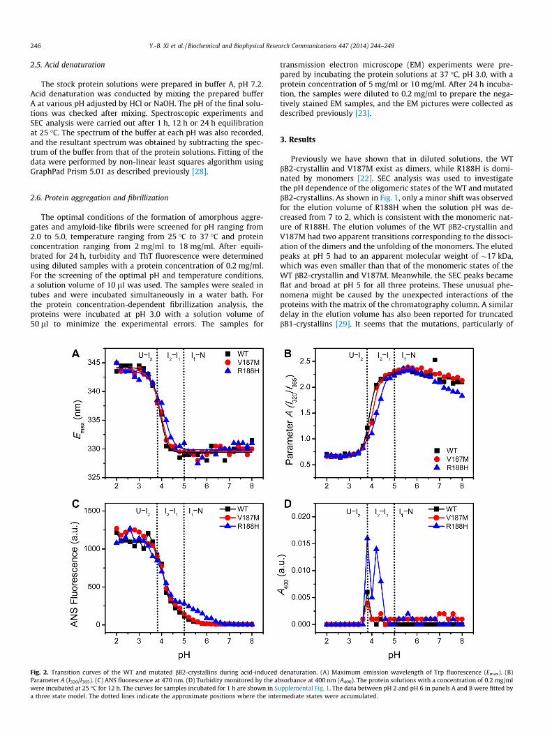
Fig. 2. Transition curves of the WT and mutated bB2-crystallins during acid-inducedParameter A (I320/I365). (C) ANS fluorescence at 470 nm. (D) Turbidity monitored by the awere incubated at 25 �C for 12 h. The curves for samples incubated for 1 h are shown in Sa three state model. The dotted lines indicate the approximate positions where the inte
transmission electron microscope (EM) experiments were pre-pared by incubating the protein solutions at 37 �C, pH 3.0, with aprotein concentration of 5 mg/ml or 10 mg/ml. After 24 h incuba-tion, the samples were diluted to 0.2 mg/ml to prepare the nega-tively stained EM samples, and the EM pictures were collected asdescribed previously [23].
3. Results
Previously we have shown that in diluted solutions, the WTbB2-crystallin and V187M exist as dimers, while R188H is domi-nated by monomers [22]. SEC analysis was used to investigatethe pH dependence of the oligomeric states of the WT and mutatedbB2-crystallins. As shown in Fig. 1, only a minor shift was observedfor the elution volume of R188H when the solution pH was de-creased from 7 to 2, which is consistent with the monomeric nat-ure of R188H. The elution volumes of the WT bB2-crystallin andV187M had two apparent transitions corresponding to the dissoci-ation of the dimers and the unfolding of the monomers. The elutedpeaks at pH 5 had to an apparent molecular weight of �17 kDa,which was even smaller than that of the monomeric states of theWT bB2-crystallin and V187M. Meanwhile, the SEC peaks becameflat and broad at pH 5 for all three proteins. These unusual phe-nomena might be caused by the unexpected interactions of theproteins with the matrix of the chromatography column. A similardelay in the elution volume has also been reported for truncatedbB1-crystallins [29]. It seems that the mutations, particularly of
denaturation. (A) Maximum emission wavelength of Trp fluorescence (Emax). (B)bsorbance at 400 nm (A400). The protein solutions with a concentration of 0.2 mg/mlupplemental Fig. 1. The data between pH 2 and pH 6 in panels A and B were fitted byrmediate states were accumulated.

Y.-B. Xi et al. / Biochemical and Biophysical Research Communications 447 (2014) 244–249 247
R188H, reduced the strong interactions of bB2-crystallin with thecolumn matrix, reflecting that the mutations might modify the bio-physical properties of the intermediate state appeared at pH 5.
The structural changes induced by acid denaturation were mon-itored by the intrinsic and extrinsic fluorescence of the proteinsincubated under various pH conditions for 1 h (SupplementalFig. 1) or 12 h (Fig. 2) at 25 �C. A comparison between the two setsof transition curves indicated that a longer incubation time wasneeded to fully denature the proteins in solutions with a pH be-tween 3.6 and 4.6. A small portion of proteins tended to formaggregates when incubated at around pH 4 for 12 h (Fig. 2D andSupplemental Fig. 2), while no aggregates were observed for sam-ples incubated for 1 h. Nonetheless, the acid-denaturation of thethree proteins could be best fitted by a three-state model underpH 5, while an additional transition between pH 5 and 7 couldbe observed from the changes of the ANS fluorescence (Fig. 2C)as well as SEC analysis (Fig. 1). Thus the acid-induced unfoldingof bB2-crystallin could be described by the following four-statemodel involving two unfolding intermediates (Scheme 1):
Intermediate I1 was a monomeric state that maintained most ofthe native structures of bB2-crystallin and possessed a few ANS-accessible hydrophobic sites, while intermediate I2 was partiallyunfolded and was prone to aggregate. The unfolded state U wasdominated by disordered structures with all Trp fluorophoresaccessible by the solvent and contained some residual b-sheetstructures (Supplemental Fig. 3).
The transition curves of V187M were almost superimposed tothose of the WT protein. As for the R188H mutation, the most
2U . I2I2N 212 ⇔⇔⇔
Scheme 1.
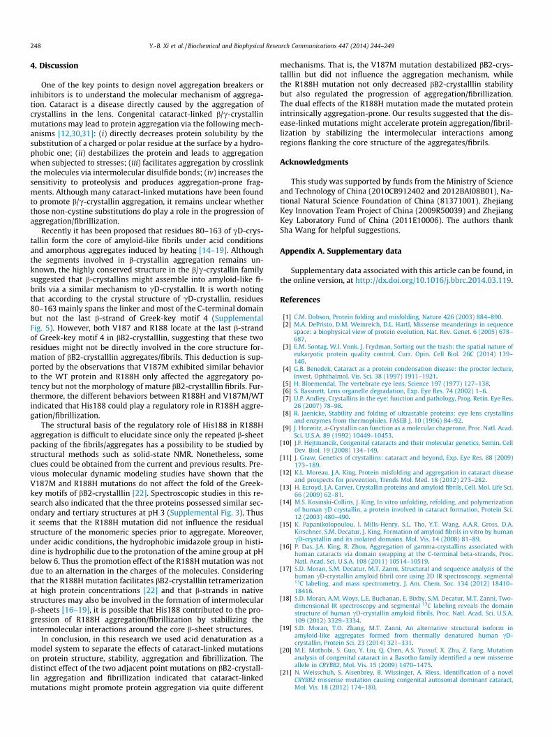
Fig. 3. Aggregation and fibrillization of the WT and mutated bB2-crystallin. (A) pH-depeconcentration of 0.2 mg/ml were incubated at 37 �C for 24 h. (B) Concentration-depenincubated at 37 �C and pH 3 for 24 h. The joint-position of the two straight lines indicat7 mg/ml for the WT protein and V187M, respectively. (C) Negatively stained transmission(D) Negatively stained transmission EM pictures of 10 mg/ml proteins incubated at 37 �
significant effect was the introduction of extra ANS-binding sitesduring the N–I1 transition reflected by the slight increase of theANS fluorescence (Fig. 2C and Supplemental Fig. 1C). It is worthnoting that the hydrophobic imidazole group at His side-chain be-comes positively charged at pH below 6. Considering that theR188H mutation dissociates bB2-crystallin into monomers in di-luted solutions [22], the extra ANS-accessible sites were morelikely to be introduced by the exposure of the hydrophobic sitesat the subunit interface (Supplemental Fig. 4). Another impairingeffect caused by R188H mutation is the destabilization of I1 re-vealed by the shift of the I1–I2 transition curve to higher pH valeswhen compared to the WT protein.
To further evaluate the effect of mutations on the aggregatorypotency of bB2-crystallin during acid denaturation, the turbiditydata were recorded for samples incubated at 37 �C for 24 h(Fig. 3A). Compared to the results in Fig. 2D, the most striking dif-ference is the serious aggregation of R188H when incubated at37 �C under a rather wide pH range (from pH 3.2 to pH 6.4). AtpH 3, all three proteins did not aggregate in diluted solutions witha protein concentration of 0.2 mg/ml. At high protein concentra-tions, all three proteins could form amyloid-like fibrils when incu-bated at 37 �C, pH 3, for 24 h (Fig. 3B). It seems that V187M hadsimilar fibrillization properties to the WT protein. The R188Hmutation slightly decreased the critical concentration of bB2-crys-tallin fibrillization and promoted the growth of the fibrils. Similarobservation could also be seen from the EM measurements. At aprotein concentration of 5 mg/ml, the WT protein only formedspherical assemblies or stick-like structures, while fibrillar orannular structures could be found for R188H. When the proteinconcentration was increased to 10 mg/ml, both the WT proteinand R188H were extensively fibrilized, and the fibrils formed byR188H were more dense and lengthy.
ndence of protein aggregation monitored by turbidity. The solutions with a proteindence of protein fibrillization monitored by ThT fluorescence. The proteins were
es the critical concentration of fibrillization, which is about 6 mg/ml for R188H andEM pictures of 5 mg/ml WT protein and R188H incubated at 37 �C and pH 3 for 24 h.C and pH 3 for 24 h.
248 Y.-B. Xi et al. / Biochemical and Biophysical Research Communications 447 (2014) 244–249
4. Discussion
One of the key points to design novel aggregation breakers orinhibitors is to understand the molecular mechanism of aggrega-tion. Cataract is a disease directly caused by the aggregation ofcrystallins in the lens. Congenital cataract-linked b/c-crystallinmutations may lead to protein aggregation via the following mech-anisms [12,30,31]: (i) directly decreases protein solubility by thesubstitution of a charged or polar residue at the surface by a hydro-phobic one; (ii) destabilizes the protein and leads to aggregationwhen subjected to stresses; (iii) facilitates aggregation by crosslinkthe molecules via intermolecular disulfide bonds; (iv) increases thesensitivity to proteolysis and produces aggregation-prone frag-ments. Although many cataract-linked mutations have been foundto promote b/c-crystallin aggregation, it remains unclear whetherthose non-cystine substitutions do play a role in the progression ofaggregation/fibrillization.
Recently it has been proposed that residues 80–163 of cD-crys-tallin form the core of amyloid-like fibrils under acid conditionsand amorphous aggregates induced by heating [14–19]. Althoughthe segments involved in b-crystallin aggregation remains un-known, the highly conserved structure in the b/c-crystallin familysuggested that b-crystallins might assemble into amyloid-like fi-brils via a similar mechanism to cD-crystallin. It is worth notingthat according to the crystal structure of cD-crystallin, residues80–163 mainly spans the linker and most of the C-terminal domainbut not the last b-strand of Greek-key motif 4 (SupplementalFig. 5). However, both V187 and R188 locate at the last b-strandof Greek-key motif 4 in bB2-crystalllin, suggesting that these tworesidues might not be directly involved in the core structure for-mation of bB2-crystalllin aggregates/fibrils. This deduction is sup-ported by the observations that V187M exhibited similar behaviorto the WT protein and R188H only affected the aggregatory po-tency but not the morphology of mature bB2-crystalllin fibrils. Fur-thermore, the different behaviors between R188H and V187M/WTindicated that His188 could play a regulatory role in R188H aggre-gation/fibrillization.
The structural basis of the regulatory role of His188 in R188Haggregation is difficult to elucidate since only the repeated b-sheetpacking of the fibrils/aggregates has a possibility to be studied bystructural methods such as solid-state NMR. Nonetheless, someclues could be obtained from the current and previous results. Pre-vious molecular dynamic modeling studies have shown that theV187M and R188H mutations do not affect the fold of the Greek-key motifs of bB2-crystalllin [22]. Spectroscopic studies in this re-search also indicated that the three proteins possessed similar sec-ondary and tertiary structures at pH 3 (Supplemental Fig. 3). Thusit seems that the R188H mutation did not influence the residualstructure of the monomeric species prior to aggregate. Moreover,under acidic conditions, the hydrophobic imidazole group in histi-dine is hydrophilic due to the protonation of the amine group at pHbelow 6. Thus the promotion effect of the R188H mutation was notdue to an alternation in the charges of the molecules. Consideringthat the R188H mutation facilitates bB2-crystalllin tetramerizationat high protein concentrations [22] and that b-strands in nativestructures may also be involved in the formation of intermolecularb-sheets [16–19], it is possible that His188 contributed to the pro-gression of R188H aggregation/fibrillization by stabilizing theintermolecular interactions around the core b-sheet structures.
In conclusion, in this research we used acid denaturation as amodel system to separate the effects of cataract-linked mutationson protein structure, stability, aggregation and fibrillization. Thedistinct effect of the two adjacent point mutations on bB2-crystall-lin aggregation and fibrillization indicated that cataract-linkedmutations might promote protein aggregation via quite different
mechanisms. That is, the V187M mutation destabilized bB2-crys-talllin but did not influence the aggregation mechanism, whilethe R188H mutation not only decreased bB2-crystalllin stabilitybut also regulated the progression of aggregation/fibrillization.The dual effects of the R188H mutation made the mutated proteinintrinsically aggregation-prone. Our results suggested that the dis-ease-linked mutations might accelerate protein aggregation/fibril-lization by stabilizing the intermolecular interactions amongregions flanking the core structure of the aggregates/fibrils.
Acknowledgments
This study was supported by funds from the Ministry of Scienceand Technology of China (2010CB912402 and 2012BAI08B01), Na-tional Natural Science Foundation of China (81371001), ZhejiangKey Innovation Team Project of China (2009R50039) and ZhejiangKey Laboratory Fund of China (2011E10006). The authors thankSha Wang for helpful suggestions.
Appendix A. Supplementary data
Supplementary data associated with this article can be found, inthe online version, at http://dx.doi.org/10.1016/j.bbrc.2014.03.119.
References
[1] C.M. Dobson, Protein folding and misfolding, Nature 426 (2003) 884–890.[2] M.A. DePristo, D.M. Weinreich, D.L. Hartl, Missense meanderings in sequence
space: a biophysical view of protein evolution, Nat. Rev. Genet. 6 (2005) 678–687.
[3] E.M. Sontag, W.I. Vonk, J. Frydman, Sorting out the trash: the spatial nature ofeukaryotic protein quality control, Curr. Opin. Cell Biol. 26C (2014) 139–146.
[4] G.B. Benedek, Cataract as a protein condensation disease: the proctor lecture,Invest. Ophthalmol. Vis. Sci. 38 (1997) 1911–1921.
[5] H. Bloemendal, The vertebrate eye lens, Science 197 (1977) 127–138.[6] S. Bassnett, Lens organelle degradation, Exp. Eye Res. 74 (2002) 1–6.[7] U.P. Andley, Crystallins in the eye: function and pathology, Prog. Retin. Eye Res.
26 (2007) 78–98.[8] R. Jaenicke, Stability and folding of ultrastable proteins: eye lens crystallins
and enzymes from thermophiles, FASEB J. 10 (1996) 84–92.[9] J. Horwitz, a-Crystallin can function as a molecular chaperone, Proc. Natl. Acad.
Sci. U.S.A. 89 (1992) 10449–10453.[10] J.F. Hejtmancik, Congenital cataracts and their molecular genetics, Semin. Cell
Dev. Biol. 19 (2008) 134–149.[11] J. Graw, Genetics of crystallins: cataract and beyond, Exp. Eye Res. 88 (2009)
173–189.[12] K.L. Moreau, J.A. King, Protein misfolding and aggregation in cataract disease
and prospects for prevention, Trends Mol. Med. 18 (2012) 273–282.[13] H. Ecroyd, J.A. Carver, Crystallin proteins and amyloid fibrils, Cell. Mol. Life Sci.
66 (2009) 62–81.[14] M.S. Kosinski-Collins, J. King, In vitro unfolding, refolding, and polymerization
of human cD crystallin, a protein involved in cataract formation, Protein Sci.12 (2003) 480–490.
[15] K. Papanikolopoulou, I. Mills-Henry, S.L. Tho, Y.T. Wang, A.A.R. Gross, D.A.Kirschner, S.M. Decatur, J. King, Formation of amyloid fibrils in vitro by humancD-crystallin and its isolated domains, Mol. Vis. 14 (2008) 81–89.
[16] P. Das, J.A. King, R. Zhou, Aggregation of gamma-crystallins associated withhuman cataracts via domain swapping at the C-terminal beta-strands, Proc.Natl. Acad. Sci. U.S.A. 108 (2011) 10514–10519.
[17] S.D. Moran, S.M. Decatur, M.T. Zanni, Structural and sequence analysis of thehuman cD-crystallin amyloid fibril core using 2D IR spectroscopy, segmental13C labeling, and mass spectrometry, J. Am. Chem. Soc. 134 (2012) 18410–18416.
[18] S.D. Moran, A.M. Woys, L.E. Buchanan, E. Bixby, S.M. Decatur, M.T. Zanni, Two-dimensional IR spectroscopy and segmental 13C labeling reveals the domainstructure of human cD-crystallin amyloid fibrils, Proc. Natl. Acad. Sci. U.S.A.109 (2012) 3329–3334.
[19] S.D. Moran, T.O. Zhang, M.T. Zanni, An alternative structural isoform inamyloid-like aggregates formed from thermally denatured human cD-crystallin, Protein Sci. 23 (2014) 321–331.
[20] M.E. Mothobi, S. Guo, Y. Liu, Q. Chen, A.S. Yussuf, X. Zhu, Z. Fang, Mutationanalysis of congenital cataract in a Basotho family identified a new missenseallele in CRYBB2, Mol. Vis. 15 (2009) 1470–1475.
[21] N. Weisschuh, S. Aisenbrey, B. Wissinger, A. Riess, Identification of a novelCRYBB2 missense mutation causing congenital autosomal dominant cataract,Mol. Vis. 18 (2012) 174–180.
Y.-B. Xi et al. / Biochemical and Biophysical Research Communications 447 (2014) 244–249 249
[22] K. Zhang, W.-J. Zhao, X.-Y. Leng, S. Wang, K. Yao, Y.-B. Yan, The importance ofthe last strand at the C-terminus in bB2-crystallin stability and assembly,Biochim. Biophys. Acta Mol. Basis Dis. 2014 (1842) 44–55.
[23] J. Xu, S. Wang, W.-J. Zhao, Y.-B. Xi, Y.-B. Yan, K. Yao, The congenital cataract-linked A2V mutation impairs tetramer formation and promotes aggregation ofbB2-crystallin, PLoS ONE 7 (2012) e51200.
[24] M.M. Bradford, A rapid and sensitive method for the quantitation ofmicrogram quantities of protein utilizing the principle of protein–dyebinding, Anal. Biochem. 72 (1976) 248–254.
[25] W. Zhang, H.-C. Cai, F.-F. Li, Y.-B. Xi, X. Ma, Y.-B. Yan, The congenital cataract-linked G61C mutation destabilizes cD-crystallin and promotes non-nativeaggregation, PLoS ONE 6 (2011) e20564.
[26] K.K. Turoverov, S.Y. Haitlina, G.P. Pinaev, Ultra-violet fluorescence of actin.Determination of native actin content in actin preparations, FEBS Lett. 62(1976) 4–6.
[27] G.-J. He, A. Zhang, W.-F. Liu, Y. Cheng, Y.-B. Yan, Conformational stability andmultistate unfolding of poly(A)-specific ribonuclease, FEBS J. 276 (2009) 2849–2860.
[28] S. Wang, X.-Y. Leng, Y.-B. Yan, The benefits of being b-crystallin heteromers:bB1-crystallin protects bA3-crystallin against aggregation during co-refolding,Biochemistry 50 (2011) 10451–10461.
[29] O.A. Bateman, N.H. Lubsen, C. Slingsby, Association behaviour of human bB1-crystallin and its truncated forms, Exp. Eye Res. 73 (2001) 321–331.
[30] V.P.R. Vendra, G. Agarwal, S. Chandani, V. Talla, N. Srinivasan, D.Balasubramanian, Structural integrity of the Greek key motif in bc-crystallins is vital for central eye lens transparency, PLoS ONE 8 (2013) e70336.
[31] S. Wang, W.-J. Zhao, H. Liu, H. Gong, Y.-B. Yan, Increasing bB1-crystallinsensitivity to proteolysis caused by the congenital cataract-microcorneasyndrome mutation S129R, Biochim. Biophys. Acta Mol. Basis Dis. 2013(1832) 302–311.


![Characterization of an antibody that recognizes peptides ... · in αA-crystallin (Asp 58 and Asp 151) [3], αB-crystallin (Asp 36 and Asp 62) [4], and βB2-crsytallin (Asp 4) [5]](https://static.fdocument.pub/doc/165x107/5ff1e68e89243b57b64135f8/characterization-of-an-antibody-that-recognizes-peptides-in-a-crystallin-asp.jpg)