Case Report Recently proposed oncocytic variant of ... · of perinuclear halo, (iii) diffuse...
8
Int J Clin Exp Pathol 2016;9(2):2374-2381 www.ijcep.com /ISSN:1936-2625/IJCEP0019351 Case Report Recently proposed oncocytic variant of chromophobe renal cell carcinoma (RCC) expressing BSND and ATP6V1G3: two new immunohistochemical markers differentiating chromophobe RCC from other RCC subtypes Shogo Tajima 1* , Yoichi Mori 2* , Yoshihiro Mukaiyama 2 , Kazuya Shinmura 1 , Kenji Koda 1 , Naoto Kuroda 3 , Shinsuke Hayami 2 , Yukio Homma 4 Departments of 1 Pathology, 2 Urology, Fujieda Municipal General Hospital, Shizuoka, Japan; 3 Department of Diagnostic Pathology, Kochi Red Cross Hospital, Kochi, Japan; 4 Department of Urology, Graduate School of Medicine, The University of Tokyo, Tokyo, Japan. * Equal contributors. Received November 7, 2015; Accepted January 3, 2016; Epub February 1, 2016; Published February 15, 2016 Abstract: Eosinophilic/oncocytic renal cell neoplasms constitute a certain proportion of renal tumors. Low-grade eosinophilic renal cell carcinomas (RCCs) with nested growth, which do not meet the criteria of oncocytoma or eo- sinophilic variant of chromophobe RCC, may be categorized as eosinophilic unclassified RCCs. Recent advances in immunohistochemical discrimination between various RCCs have made it possible to classify these tumors more accurately. BSND and ATP6V1G3 have been shown to be highly sensitive and specific immunohistochemical mark- ers for chromophobe RCC and oncocytoma. We encountered a 67-year-old Japanese woman with a newly proposed oncocytic variant of chromophobe RCC, which originated from the upper part of her right kidney. It was morphologi- cally different from oncocytoma and eosinophilic variant of chromophobe RCC; it seemed that cases like this one would be classified as eosinophilic unclassified RCCs with use of commonly available immunohistochemical mark- ers. Immunohistochemistry using BSND and ATP6V1G3 proved helpful in making the diagnosis with confidence in this case. The diagnosis was also confirmed by the loss of chromosomes 10 and 17, which is typically observed in chromophobe RCC. Further exploration of new immunohistochemical markers is needed for more accurately clas- sifying RCCs than ever before. Keywords: ATP6V1G3, BSND, chromophobe renal cell carcinoma, eosinophilic variant, oncocytic variant, oncocy- toma Introduction Recent advances in immunohistochemical dis- crimination between various renal cell carcino- mas (RCCs) have made it possible to classify them with greater accuracy [1]. However, new immunohistochemical markers that are more sensitive and specific to a certain tumor type are being actively explored. Such markers are especially important when diagnosing cases that are difficult to differentiate based on their morphology. Despite recent advancements in marker discovery, unclassified RCCs represent 1-5% of all RCCs [2]. Eosinophilic/oncocytic renal cell neoplasms constitute a certain proportion of all renal tumors [3]. Low-grade eosinophilic RCCs with nested growth, which do not meet the criteria of oncocytoma or eosinophilic variant of chromo- phobe RCC, may be categorized as unclassified RCC. Oncocytomas or eosinophilic variants of chromophobe RCC may be diagnosed with con- fidence using immunohistochemical markers such as CK7, MOC31, CD82, and c-kit, in addi- tion to their morphology [4]. However, more sensitive and specific markers for them are required, as some cases categorized as eosino- philic unclassified RCC may in fact represent oncocytomas or eosinophilic variants of chro- mophobe RCC that are particularly difficult to diagnose. More recently, BSND and ATP6V1G3 have been shown to be highly sensitive and specific immu-
Transcript of Case Report Recently proposed oncocytic variant of ... · of perinuclear halo, (iii) diffuse...
Int J Clin Exp Pathol 2016;9(2):2374-2381www.ijcep.com /ISSN:1936-2625/IJCEP0019351
Case Report Recently proposed oncocytic variant of chromophobe renal cell carcinoma (RCC) expressing BSND and ATP6V1G3: two new immunohistochemical markers differentiating chromophobe RCC from other RCC subtypes
Shogo Tajima1*, Yoichi Mori2*, Yoshihiro Mukaiyama2, Kazuya Shinmura1, Kenji Koda1, Naoto Kuroda3, Shinsuke Hayami2, Yukio Homma4
Departments of 1Pathology, 2Urology, Fujieda Municipal General Hospital, Shizuoka, Japan; 3Department of Diagnostic Pathology, Kochi Red Cross Hospital, Kochi, Japan; 4Department of Urology, Graduate School of Medicine, The University of Tokyo, Tokyo, Japan. *Equal contributors.
Received November 7, 2015; Accepted January 3, 2016; Epub February 1, 2016; Published February 15, 2016
Abstract: Eosinophilic/oncocytic renal cell neoplasms constitute a certain proportion of renal tumors. Low-grade eosinophilic renal cell carcinomas (RCCs) with nested growth, which do not meet the criteria of oncocytoma or eo-sinophilic variant of chromophobe RCC, may be categorized as eosinophilic unclassified RCCs. Recent advances in immunohistochemical discrimination between various RCCs have made it possible to classify these tumors more accurately. BSND and ATP6V1G3 have been shown to be highly sensitive and specific immunohistochemical mark-ers for chromophobe RCC and oncocytoma. We encountered a 67-year-old Japanese woman with a newly proposed oncocytic variant of chromophobe RCC, which originated from the upper part of her right kidney. It was morphologi-cally different from oncocytoma and eosinophilic variant of chromophobe RCC; it seemed that cases like this one would be classified as eosinophilic unclassified RCCs with use of commonly available immunohistochemical mark-ers. Immunohistochemistry using BSND and ATP6V1G3 proved helpful in making the diagnosis with confidence in this case. The diagnosis was also confirmed by the loss of chromosomes 10 and 17, which is typically observed in chromophobe RCC. Further exploration of new immunohistochemical markers is needed for more accurately clas-sifying RCCs than ever before.
Keywords: ATP6V1G3, BSND, chromophobe renal cell carcinoma, eosinophilic variant, oncocytic variant, oncocy-toma
Introduction
Recent advances in immunohistochemical dis-crimination between various renal cell carcino-mas (RCCs) have made it possible to classify them with greater accuracy [1]. However, new immunohistochemical markers that are more sensitive and specific to a certain tumor type are being actively explored. Such markers are especially important when diagnosing cases that are difficult to differentiate based on their morphology. Despite recent advancements in marker discovery, unclassified RCCs represent 1-5% of all RCCs [2].
Eosinophilic/oncocytic renal cell neoplasms constitute a certain proportion of all renal tumors [3]. Low-grade eosinophilic RCCs with
nested growth, which do not meet the criteria of oncocytoma or eosinophilic variant of chromo-phobe RCC, may be categorized as unclassified RCC. Oncocytomas or eosinophilic variants of chromophobe RCC may be diagnosed with con-fidence using immunohistochemical markers such as CK7, MOC31, CD82, and c-kit, in addi-tion to their morphology [4]. However, more sensitive and specific markers for them are required, as some cases categorized as eosino-philic unclassified RCC may in fact represent oncocytomas or eosinophilic variants of chro-mophobe RCC that are particularly difficult to diagnose.
More recently, BSND and ATP6V1G3 have been shown to be highly sensitive and specific immu-

Oncocytic variant of chromophobe RCC
2375 Int J Clin Exp Pathol 2016;9(2):2374-2381
nohistochemical markers for chromophobe RCC and oncocytoma [5]. These proteins are involved in the regulation of membrane trans-port and are expressed in the distal nephron, including in the collecting duct, of the normal kidney [5].
We encountered a case of newly proposed oncocytic variant of chromophobe RCC [6, 7]. In order to confirm this diagnosis and to deter-mine whether this tumor is truly a variant of
chromophobe RCC, immunohistochemistry of BSND and ATP6V1G3 was performed.
Clinical summary
A 67-year-old Japanese woman was referred to our hospital due to a mass in her right kidney, which was found by abdominal ultrasonography during check-up. She had no other complaints and laboratory tests revealed no abnormality. Computed tomography (CT) was performed and a mass measuring 82×68×62 mm with a cystic part was found in the upper part of the right kidney. The mass was moderately enhanced in appearance with contrast-enhanced CT (Figure 1A, 1B). No lymph node swelling and meta- stasis were identified. RCC was suspected and tumor stage was clinically evaluated as pT2aN0M0. Subsequently, laparoscopic right nephrectomy and lymph node dissection were performed. The patient has been recurrence-free for 3 months. Of note, she had no familial history of Birt-Hogg-Dube syndrome, and no previous history of neuroblastoma and hemo- dialysis.
Pathological findings
The surgically resected specimen revealed a brownish tumor, measuring 84×66×62 mm, in the upper part of the right kidney. It was well circumscribed but not encapsulated. A cystic part was present and this was considered to indicate cystic degeneration of the tumor (Figure 2). Central scar and necrosis were not apparent.
Figure 1. Computed tomography findings. A. Axial; B. Coronal. A mass was found in the upper part of the right kid-ney. The mass was moderately enhanced with contrast material administration. Cystic part was observed within the mass.
Figure 2. Macroscopic findings. The surgically resect-ed specimen revealed a brownish tumor in the upper part of the right kidney. It was well circumscribed but not encapsulated. Cystic degeneration was present within the tumor.
Oncocytic variant of chromophobe RCC
2376 Int J Clin Exp Pathol 2016;9(2):2374-2381
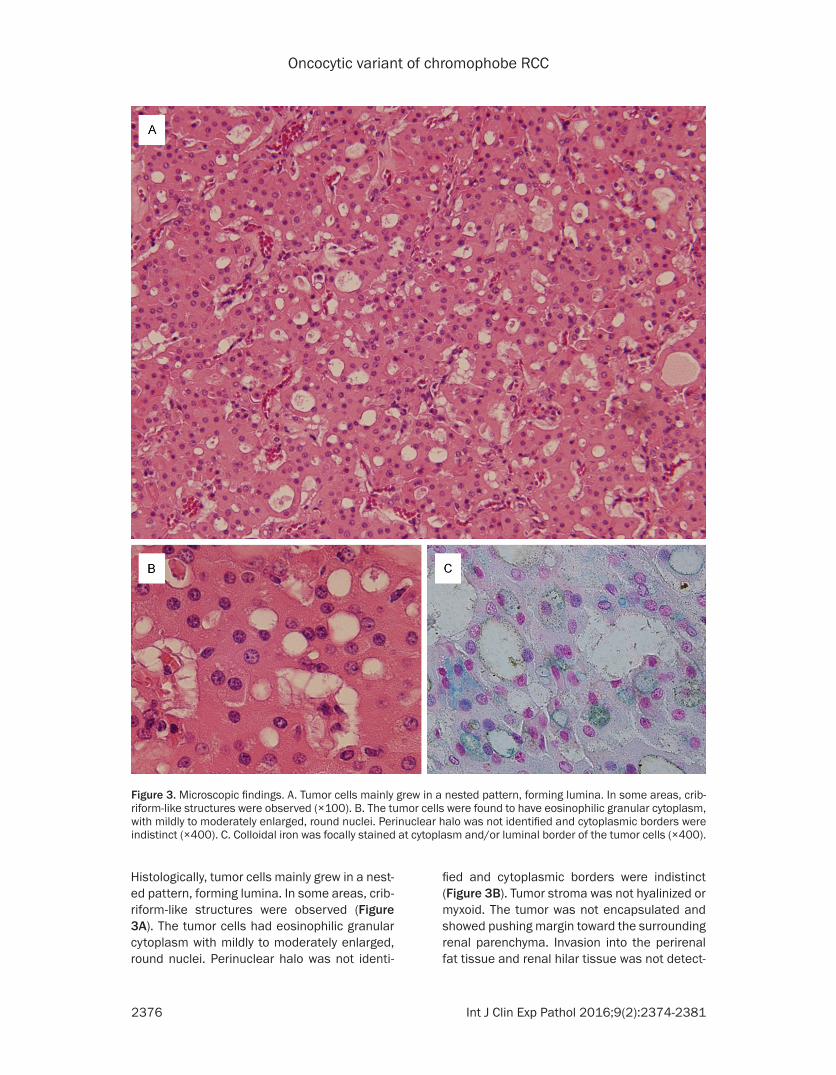
Histologically, tumor cells mainly grew in a nest-ed pattern, forming lumina. In some areas, crib-riform-like structures were observed (Figure 3A). The tumor cells had eosinophilic granular cytoplasm with mildly to moderately enlarged, round nuclei. Perinuclear halo was not identi-
fied and cytoplasmic borders were indistinct (Figure 3B). Tumor stroma was not hyalinized or myxoid. The tumor was not encapsulated and showed pushing margin toward the surrounding renal parenchyma. Invasion into the perirenal fat tissue and renal hilar tissue was not detect-
Figure 3. Microscopic findings. A. Tumor cells mainly grew in a nested pattern, forming lumina. In some areas, crib-riform-like structures were observed (×100). B. The tumor cells were found to have eosinophilic granular cytoplasm, with mildly to moderately enlarged, round nuclei. Perinuclear halo was not identified and cytoplasmic borders were indistinct (×400). C. Colloidal iron was focally stained at cytoplasm and/or luminal border of the tumor cells (×400).

Oncocytic variant of chromophobe RCC
2377 Int J Clin Exp Pathol 2016;9(2):2374-2381
ed. Lymphatic invasion and vascular invasion were not observed. Surgical margins were free of the tumor. No lymph node metastasis was detected.
Histochemically, colloidal iron was stained in the cytoplasm and/or luminal border of the tumor cells; this staining was focally observed (Figure 3C).
Immunohistochemically, the tumor cells were diffusely positive for CK7 (OV-TL 12/30, 1:80; Dako, Glostrup, Denmark) (Figure 4A), MOC31 (MOC31, 1:100; Dako) (Figure 4B), CD82 (G-2, 1:200; Santa Cruz Biotechnology, Santa Cruz, CA) (Figure 4C), BSND (polyclonal, 1:1000; Sigma-Aldrich, St. Louis, MO) (Figure 4D), ATP6V1G3 (polyclonal, 1:2000; Sigma-Aldrich) (Figure 4E), and mitochondrial antigen (MTC02,
Figure 4. Immunohistochemical findings. A. Diffuse Immunopositivity for CK7 (×400). B. Diffuse Immunopositivity for MOC31 (×400). C. Diffuse Immunopositivity for CD82 (×400). D. Diffuse Immunopositivity for BSND (×400). E. Diffuse Immunopositivity for ATP6V1G3 (×400). F. Diffuse immunopositivity for mitochondrial antigen (×400).
Oncocytic variant of chromophobe RCC
2378 Int J Clin Exp Pathol 2016;9(2):2374-2381
1:100; Epitomics, Burlingame, CA) (Figure 4F). They were focally positive for c-kit (polyclonal, 1:400; Dako). However, they were negative for carboanhydrase IX (D47G3, 1:200; Cell Sig- naling Technology, Beverly, MA), RCC marker (PN-15, prediluted; Cell Marque, Rocklin, CA), CD10 (56C6, prediluted; Novocastra Labora-tories, Newcastle Upon Tyne, UK), Melan A (A103, 1:100; Novocastra), αSMA (1A4, 1:200; DAKO), and TFE3 (polyclonal, 1:3600; Santa Cruz Biotechnology). Ki-67 (MIB-1, 1:100; Dako) labeling index was 4.6%, counting 1000 nuclei.
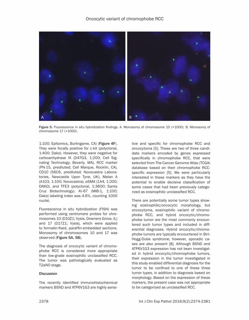
Fluorescence in situ hybridization (FISH) was performed using centromere probes for chro-mosomes 10 (D10Z1; Vysis, Downers Grove, IL) and 17 (D17Z1; Vysis), which were applied to formalin-fixed, paraffin-embedded sections. Monosomy of chromosomes 10 and 17 was observed (Figure 5A, 5B).
The diagnosis of oncocytic variant of chromo-phobe RCC is considered more appropriate than low-grade eosinophilic unclassified RCC. The tumor was pathologically evaluated as T2aN0 stage.
Discussion
The recently identified immunohistochemical markers BSND and ATP6V1G3 are highly sensi-
tive and specific for chromophobe RCC and oncocytoma [5]. These are two of three candi-date markers encoded by genes expressed specifically in chromophobe RCC, that were selected from The Cancer Genome Atlas (TCGA) database based on their chromophobe RCC-specific expression [5]. We were particularly interested in these markers as they have the potential to enable decisive classification of some cases that had been previously catego-rized as eosinophilic unclassified RCC.
There are potentially some tumor types show-ing eosinophilic/oncocytic morphology, but oncocytoma, eosinophilic variant of chromo-phobe RCC, and hybrid oncocytic/chromo-phobe tumor are the most commonly encoun-tered such tumor types and included in diff- erential diagnoses. Hybrid oncocytic/chromo-phobe tumors are typically encountered in Birt-Hogg-Dube syndrome; however, sporadic ca- ses are also present [8]. Although BSND and ATP6V1G3 expression has not been investigat-ed in hybrid oncocytic/chromophobe tumors, their expression in the tumor investigated in this study enabled differential diagnosis for the tumor to be confined to one of these three tumor types, in addition to diagnosis based on morphology. Based on the expression of these markers, the present case was not appropriate to be categorized as unclassified RCC.
Figure 5. Fluorescence in situ hybridization findings. A. Monosomy of chromosome 10 (×1000). B. Monosomy of chromosome 17 (×1000).
Oncocytic variant of chromophobe RCC
2379 Int J Clin Exp Pathol 2016;9(2):2374-2381
When considering differential diagnoses for the aforementioned three tumors, it must be noted that eosinophilic variants of chromophobe RCC and hybrid oncocytic/chromophobe tumor are morphologically different from the present case. The former have wrinkled nuclei and peri-nuclear halo in tumor cells [9]; while the latter exhibit coexistence of oncocytoma-like tumor cells and chromophobe RCC-like tumor cells, and/or tumor cells with round nuclei and peri-nuclear halos [10]. These findings were not observed in our case. The immunophenotype of the tumor in this case is as follows: CK7-, MOC31+, CD82+, and c-kit focal +. This immu-nophenotype is typically observed in chromo-phobe RCC, but not in oncocytoma [4]. Th- erefore, the possibility of the tumor being an oncocytoma was ruled out. Observation of dif-fuse expression of BSND and ATP6V1G3 and the aforementioned immunophenotype strong-ly suggested that the tumor was within the spectrum of chromophobe RCC; however, the morphology of the tumor cells was found to be oncocyte-like, in that the tumor cells had eosin-ophilic granular cytoplasm with round nuclei.
Oncocytic variant of chromophobe RCC is a recently proposed tumor type [6, 7]. The first such case was reported in 2010 [6], and, in 2013, a more comprehensive study composed of five such cases, including the case reported in 2010, was documented [7]. Five characteris-tic findings were pointed out as follows: (i) tubu-lar and/or solid growth pattern, (ii) oncocytic cytoplasm with round nuclei and the absence of perinuclear halo, (iii) diffuse immunopositivi-ty for CK7 and mitochondrial antigen, and (iv) chromosomal abnormalities observed in chro-mophobe RCC [7]. The morphology of the tumor in the present case, as well as its immunophe-notype, indicated the possibility of the tumor being an oncocytic variant of chromophobe RCC.
Chromosomal analysis was used to confirm this diagnosis. It has been shown by SNP array anal-ysis that a loss of one copy of the entire chro-mosome, for most or all of chromosomes 1, 2, 6, 10, 13, and 17, was detected in the majority of cases of chromophobe RCC, including eosin-ophilic variants [11]. The genomic profile of oncocytoma and hybrid oncocytic/chromoph- obe tumor was balanced or showed a limited number of random imbalances, and chromo-
some losses such as those characteristic of chromophobe RCC were not observed [12]. Thus, monosomy of chromosome 10 and 17 observed in our case supported the diagnosis of oncocytic variant of chromophobe RCC.
Recently, the discovery of an oncocytic variant of papillary RCC has been reported. This tumor type should be considered when making differ-ential diagnoses, as a solid variant has also been documented [13, 14]. The oncocytic vari-ant of papillary RCC is reported to mimic onco-cytoma [13]. In addition, CK7 is expressed in this tumor type [13, 14]. These findings are similar to those observed in the present case. Had a focal papillary growth pattern or the prev-alence of foamy macrophages in the stroma of the tumor investigated in this study been observed, the tumor may have potentially been considered a variant of oncocytic papillary RCC [13, 15]. However, these features were not observed. Additionally, chromosomal analysis of some oncocytic variants of papillary RCCs has revealed gain of chromosomes 7 and 17, as is observed in the standard papillary RCCs [14]. In our case, loss of chromosome 17 was demonstrated and therefore, the possibility of the tumor being a solid variant of oncocytic papillary RCC was ruled out. Furthermore, dif-fuse expression of BSND and ATP6V1G3, which was observed in our case, was helpful for excluding this possibility, as these markers do not exhibit staining in papillary RCC [5].
Prognosis of chromophobe RCC does not sig-nificantly change for the eosinophilic variant; however, once sarcomatoid changes occur, the prognosis worsens [9]. Reliable prognostic data is not available for the oncocytic variant of chro-mophobe RCC. Considering that mitochondria are thought to accumulate only in tumor cells that are not actively dividing, the observation of mitochondrial hyperplasia in oncocytic tumors may suggest a low malignant potential [16, 17].
In conclusion, our report presents a newly proposed oncocytic variant of chromophobe RCC. Recently established immunohistochemi-cal markers of chromophobe RCC, BSND and ATP6V1G3, were helpful for the differential diagnosis of this tumor type, and diagnosis was also confirmed by chromosomal analysis. Fur- ther studies are required to gain an under-standing of the prognosis associated with
Oncocytic variant of chromophobe RCC
2380 Int J Clin Exp Pathol 2016;9(2):2374-2381
oncocytic variant of chromophobe RCC. In order to investigate this tumor type, it must be recog-nized and diagnosed correctly. The immunohis-tochemical markers BSND and ATP6V1G3 are highly useful to avoid facilely categorizing this tumor type as eosinophilic unclassified RCC.
Disclosure of conflict of interest
None.
Address correspondence to: Dr. Shogo Tajima, Department of Pathology, Fujieda Municipal General Hospital, Shizuoka 426-8677, Japan. Tel: +81-54-646-1111; Fax: +81-54-646-1122; E-mail: [email protected]
References
[1] Kuroda N, Tanaka A, Ohe C and Nagashima Y. Recent advances of immunohistochemistry for diagnosis of renal tumors. Pathol Int 2013; 63: 381-390.
[2] Lopez-Beltran A, Kirkali Z, Montironi R, Blanca A, Algaba F, Scarpelli M, Yorukoglu K, Hartmann A and Cheng L. Unclassified renal cell carcino-ma: a report of 56 cases. BJU Int 2012; 110: 786-793.
[3] Kryvenko ON, Jorda M, Argani P and Epstein JI. Diagnostic approach to eosinophilic renal neo-plasms. Arch Pathol Lab Med 2014; 138: 1531-1541.
[4] Ohe C, Kuroda N, Takasu K, Senzaki H, Shikata N, Yamaguchi T, Miyasaka C, Nakano Y, Sakaida N and Uemura Y. Utility of immunohis-tochemical analysis of KAI1, epithelial-specific antigen, and epithelial-related antigen for dis-tinction of chromophobe renal cell carcinoma, an eosinophilic variant from renal oncocytoma. Med Mol Morphol 2012; 45: 98-104.
[5] Shinmura K, Igarashi H, Kato H, Koda K, Ogawa H, Takahashi S, Otsuki Y, Yoneda T, Kawanishi Y, Funai K, Takayama T, Ozono S and Sugimura H. BSND and ATP6V1G3: Novel Immunohistochemical Markers for Chromo- phobe Renal Cell Carcinoma. Medicine (Balti- more) 2015; 94: e989.
[6] Yamaguchi T, Kuroda N, Imamura Y, Hes O, Michal M, Sima R, Nakayama K and Sato N. Imprint cytologic features of chromophobe re-nal cell carcinoma morphologically resembling renal oncocytoma: is this an oncocytic variant of chromophobe renal cell carcinoma? Diagn Cytopathol 2010; 38: 509-513.
[7] Kuroda N, Tanaka A, Yamaguchi T, Kasahara K, Naruse K, Yamada Y, Hatanaka K, Shinohara N, Nagashima Y, Mikami S, Oya M, Hamashima T, Michal M and Hes O. Chromophobe renal
cell carcinoma, oncocytic variant: a proposal of a new variant giving a critical diagnostic pitfall in diagnosing renal oncocytic tumors. Med Mol Morphol 2013; 46: 49-55.
[8] Petersson F, Gatalica Z, Grossmann P, Perez Montiel MD, Alvarado Cabrero I, Bulimbasic S, Swatek A, Straka L, Tichy T, Hora M, Kuroda N, Legendre B, Michal M and Hes O. Sporadic hy-brid oncocytic/chromophobe tumor of the kid-ney: a clinicopathologic, histomorphologic, im-munohistochemical, ultrastructural, and mo-lecular cytogenetic study of 14 cases. Virchows Arch 2010; 456: 355-365.
[9] Amin MB, Paner GP, Alvarado-Cabrero I, Young AN, Stricker HJ, Lyles RH and Moch H. Ch- romophobe renal cell carcinoma: histomorpho-logic characteristics and evaluation of conven-tional pathologic prognostic parameters in 145 cases. Am J Surg Pathol 2008; 32: 1822-1834.
[10] Hes O, Petersson F, Kuroda N, Hora M and Michal M. Renal hybrid oncocytic/chromo-phobe tumors-a review. Histol Histopathol 2013; 28: 1257-1264.
[11] Davis CF, Ricketts CJ, Wang M, Yang L, Cherniack AD, Shen H, Buhay C, Kang H, Kim SC, Fahey CC, Hacker KE, Bhanot G, Gordenin DA, Chu A, Gunaratne PH, Biehl M, Seth S, Kaipparettu BA, Bristow CA, Donehower LA, Wallen EM, Smith AB, Tickoo SK, Tamboli P, Reuter V, Schmidt LS, Hsieh JJ, Choueiri TK, Hakimi AA, Chin L, Meyerson M, Kucherlapati R, Park WY, Robertson AG, Laird PW, Henske EP, Kwiatkowski DJ, Park PJ, Morgan M, Shuch B, Muzny D, Wheeler DA, Linehan WM, Gibbs RA, Rathmell WK and Creighton CJ. The somat-ic genomic landscape of chromophobe renal cell carcinoma. Cancer Cell 2014; 26: 319-330.
[12] Pote N, Vieillefond A, Couturier J, Arrufat S, Metzger I, Delongchamps NB, Camparo P, Mege-Lechevallier F, Molinie V and Sibony M. Hybrid oncocytic/chromophobe renal cell tu-mours do not display genomic features of chro-mophobe renal cell carcinomas. Virchows Arch 2013; 462: 633-638.
[13] Mai KT, Kohler DM, Robertson SJ, Belanger EC and Marginean EC. Oncocytic papillary renal cell carcinoma with solid architecture: mimic of renal oncocytoma. Pathol Int 2008; 58: 164-168.
[14] Hes O, Brunelli M, Michal M, Cossu Rocca P, Hora M, Chilosi M, Mina M, Boudova L, Menestrina F and Martignoni G. Oncocytic papillary renal cell carcinoma: a clinicopatho-logic, immunohistochemical, ultrastructural, and interphase cytogenetic study of 12 cases. Ann Diagn Pathol 2006; 10: 133-139.
Oncocytic variant of chromophobe RCC
2381 Int J Clin Exp Pathol 2016;9(2):2374-2381
[15] Srigley JR and Delahunt B. Uncommon and re-cently described renal carcinomas. Mod Pathol 2009; 22 Suppl 2: S2-S23.
[16] Sobrinho-Simoes M and Maximo V. Warthin’s tumour. Virchows Arch 2006; 448: 877-878.
[17] Teymoortash A and Werner JA. Tissue that has lost its track: Warthin’s tumour. Virchows Arch 2005; 446: 585-588.