Carcinoma de células renales -...
19
Carcinoma de células renales Conceptos generales Creado para curso CES 2013
Transcript of Carcinoma de células renales -...
Carcinoma de células renales Conceptos generales
Creado para curso CES 2013
YOUR LOGO Page 2
Carcinoma de células renals
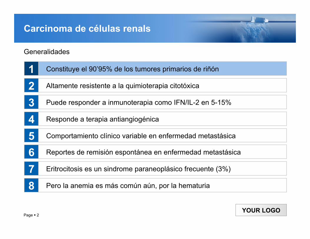
Generalidades
Constituye el 90’95% de los tumores primarios de riñón
Altamente resistente a la quimioterapia citotóxica
Puede responder a inmunoterapia como IFN/IL-2 en 5-15%
Responde a terapia antiangiogénica
Comportamiento clínico variable en enfermedad metastásica
1 2 3 4 5
Reportes de remisión espontánea en enfermedad metastásica
Eritrocitosis es un sindrome paraneoplásico frecuente (3%)
Pero la anemia es más común aún, por la hematuria
6 7 8
YOUR LOGO Page 3
Carcinoma de células renals
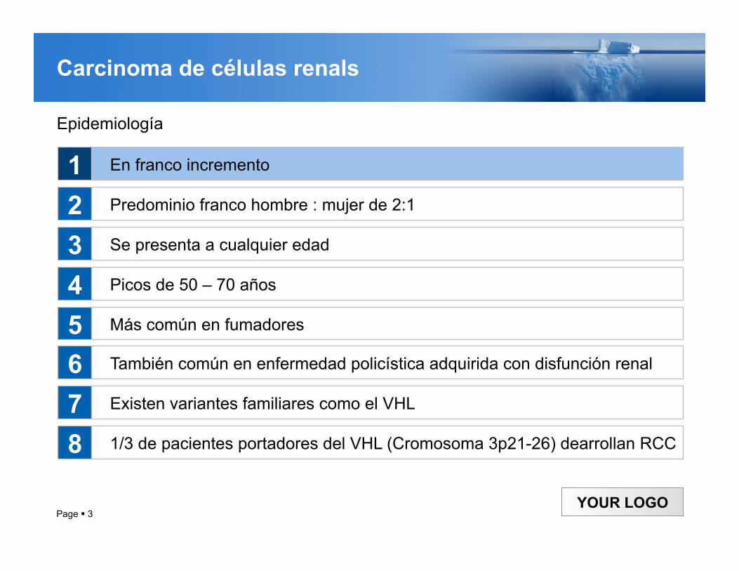
Epidemiología
En franco incremento
Predominio franco hombre : mujer de 2:1
Se presenta a cualquier edad
Picos de 50 – 70 años
Más común en fumadores
1 2 3 4 5
También común en enfermedad policística adquirida con disfunción renal
Existen variantes familiares como el VHL
1/3 de pacientes portadores del VHL (Cromosoma 3p21-26) dearrollan RCC
6 7 8
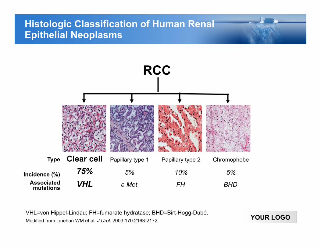
YOUR LOGO VHL=von Hippel-Lindau; FH=fumarate hydratase; BHD=Birt-Hogg-Dubé. Modified from Linehan WM et al. J Urol. 2003;170:2163-2172.
RCC
Clear cell 75%
Type
Incidence (%) Associated
mutations VHL
Papillary type 1
5%
c-Met
Papillary type 2
10%
FH
Chromophobe
5%
BHD
Histologic Classification of Human Renal Epithelial Neoplasms
YOUR LOGO Page 5
Carcinoma de células renals
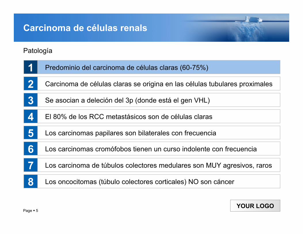
Patología
Predominio del carcinoma de células claras (60-75%)
Carcinoma de células claras se origina en las células tubulares proximales
Se asocian a deleción del 3p (donde está el gen VHL)
El 80% de los RCC metastásicos son de células claras
Los carcinomas papilares son bilaterales con frecuencia
1 2 3 4 5
Los carcinomas cromófobos tienen un curso indolente con frecuencia
Los carcinoma de túbulos colectores medulares son MUY agresivos, raros
Los oncocitomas (túbulo colectores corticales) NO son cáncer
6 7 8
YOUR LOGO
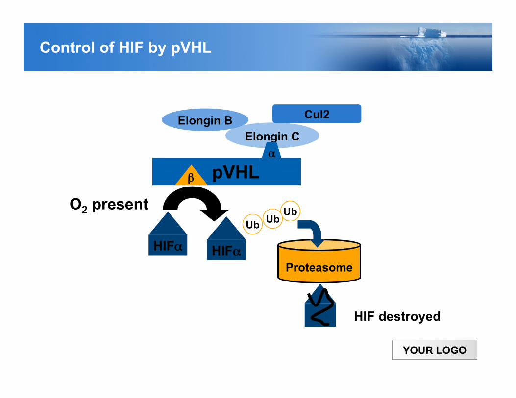
Ub
Elongin C α!
β! pVHL
Ub
Proteasome
HIF destroyed
O2 present
HIFα!
Ub
HIFα!
Cul2 Elongin B
Control of HIF by pVHL
YOUR LOGO
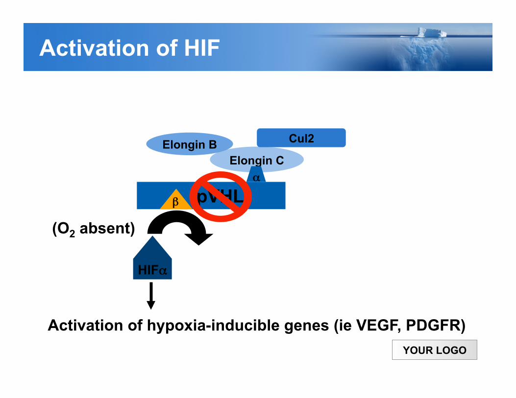
Elongin C α!
β! pVHL (O2 absent)
HIFα!
Cul2 Elongin B
Activation of hypoxia-inducible genes (ie VEGF, PDGFR)
Activation of HIF

YOUR LOGO Page 8
Carcinoma de células renales
Clásica (10-20%) Hematuria Dolor abdominal Masa en flanco
Otros síntomas Fiebre Pérdida de peso Varicocele
Cómo se detecta?
La forma de detección más común es hallazgo incidental en imágenes (CT, ecografía o RM)
Esto ha mejorado el pronóstico (por migración de estadío)
Paraneoplásicos
Eritrocitosis Hipercalcemia Disfunción hepática no metastásica (sindrome de Stauffer) Disfibrogenemia adquirida
Presentación Clínica
YOUR LOGO Page 9
Carcinoma de células renales
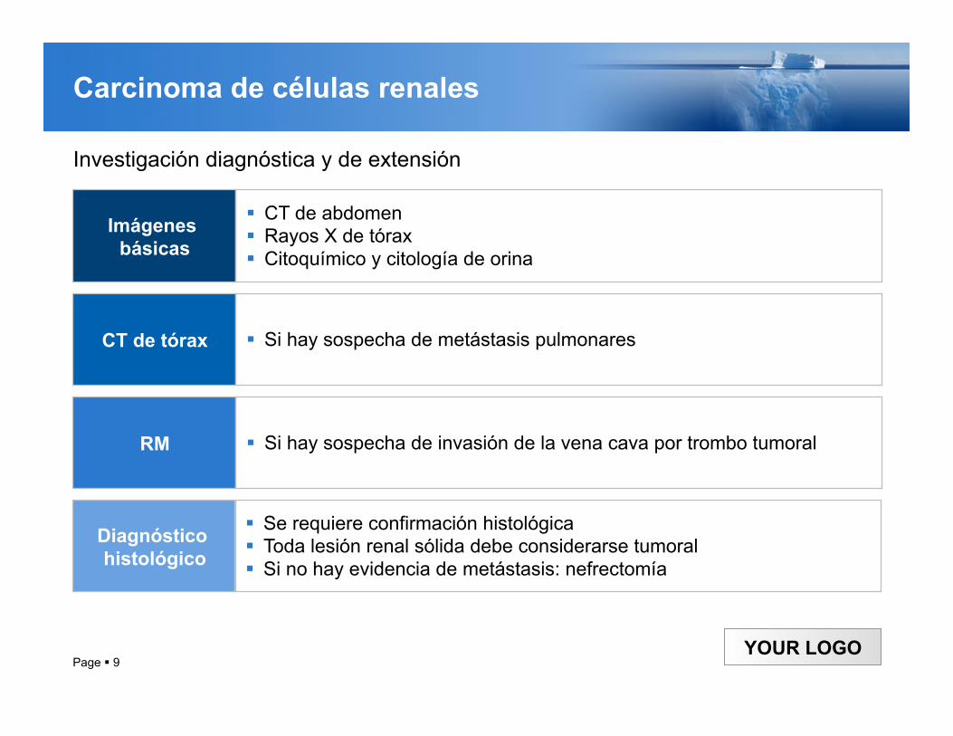
Imágenes básicas
CT de abdomen Rayos X de tórax Citoquímico y citología de orina
CT de tórax Si hay sospecha de metástasis pulmonares
RM Si hay sospecha de invasión de la vena cava por trombo tumoral
Diagnóstico histológico
Se requiere confirmación histológica Toda lesión renal sólida debe considerarse tumoral Si no hay evidencia de metástasis: nefrectomía
Investigación diagnóstica y de extensión
YOUR LOGO Page 10
Carcinoma de células renals
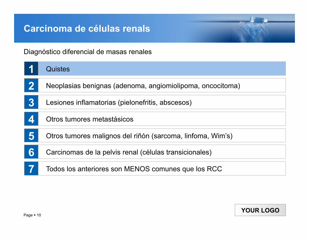
Diagnóstico diferencial de masas renales
Quistes
Neoplasias benignas (adenoma, angiomiolipoma, oncocitoma)
Lesiones inflamatorias (pielonefritis, abscesos)
Otros tumores metastásicos
Otros tumores malignos del riñón (sarcoma, linfoma, Wim’s)
1 2 3 4 5
Carcinomas de la pelvis renal (células transicionales)
Todos los anteriores son MENOS comunes que los RCC
6 7
YOUR LOGO
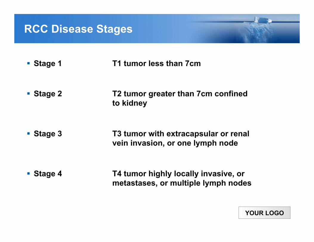
RCC Disease Stages
Stage 1 T1 tumor less than 7cm
Stage 2 T2 tumor greater than 7cm confined to kidney
Stage 3 T3 tumor with extracapsular or renal vein invasion, or one lymph node
Stage 4 T4 tumor highly locally invasive, or metastases, or multiple lymph nodes
YOUR LOGO
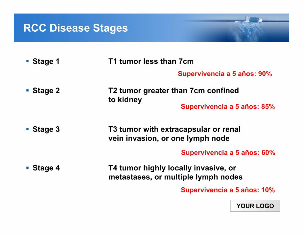
RCC Disease Stages
Stage 1 T1 tumor less than 7cm
Stage 2 T2 tumor greater than 7cm confined to kidney
Stage 3 T3 tumor with extracapsular or renal vein invasion, or one lymph node
Stage 4 T4 tumor highly locally invasive, or metastases, or multiple lymph nodes
Supervivencia a 5 años: 90%
Supervivencia a 5 años: 85%
Supervivencia a 5 años: 60%
Supervivencia a 5 años: 10%
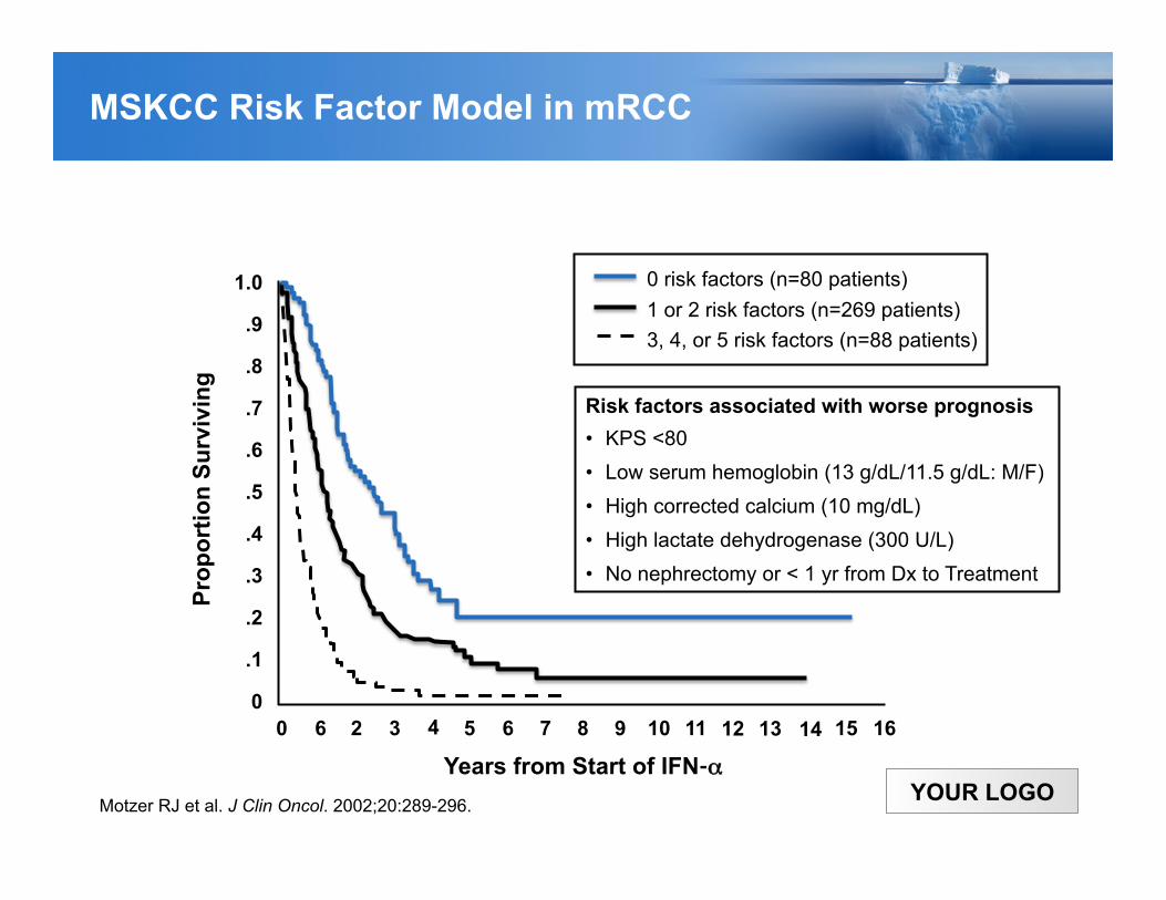
YOUR LOGO Motzer RJ et al. J Clin Oncol. 2002;20:289-296.
0 risk factors (n=80 patients) 1 or 2 risk factors (n=269 patients) 3, 4, or 5 risk factors (n=88 patients)
Risk factors associated with worse prognosis • KPS <80 • Low serum hemoglobin (13 g/dL/11.5 g/dL: M/F) • High corrected calcium (10 mg/dL) • High lactate dehydrogenase (300 U/L) • No nephrectomy or < 1 yr from Dx to Treatment
Years from Start of IFN-α
Prop
ortio
n Su
rviv
ing
0
.1
.2
.3
.4
.5
.6
.7
.8
.9
1.0
0 2 16 14 13 11 9 5 4 3 6 15 12 10 8 7 6
MSKCC Risk Factor Model in mRCC
YOUR LOGO
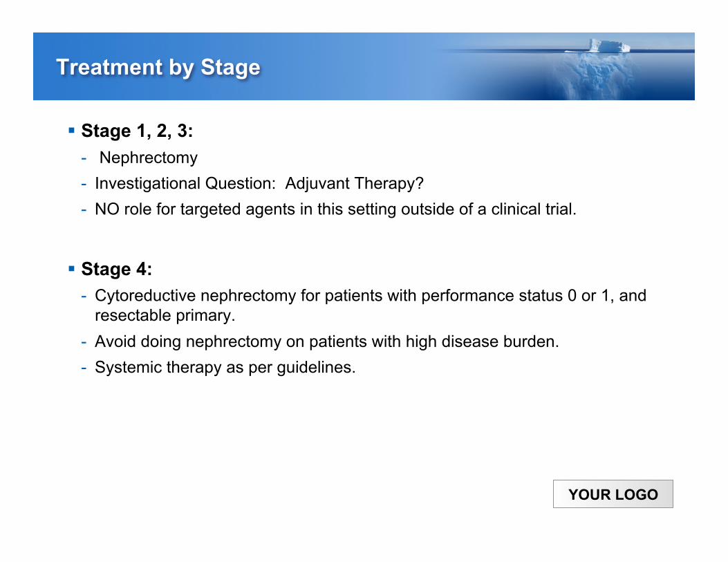
Treatment by Stage
Stage 1, 2, 3: - Nephrectomy - Investigational Question: Adjuvant Therapy? - NO role for targeted agents in this setting outside of a clinical trial.
Stage 4: - Cytoreductive nephrectomy for patients with performance status 0 or 1, and
resectable primary. - Avoid doing nephrectomy on patients with high disease burden. - Systemic therapy as per guidelines.
YOUR LOGO
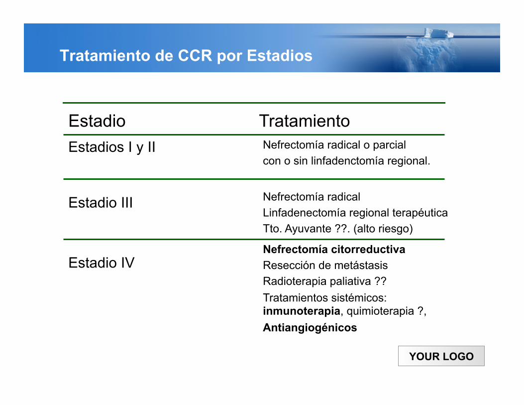
Tratamiento de CCR por Estadios
Nefrectomía citorreductiva Resección de metástasis Radioterapia paliativa ?? Tratamientos sistémicos: inmunoterapia, quimioterapia ?, Antiangiogénicos
Estadio IV
Nefrectomía radical Linfadenectomía regional terapéutica Tto. Ayuvante ??. (alto riesgo)
Estadio III
Nefrectomía radical o parcial con o sin linfadenctomía regional.
Estadios I y II
Tratamiento Estadio
YOUR LOGO
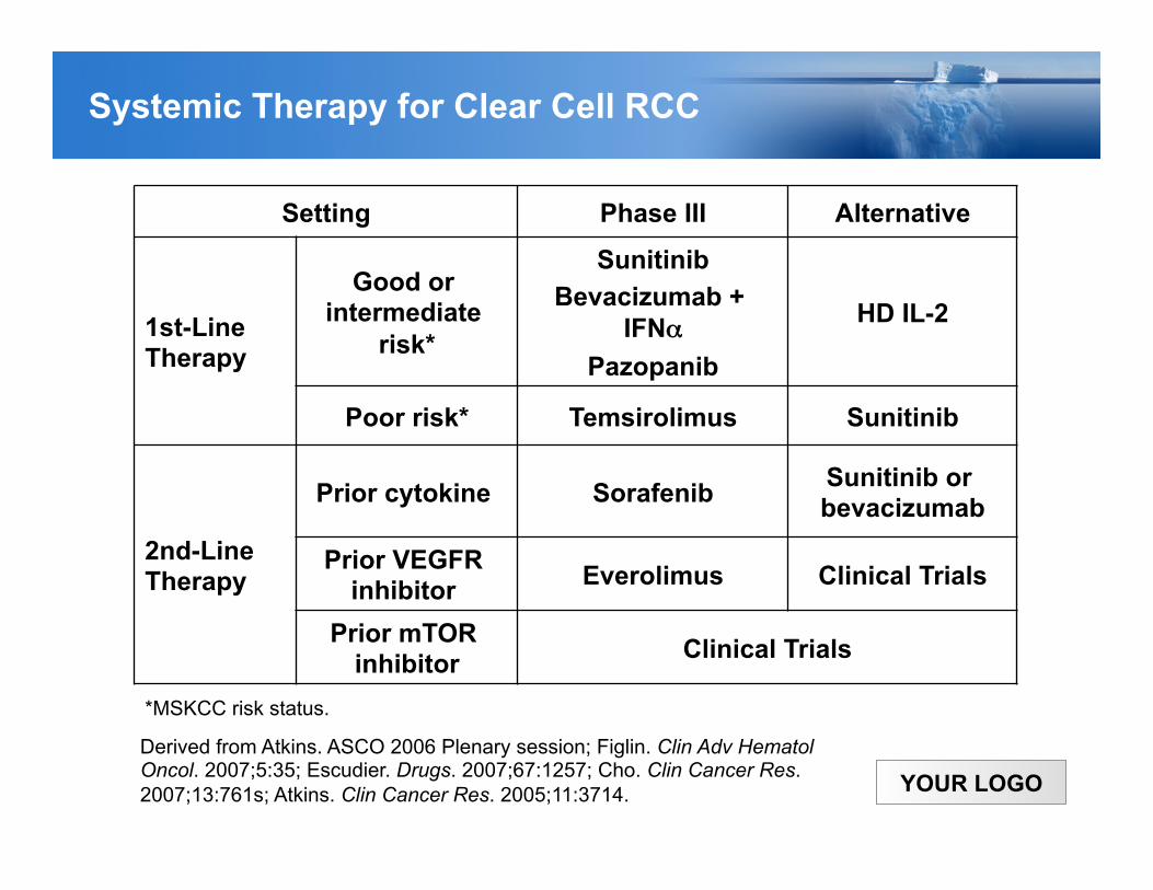
Setting Phase III Alternative
1st-Line Therapy
Good or intermediate
risk*
Sunitinib Bevacizumab +
IFNα Pazopanib
HD IL-2
Poor risk* Temsirolimus Sunitinib
2nd-Line Therapy
Prior cytokine Sorafenib Sunitinib or bevacizumab
Prior VEGFR inhibitor Everolimus Clinical Trials
Prior mTOR inhibitor Clinical Trials
Derived from Atkins. ASCO 2006 Plenary session; Figlin. Clin Adv Hematol Oncol. 2007;5:35; Escudier. Drugs. 2007;67:1257; Cho. Clin Cancer Res. 2007;13:761s; Atkins. Clin Cancer Res. 2005;11:3714.
*MSKCC risk status.
Systemic Therapy for Clear Cell RCC
YOUR LOGO 5
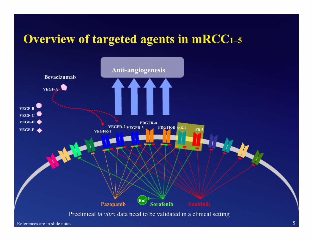
VEGFR-2 VEGFR-1
PDGFR-α VEGFR-3 PDGFR-ß c-Kit Flt-3
Overview of targeted agents in mRCC1–5 Anti-angiogenesis Bevacizumab VEGF-A
VEGF-B
VEGF-C
VEGF-D VEGF-E
Pazopanib Sorafenib Raf
Sunitinib
Preclinical in vitro data need to be validated in a clinical setting References are in slide notes
YOUR LOGO
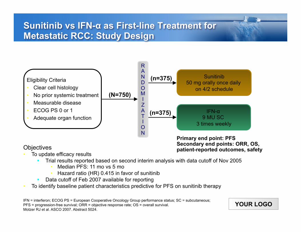
Sunitinib vs IFN-α as First-line Treatment for Metastatic RCC: Study Design
Primary end point: PFS Secondary end points: ORR, OS, patient-reported outcomes, safety
(N=750)
(n=375)
(n=375)
Sunitinib 50 mg orally once daily
on 4/2 schedule
IFN-α 9 MU SC
3 times weekly
Eligibility Criteria • Clear cell histology • No prior systemic treatment • Measurable disease • ECOG PS 0 or 1 • Adequate organ function
IFN = interferon; ECOG PS = European Cooperative Oncology Group performance status; SC = subcutaneous; PFS = progression-free survival; ORR = objective response rate; OS = overall survival. Motzer RJ et al. ASCO 2007. Abstract 5024.
Objectives • To update efficacy results
Trial results reported based on second interim analysis with data cutoff of Nov 2005 • Median PFS: 11 mo vs 5 mo • Hazard ratio (HR) 0.415 in favor of sunitinib
Data cutoff of Feb 2007 available for reporting • To identify baseline patient characteristics predictive for PFS on sunitinib therapy
RANDOM I Z A T I ON
YOUR LOGO
0 0,1 0,2 0,3 0,4 0,5 0,6 0,7 0,8 0,9 1,0
0 5 10 15 20 25 30
PFS
Pro
babi
lity
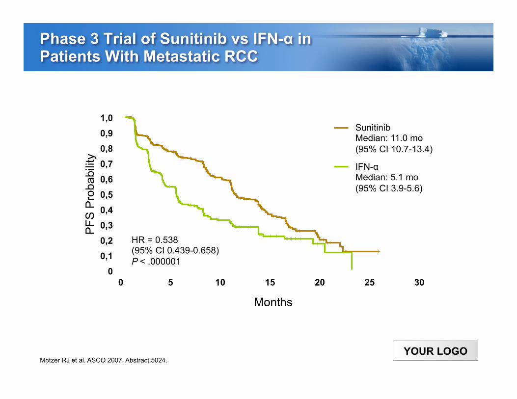
HR = 0.538 (95% CI 0.439-0.658) P < .000001
Months
Sunitinib Median: 11.0 mo (95% CI 10.7-13.4) IFN-α Median: 5.1 mo (95% CI 3.9-5.6)
Phase 3 Trial of Sunitinib vs IFN-α in Patients With Metastatic RCC
Motzer RJ et al. ASCO 2007. Abstract 5024.