Can the Number of Radiofrequency Activations Predict ...correlated with a decrease in FEV 1 (R2 =...
13
ORIGINAL RESEARCH Can the Number of Radiofrequency Activations Predict Serious Adverse Events after Bronchial Thermoplasty? A Retrospective Case-Control Study Shota Yamamoto . Motoyasu Iikura . Tamaki Kakuwa . Yoshie Tsujimoto . Sachi Matsubayashi . Naoko Nagano . Tomoyuki Suzuki . Keita Sakamoto . Konomi Kobayashi . Ayako Shiozawa . Masao Hashimoto . Satoru Ishii . Manabu Suzuki . Shinyu Izumi . Masayuki Hojo . Terumitsu Hasebe . Haruhito Sugiyama Received: September 25, 2019 / Published online: November 7, 2019 Ó The Author(s) 2019 ABSTRACT Introduction: Bronchial thermoplasty (BT) is a bronchoscopic procedure that involves the delivery of thermal radiofrequency energy to the bronchial wall for treating severe asthma. It has been suggested that too many radiofre- quency activations could induce serious adverse events (SAEs) at an early stage. We aimed to examine the number of radiofrequency activa- tions at each session and early lung function changes from baseline to determine whether these are related to SAEs. Methods: We retrospectively investigated 13 consecutive patients who underwent three ses- sions each of BT for severe asthma from Febru- ary 2015 to January 2016. Lung function tests were performed on the day before and after each BT procedure. Since we compared the number of activations and lung function changes from baseline after each session, a total of 39 sessions were reviewed. The relationship between the number of radiofrequency activa- tions and each lung function change from baseline was also examined by linear regression analysis. Results: A total of 10 SAEs (4 of pneumonia, 3 of atelectasis, 2 of bronchial asthma exacerba- tion and 1 of hemoptysis) were observed fol- lowing the 39 BT sessions. When we compared sessions with and without SAEs, there were no differences in the number of activations (mean ± SD, 71.5 ± 28.6 times in sessions with SAEs; 66.5 ± 25.1 times in sessions without SAEs; p = 0.772) and lung function changes (mean changes in FVC/%FVC/FEV 1 /%FEV 1 / %PEF from baseline; - 0.49 l/- 14.2%/- 0.36 l/- 11.7%/- 9.6% in sessions with SAEs; - 0.43 l/- 13.3%/- 0.34 l/- 12.1%/- 9.4% in sessions Enhanced digital features To view enhanced digital features for this article go to https://doi.org/10.6084/ m9.figshare.10043030. S. Yamamoto (&) Á M. Iikura Á T. Kakuwa Á Y. Tsujimoto Á S. Matsubayashi Á N. Nagano Á T. Suzuki Á K. Sakamoto Á K. Kobayashi Á A. Shiozawa Á M. Hashimoto Á S. Ishii Á M. Suzuki Á S. Izumi Á H. Sugiyama Department of Respiratory Medicine, National Center for Global Health and Medicine, 1-21-1 Toyama, Shinjuku-ku, Tokyo 162-8655, Japan e-mail: [email protected] S. Yamamoto Á T. Hasebe Department of Radiology, Tokai University Hachioji Hospital, Tokai University School of Medicine, 1838 Ishikawa-machi, Hachioji, Tokyo 192-0032, Japan M. Hojo Respiratory Disease Center, NTT Medical Center Tokyo, 5-9-22 Higashi-Gotanda, Shinagawa-ku, Tokyo 141-8625, Japan Pulm Ther (2019) 5:221–233 https://doi.org/10.1007/s41030-019-00103-7
Transcript of Can the Number of Radiofrequency Activations Predict ...correlated with a decrease in FEV 1 (R2 =...

ORIGINAL RESEARCH
Can the Number of Radiofrequency ActivationsPredict Serious Adverse Events after BronchialThermoplasty? A Retrospective Case-Control Study
Shota Yamamoto . Motoyasu Iikura . Tamaki Kakuwa . Yoshie Tsujimoto . Sachi Matsubayashi .
Naoko Nagano . Tomoyuki Suzuki . Keita Sakamoto . Konomi Kobayashi . Ayako Shiozawa .
Masao Hashimoto . Satoru Ishii . Manabu Suzuki . Shinyu Izumi . Masayuki Hojo .
Terumitsu Hasebe . Haruhito Sugiyama
Received: September 25, 2019 / Published online: November 7, 2019� The Author(s) 2019
ABSTRACT
Introduction: Bronchial thermoplasty (BT) is abronchoscopic procedure that involves thedelivery of thermal radiofrequency energy tothe bronchial wall for treating severe asthma. Ithas been suggested that too many radiofre-quency activations could induce serious adverseevents (SAEs) at an early stage. We aimed to
examine the number of radiofrequency activa-tions at each session and early lung functionchanges from baseline to determine whetherthese are related to SAEs.Methods: We retrospectively investigated 13consecutive patients who underwent three ses-sions each of BT for severe asthma from Febru-ary 2015 to January 2016. Lung function testswere performed on the day before and aftereach BT procedure. Since we compared thenumber of activations and lung functionchanges from baseline after each session, a totalof 39 sessions were reviewed. The relationshipbetween the number of radiofrequency activa-tions and each lung function change frombaseline was also examined by linear regressionanalysis.Results: A total of 10 SAEs (4 of pneumonia, 3of atelectasis, 2 of bronchial asthma exacerba-tion and 1 of hemoptysis) were observed fol-lowing the 39 BT sessions. When we comparedsessions with and without SAEs, there were nodifferences in the number of activations(mean ± SD, 71.5 ± 28.6 times in sessions withSAEs; 66.5 ± 25.1 times in sessions withoutSAEs; p = 0.772) and lung function changes(mean changes in FVC/%FVC/FEV1/%FEV1/%PEF from baseline; - 0.49 l/- 14.2%/- 0.36l/- 11.7%/- 9.6% in sessions with SAEs; - 0.43l/- 13.3%/- 0.34 l/- 12.1%/- 9.4% in sessions
Enhanced digital features To view enhanced digitalfeatures for this article go to https://doi.org/10.6084/m9.figshare.10043030.
S. Yamamoto (&) � M. Iikura � T. Kakuwa �Y. Tsujimoto � S. Matsubayashi � N. Nagano �T. Suzuki � K. Sakamoto � K. Kobayashi �A. Shiozawa � M. Hashimoto � S. Ishii � M. Suzuki �S. Izumi � H. SugiyamaDepartment of Respiratory Medicine, NationalCenter for Global Health and Medicine, 1-21-1Toyama, Shinjuku-ku, Tokyo 162-8655, Japane-mail: [email protected]
S. Yamamoto � T. HasebeDepartment of Radiology, Tokai University HachiojiHospital, Tokai University School of Medicine, 1838Ishikawa-machi, Hachioji, Tokyo 192-0032, Japan
M. HojoRespiratory Disease Center, NTT Medical CenterTokyo, 5-9-22 Higashi-Gotanda, Shinagawa-ku,Tokyo 141-8625, Japan
Pulm Ther (2019) 5:221–233
https://doi.org/10.1007/s41030-019-00103-7

without SAEs; p[0.05 for all the above).Increase in the number of activations correlatedwith decreased FEV1 (R
2 = 0.17, p = 0.0088) and%FEV1 (R2 = 0.11, p = 0.0357).Conclusions: Increase in the number ofradiofrequency activations during BT is relatedto a decrease in FEV1 and %FEV1 from baseline.The number of radiofrequency activations,however, is not associated with SAEs after BT.
Keywords: Bronchial asthma; Bronchialthermoplasty; Lung function tests; Seriousadverse events
PLAIN LANGUAGE SUMMARY
Bronchial thermoplasty (BT) is a bronchoscopicprocedure that involves the delivery of thermalradiofrequency energy to the bronchial wall fortreating severe asthma. BT is performed as threesessions at 3-week intervals. A previous in vitrostudy and a case report demonstrated that toomuch thermal energy caused irreversibleepithelial cell necrosis. It might induce earlyserious adverse events (SAEs). This study evalu-ated the number of radiofrequency activationsduring successive BT sessions and early lungfunction changes from baseline to determinewhether these are related to SAEs. We retro-spectively investigated 13 consecutive patients(mean age, 55.2 years; 7 males) who underwentthe BT procedure for severe asthma fromFebruary 2015 to January 2016 at the NationalCenter for Global Health and Medicine (Tokyo,Japan). Lung function tests were performed onthe day before and after each BT procedure. Atotal of 10 SAEs (4 of pneumonia, 3 of atelec-tasis, 2 of bronchial asthma exacerbation and 1of hemoptysis) were observed following 39 BTsessions. Comparison of sessions with andwithout SAE indicated no difference in thenumber of activations (mean ± SD, 71.5 ± 28.6times during sessions with SAEs; 66.5 ± 25.1times during sessions without SAEs; p = 0.31)and lung function changes from baseline.Increase in the number of radiofrequency acti-vations correlated with reduction in FEV1 and%FEV1.
Key Summary Points
Why carry out this study?
Bronchial thermoplasty (BT) is a novelbronchoscopic procedure that involvesthe delivery of thermal radiofrequencyenergy to the bronchial wall for treatingsevere asthma
It is hypothesized that too manyradiofrequency activations could induceearly serious adverse events (SAEs)
What was learned from the study?
When we compared BT sessions with andwithout SAEs, there was no difference inthe number of radiofrequency activations(mean ± SD, 71.5 ± 28.6 times in sessionswith SAEs; 66.5 ± 25.1 times in sessionswithout SAEs; p = 0.77)
Increase in the number of activationscorrelated with a decrease in FEV1
(R2 = 0.17, p = 0.0088) and %FEV1
(R2 = 0.11, p = 0.0357)
This study revealed that the number ofradiofrequency activations is notassociated with SAEs; further studieselucidating the predictive factors for SAEsare required to enhance patient safety
INTRODUCTION
Asthma remains an important health problemwith significant morbidity, mortality and eco-nomic burden. Airway smooth muscle contrac-tion and the resultant bronchoconstrictioninduce the many variable symptoms of asthma.Increased airway smooth muscle mass is acharacteristic feature of asthma, especiallyamong patients with severe asthma. Bronchialthermoplasty (BT) is a novel intervention thatinvolves the delivery of thermal radiofrequencyenergy to the bronchial wall during a series ofbronchoscopies, resulting in prolonged
222 Pulm Ther (2019) 5:221–233
reduction of airway smooth muscle mass. Inprevious clinical studies, treatment of patientswith airways between 3 and 10 mm in diameterled to clinically meaningful reduction in mus-cle-mediated narrowing of the airway and toimprovement of asthma symptoms [1–3].
The most common complications of BT aretransient adverse respiratory events that occur amedian of 1 day after the procedure and rarelyrequire hospitalization [4]. Recently, however,serious adverse events (SAEs) involving the air-ways, which required prolonged hospitalizationor threatened the patient’s life, have beenreported after the BT procedure [5–8]. Hence, itis necessary to determine early predictors ofSAEs after BT to enhance patient safety.
Even a single radiofrequency activation canaffect the bronchial mucosa and cause epithelialsloughing [9]. A previous in vitro study and acase report demonstrated that too much ther-mal energy caused irreversible epithelial cellnecrosis [10, 11]. Several studies suggested thatintense thermal stimulation of the bronchialmucosa could represent a strong boost forinflammation, with microvascular alterationbeing directly induced by the heat or throughthe release of chemical mediators causingmucosal exudation and edema [5, 12]. Since thenumber of activations is directly related to theamount of thermal energy, we hypothesizedthat the number of activations was related toSAEs in the early post-procedure stage. Gener-ally, lung function tests are used to evaluateairway obstruction in persons with asthma.Hence, we hypothesized that early changes inlung function following BT might be useful topredict SAEs.
In this study, we aimed to examine thenumber of radiofrequency activations duringeach BT session and early lung function changesfrom baseline to determine whether these arerelated to SAEs following BT.
METHODS
Patients
We retrospectively investigated adult asthmapatients aged 18 years or older who underwent
three sequential BT procedures at the NationalCenter for Global Health and Medicine, Tokyo,Japan, from February 2015 to January 2016. Allof them performed lung function tests on theday before and after each BT procedure. Aspreviously described [3], BT is indicated inpatients aged [ 18 years and with severeuncontrolled asthma despite high-dose inhala-tional corticosteroid therapy. The contraindi-cations to BT include [3]: (1) presence of apacemaker, internal defibrillator or otherimplantable electronic devices; (2) known sen-sitivity to the medications used during bron-choscopy; (3) previous treatment with BT at thesame area; (4) active respiratory infection; (5)pulmonary emphysema or any cystic disease;(6) asthma exacerbation or change in the doseof systemic corticosteroids for asthma in thepast 14 days; (7) known coagulopathy; (8)therapy with anticoagulants, antiplatelet agentsand non-steroidal anti-inflammatory drugsbefore the procedure.
Study Design
We conducted a retrospective case-controlstudy. Patient factors (age, sex, height, bodyweight, smoking status, nasal comorbidities,laboratory data, Global Initiative for Asthmatreatment step, quality of life, medications andlung function tests) and BT procedure factors(number of radiofrequency activations, proce-dure time and time/activation ratio) wereinvestigated from the patients’ charts. SAEswere defined as undesirable adverse experiencesoccurring within 30 days after the BT procedure,as previously described [13]. They includeddeath, life-threatening conditions, prolongedhospitalization, disability and conditionsrequiring intervention (e.g., antibiotics, sys-temic steroid, oxygen therapy and artificialventilator). The primary aim of this study was toexamine the number of radiofrequency activa-tions during each session and early lung func-tion changes from baseline following BT and todetermine whether these are related to SAEs.The secondary aim was to evaluate the rela-tionship between the number of activations andeach lung function change from baseline. Each
Pulm Ther (2019) 5:221–233 223

treatment session was divided into two groups:the SAE group, which represented sessions withSAEs, and non-SAE group, which representedsessions without SAEs. This study followed theprinciples of the Declaration of Helsinki andwas approved by the institutional review boardof the National Center for Global Health andMedicine, Tokyo, Japan (approval no. NCGM-G-001801-00) on June 15, 2015. We verifiedthat written informed consent was obtainedbefore each patient’s participation in this study.
Bronchial Thermoplasty
Using the ALAIR� device (Boston ScientificJapan, Tokyo, Japan), BT was performed in threesessions at 3-week intervals. The lower lobebronchi were treated in turn during the first twosessions, beginning with the right bronchus inthe first session and the left bronchus in thesecond session, and both upper lobe bronchi inthe third BT procedure. The right middle lobebronchus was not treated in order to avoid thetheoretical risk of obstruction and right middlelobe syndrome. The system was adjusted todeliver a constant temperature of 65�C for 10 sat each treatment site from the small 3-mmbronchi to the lobar bronchi. The procedurewas performed under intravenous sedation withmidazolam. The patients underwent close clin-ical monitoring immediately after the BT pro-cedure. If there were no SAEs, they weredischarged a few days after the BT procedure.Prednisone (50 mg/day) was systematicallyadministered orally from 3 days before the BTprocedure to 1 day after each procedure.
Lung Function Tests
BT was performed as three sessions at 3-weekintervals. We examined lung function tests byspirometry (Superior Spiro Discom-21 FXIII;Chest, Tokyo, Japan) on the day before (asbaseline) and the day after each BT procedure.Differences between lung function on the daybefore and after BT were calculated as thechange from baseline. A single experiencedclinical technologist performed all the lungfunction tests with the patients in the seated
position, the tests being performed at least twiceto verify the stability of lung function values.For quantitative evaluation of airway status, theparameters of forced vital capacity (FVC), per-cent forced vital capacity (%FVC), forced expi-ratory volume in 1 s (FEV1), percent forcedexpiratory volume in 1 s (%FEV1) and percentpeak expiratory flow (%PEF) were used. We usedeach prediction formula to predict the FVCvalue by Baldwin, FEV1 value by Berglund andPEF value by Cherniack.
Statistical Analysis
Qualitative variables are shown as numbers andpercentages. Quantitative variables are reportedas mean values and SDs, unless otherwise indi-cated. We used Mann-Whitney U test to com-pare the treated lobes, mean number ofactivations, procedure time, time/activationratio and lung function changes from baseline(FVC, %FVC, FEV1, %FEV1, %PEF) between ses-sions with and without SAEs. The transition oflung function among all bronchial thermo-plasty sessions was compared using the paired t-test. The potential associations between thenumber of activations and lung functionchanges from baseline were assessed usingPearson’s correlation coefficient (R2). All testswere two-tailed. P values \ 0.05 were consid-ered significant. Data were analyzed statisticallywith JMP� 14.0.0 software (SAS Institute Inc.,Cary, NC, USA).
RESULTS
Study Population
Patient demographics and baseline characteris-tics are shown in Table 1. A total of 13 patientswere investigated in this study. The patientswere on several medications, including high-dose inhaled corticosteroids (meanbeclomethasone-equivalent dose of1700 lg/day), systemic steroids (39% ofpatients) and molecular targeted drugs (46% ofpatients). All patients used high-dose inhaledcorticosteroids and long-acting beta-agonists.
224 Pulm Ther (2019) 5:221–233
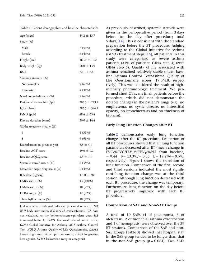
As previously described, systemic steroids weregiven in the perioperative period (from 3 daysbefore to the day after procedure; total5 days)[14]. This is consistent with the standardpreparation before the BT procedure. Judgingaccording to the Global Initiative for Asthma(GINA) treatment steps [15], all patients in thisstudy were categorized as severe asthmapatients (31% of patients: GINA step 4; 69%:GINA step 5). Quality of life associated withasthma remained relatively stable (mean base-line Asthma Control Test/Asthma Quality ofLife Questionnaire scores, 19.0/4.8, respec-tively). This was considered the result of high-intensity pharmacologic treatment. We per-formed chest CT scans in all patients before theprocedure, which did not demonstrate thenotable changes in the patient’s lungs (e.g., noemphysema, no cystic disease, no interstitialopacity, no bronchiectasis and no thickness ofbronchi).
Early Lung Function Changes after BT
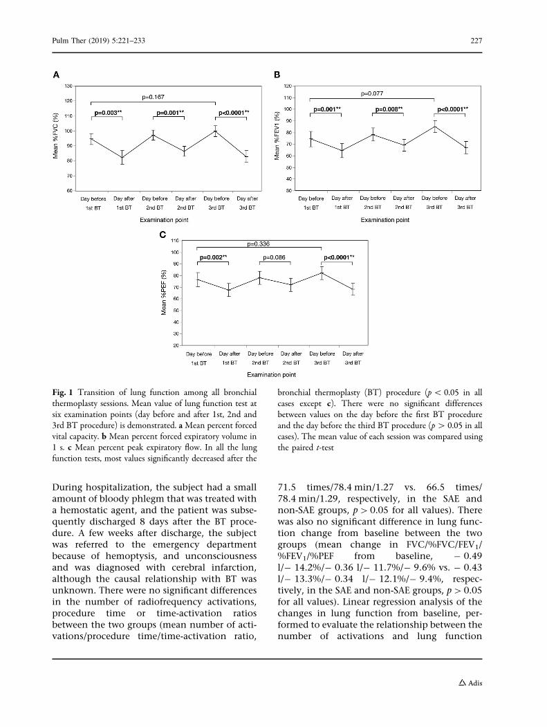
Table 2 demonstrates early lung functionchanges after the BT procedure. Evaluation ofall BT procedures showed that all lung functionparameters decreased after BT (mean change inFVC/%FVC/FEV1/%FEV1/%PEF from baseline,- 0.44 l/- 13.3%/- 0.35 l/- 12.2%/- 9.5%,respectively). Figure 1 shows the transition oflung function. Comparison of the first, secondand third sessions indicated the most signifi-cant lung function change was at the thirdsession. Although lung function decreased witheach BT procedure, the change was temporary.Furthermore, lung function on the day beforeBT progressively improved with each BTprocedure.
Comparison of SAE and Non-SAE Groups
A total of 10 SAEs (4 of pneumonia, 3 ofatelectasis, 2 of bronchial asthma exacerbationand 1 of hemoptysis) were observed over the 39BT sessions. Comparison of the SAE and non-SAE groups (Table 3) showed that hospital stayin the SAE group tended to be longer than thatin the non-SAE group (p = 0.064). Two SAEs
Table 1 Patient demographics and baseline characteristics
Age (years) 55.2 ± 13.7
Sex, n (%)
Male 7 (54%)
Female 6 (46%)
Height (cm) 160.9 ± 10.0
Body weight (kg) 58.0 ± 13.9
BMI 22.1 ± 3.6
Smoking status, n (%)
Never-smoker 9 (69%)
Ex-smoker 4 (31%)
Nasal comorbidities, n (%) 9 (69%)
Peripheral eosinophils (/ll) 205.5 ± 229.9
IgE (IU/ml) 383.5 ± 586.9
FeNO (ppb) 48.4 ± 65.4
Disease duration (years) 30.0 ± 14.4
GINA treatment step, n (%)
4 4 (31%)
5 9 (69%)
Exacerbations in previous year 6.3 ± 5.1
Baseline ACT score 19.0 ± 4.2
Baseline AQLQ score 4.8 ± 1.1
Systemic steroid use, n (%) 5 (38%)
Molecular target drug use, n (%) 6 (46%)
ICS dose (lg/day) 1700 ± 380
LABA use, n (%) 13 (100%)
LAMA use, n (%) 10 (77%)
LTRA use, n (%) 12 (92%)
Theophylline use, n (%) 10 (77%)
Unless otherwise indicated, values are presented as mean ± SD
BMI body mass index, ICS inhaled corticosteroids; ICS dose
was calculated as the beclomethasone-equivalent dose, IgE
immunoglobulin E, FeNO fractional exhaled nitric oxide,
GINA Global Initiative for Asthma, ACT Asthma Control
Test, AQLQ Asthma Quality of Life Questionnaire, LAMA
long-acting muscarinic receptor antagonist, LABA long-acting
beta agonist, LTRA leukotriene receptor antagonist
Pulm Ther (2019) 5:221–233 225

occurred within 3 weeks of the first BT session,and four each occurred within 3 weeks of thesecond and third BT sessions. Asthma exacer-bation was only seen within 3 weeks of the thirdBT session. In two cases, exacerbations ofasthma were treated with additional bron-chodilators and systemic steroids, and these
patients needed longer hospitalization thanthose with the other adverse events. Threepatients had complete atelectasis although thelung segment re-inflated without treatment,while four cases who developed pneumonianeeded standard antibiotic therapy. One of thecases had a serious course with hemoptysis.
Table 2 Early lung function changes evaluated on the day after bronchial thermoplasty
Day before BT (baseline) Day after BT Change frombaseline
First procedure at right lower lobe (13 sessions)
FVC (l) 3.23 ± 1.13 2.83 ± 1.13 - 0.39 ± 0.39
%FVC (%) 95.0 ± 12.5 82.5 ± 15.7 - 12.5 ± 11.4
FEV1 (l) 2.14 ± 1.03 1.85 ± 0.87 - 0.29 ± 0.26
%FEV1 (%) 74.5 ± 22.7 64.6 ± 20.5 - 10.0 ± 7.6
%PEF (%) 76.6 ± 20.9 67.7 ± 20.1 - 9.0 ± 8.0
Second procedure at left lower lobe (13 sessions)
FVC (l) 3.29 ± 1.11 2.94 ± 1.01 - 0.36 ± 0.27
%FVC (%) 97.5 ± 12.0 86.6 ± 11.5 - 10.8 ± 7.7
FEV1 (l) 2.21 ± 0.90 1.97 ± 0.77 - 0.25 ± 0.33
%FEV1 (%) 78.2 ± 19.5 69.2 ± 17.0 - 9.0 ± 9.9
%PEF (%) 78.1 ± 19.8 72.2 ± 19.5 - 6.0 ± 11.1
Third procedure at bilateral upper lobes (13 sessions)
FVC (l) 3.41 ± 1.29 2.82 ± 1.05 - 0.59 ± 0.40
%FVC (%) 100.1 ± 11.8 86.6 ± 11.5 - 17.0 ± 9.1
FEV1 (l) 2.45 ± 1.00 1.92 ± 0.83 - 0.53 ± 0.32
%FEV1 (%) 85.2 ± 17.1 67.1 ± 17.0 - 18.1 ± 7.6
%PEF (%) 82.2 ± 18.0 68.4 ± 17.8 - 13.9 ± 7.7
All procedures (39 sessions)
FVC (l) 3.31 ± 1.16 2.86 ± 1.07 - 0.44 ± 0.37
%FVC (%) 97.4 ± 12.3 84.1 ± 13.7 - 13.3 ± 9.9
FEV1 (l) 2.26 ± 0.98 1.91 ± 0.83 - 0.35 ± 0.33
%FEV1 (%) 79.2 ± 20.4 66.9 ± 18.4 - 12.2 ± 9.4
%PEF (%) 78.9 ± 20.0 69.4 ± 19.3 - 9.5 ± 9.6
Values are presented as mean ± SD unless otherwise indicatedBT bronchial thermoplasty, FVC forced vital capacity, %FVC percent forced vital capacity, FEV1 forced expiratory volumein 1 s, %FEV1 percent forced expiratory volume in 1 s, %PEF percent peak expiratory flow
226 Pulm Ther (2019) 5:221–233
During hospitalization, the subject had a smallamount of bloody phlegm that was treated witha hemostatic agent, and the patient was subse-quently discharged 8 days after the BT proce-dure. A few weeks after discharge, the subjectwas referred to the emergency departmentbecause of hemoptysis, and unconsciousnessand was diagnosed with cerebral infarction,although the causal relationship with BT wasunknown. There were no significant differencesin the number of radiofrequency activations,procedure time or time-activation ratiosbetween the two groups (mean number of acti-vations/procedure time/time-activation ratio,
71.5 times/78.4 min/1.27 vs. 66.5 times/78.4 min/1.29, respectively, in the SAE andnon-SAE groups, p[0.05 for all values). Therewas also no significant difference in lung func-tion change from baseline between the twogroups (mean change in FVC/%FVC/FEV1/%FEV1/%PEF from baseline, - 0.49l/- 14.2%/- 0.36 l/- 11.7%/- 9.6% vs. - 0.43l/- 13.3%/- 0.34 l/- 12.1%/- 9.4%, respec-tively, in the SAE and non-SAE groups, p[0.05for all values). Linear regression analysis of thechanges in lung function from baseline, per-formed to evaluate the relationship between thenumber of activations and lung function
Fig. 1 Transition of lung function among all bronchialthermoplasty sessions. Mean value of lung function test atsix examination points (day before and after 1st, 2nd and3rd BT procedure) is demonstrated. aMean percent forcedvital capacity. b Mean percent forced expiratory volume in1 s. c Mean percent peak expiratory flow. In all the lungfunction tests, most values significantly decreased after the
bronchial thermoplasty (BT) procedure (p\ 0.05 in allcases except c). There were no significant differencesbetween values on the day before the first BT procedureand the day before the third BT procedure (p[ 0.05 in allcases). The mean value of each session was compared usingthe paired t-test
Pulm Ther (2019) 5:221–233 227

(Fig. 2), showed that FEV1 and %FEV1 had anegative correlation with the number of acti-vations. On the other hand, FVC, %FVC and%PEF did not have any correlation.
DISCUSSION
To the best of our knowledge, this is the firstreport assessing the relationship between the
Table 3 Comparison between sessions with and without serious adverse events and details of the serious adverse eventsafter bronchial thermoplasty
SAE groupN = 10
Non-SAEgroupN = 29
p value
Hospital stay (days) 10.1 ± 6.6 6.1 ± 1.7 0.064
Serious adverse event
Pneumonia, n 4 (1 at the right lower lobe; 2 at the left lower lobe; 1 at the
right upper lobe)
– –
Atelectasis, n 3 (1 at the right lower lobe; 2 at the left lower lobe) – –
Asthma exacerbation, n 2 (2 at the bilateral upper lobe) – –
Hemoptysis, n 1 (unknown origin) – –
Treated lobe
Right lower lobe, n (%) 2 (20) 11 (38) 0.315
Left lower lobe, n (%) 4 (40) 9 (31) 0.623
Bilateral upper lobes, n (%) 4 (40) 9 (31) 0.623
BT procedure
Number of radiofrequency
activations (times)
71.5 ± 28.6 66.5 ± 25.1 0.772
Procedure time (min) 78.4 ± 20.7 78.4 ± 22.1 0.936
Time/activation ratio 1.27 ± 0.70 1.29 ± 0.46 0.489
Lung function changes from
baseline
FVC (l) - 0.49 ± 0.35 - 0.43 ± 0.38 0.479
%FVC (%) - 14.2 ± 8.6 - 13.3 ± 10.5 0.618
FEV1 (l) - 0.36 ± 0.36 - 0.34 ± 0.33 0.847
%FEV1 (%) - 11.7 ± 9.8 - 12.1 ± 9.5 0.785
%PEF (%) - 9.6 ± 10.7 - 9.4 ± 9.5 0.898
Values are presented as mean ± SD unless otherwise indicatedAll the results were analyzed using Mann-Whitney U testBT bronchial thermoplasty, SAE serious adverse event, FVC forced vital capacity, %FVC percent forced vital capacity, FEV1
forced expiratory volume in 1 s, %FEV1 percent forced expiratory volume in 1 s, %PEF percent peak expiratory flow*p\ 0.05
228 Pulm Ther (2019) 5:221–233

number of radiofrequency activations and earlySAEs after BT. We performed lung function teststo quantitatively evaluate morphologic airwaychanges to enable objective evaluation of thechanges. Generally speaking, bronchial asthmaexacerbation is involved with worsening lungfunction (FEV1 and PEF) [16], so lung functiontests are suitable for assessment of asthmaticpatients. Although Langton et al. focused onthe number of activations as a predictor oftreatment response [17], we hypothesized thatthis parameter could be a predictor of SAEs inthe early stages after BT. Actually, in the presentstudy, the number of activations was signifi-cantly related to the reduction in FEV1 and%FEV1 after the BT procedure (change in FEV1,R2 = 0.171, p = 0.009; change in %FEV1,R2 = 0.114, p = 0.036). The amount of FEV1
reduction on the day after BT and the correla-tion between the number of activations andFEV1 followed the same trend as in Langton’sreport [18]. In addition, both Langton’s and ourreports demonstrated that upper lobe treatmentcauses a greater decrease in FEV1 than lowerlobe treatment. Increase in the number of acti-vations might affect edematous changes in thebronchial mucosa and around the bronchus[4, 9, 19]. According to previous in vitroresearch, administration of thermal energy at atemperature of 37–70 �C to cultured airwayepithelial cells and airway smooth muscle cellscan cause irreversible epithelial cell necrosis[10]. In this research, we concluded that notonly the number of activations, but also lungfunction changes over a short period were notenough to predict SAE occurrence.
Our research group has previously reportedon airway morphologic and functional changesafter BT therapy. We studied asthma-relatedquality of life, lung function tests and thenumber of asthma exacerbations both 1 and12 months after the third BT session in 12Japanese patients. We found that %FEV1 sig-nificantly improved a month after the third BTsession (mean %FEV1 before first BT session:70.5%, mean FEV1 a month after third BT ses-sion: 82.2%; p\ 0.05) [20]. The report alsoconfirmed that patients maintain the lungfunction improvement for a year (mean %FEV1
within a year of the third BT session: 82.3%).
The reason why the BT procedure was capable ofmaintaining a sustained effect over a year in ourstudy is that the total number of activationsthroughout BT in our institute was more than1.3 times higher than in the AIR2 trial. Due tothe larger number of activations, more periph-eral airways could be treated. It was also previ-ously reported that the number of activationscould play a role in determining clinicalresponse to treatment [17]. In the present study,%FEV1 followed the same trend as in our pre-vious report. Moreover, %FEV1 tended toimprove the day before the second BT proce-dure (mean %FEV1 a day before first BT session:74.5%, mean %FEV1 a day before second BTsession: 78.2%). After the first BT procedure,%FEV1 decreased temporarily, although %FEV1
on the day before the third BT procedure wasbetter than that on the day before the first BTprocedure (mean %FEV1 on the day before thirdBT: 85.2%) (Fig. 1). Our findings were supportedby the recent study by Ishii et al. conducted in a68-year-old Japanese woman with severeasthma who underwent BT, which confirmeddilation of the bronchial lumen and decreasedbronchial wall thickness by comparing thechanges in the bronchial lumen and wallsbefore BT and after the first and third sessionsby three-dimensional (3D) airway analysis [21].Previous studies demonstrated the occurrenceof morphologic changes in airways right afterthe BT procedure. However, there is no proofthat the changes are related to functionalimprovement. Our reports suggest that the BTprocedure leads to an initial decrease in lungfunction, although repeated sessions lead togradual dilatation of the bronchial lumen and adecrease in bronchial wall thickness.
Previous major clinical trials have proved thesafety and efficacy of BT procedures [1–3].Compared with the incidence of SAEs in previ-ous clinical trials, including the AIR trial, AIR2trial and RISA study (7.3%, 8.4% and 26.7%,respectively), adverse events occurred relativelyfrequently among elderly and adult patientswith severe asthma in this study (25.6%). Apartfrom the number of activations, the differencesbetween the above trials and the present studyare asthma severity, age and study design. Interms of asthma severity, the AIR and AIR2 trials
Pulm Ther (2019) 5:221–233 229

230 Pulm Ther (2019) 5:221–233

targeted persons with moderate-to-severe per-sistent asthma. On the other hand, the presentstudy and the RISA study targeted persons withsevere asthma who were symptomatic despitemaximal pharmacologic treatment. Asthmaseverity might affect SAE occurrence, althoughwe could not assess the relationship betweenasthma severity and SAE occurrence because ofthe small number of subjects analyzed. Usingtwo independent data sources, Burn et al.showed that there were more reports of re-ad-missions in the group who underwent the BTprocedure in clinical practice (re-admission rate,95% CI; 19.1%, 13.2–26.2) compared with theprevious clinical trials. Further, they consideredasthma severity and age as factors affecting thecomplication rate of BT. The average age ofpatients in the AIR trial, AIR2 trial, RISA study,Burn et al’s report and this study was 39.4 years,40.7 years, 39.1 years, 42.6 years and 55.2 years,respectively [12]. Actually, the incidence ofadverse events in elderly patients in the presentstudy (25.6%) is slightly high compared withthe other studies. The difference could bebecause this study reflects real-world data thatwere generated during routine clinical practiceand enrolled many elderly and/or adult patientswith severe asthma. Among the SAE group,additional needs for antibiotics or corticosteroidtherapy were observed but significant prolon-gations of hospital stays were not observed (SAEgroup, 10.1 days; non-SAE group 6.1 days;p = 0.064) (Table 3). The previously reportedtreatments for adverse events after BT included
fibrin plug removal by bronchoscopy foratelectasis secondary to occlusion by the plug[5], chest tube insertion for pneumothorax afterpartial atelectasis [7] and bronchial arterythrombosis and intubation for bronchial arteryaneurysm rupture [6]. Such adverse events arethe leading causes of death without propertreatment. Since a significant therapeutic effectof BT has been reported, the number of eligiblepatients for BT are expected to increase. How-ever, the determinant factors related to SAEsremain unclear. This study is the first step toproving whether the number of activations andchanges in lung function after the BT procedureare related to SAEs. Future studies should con-sider asthma severity and include an age-mat-ched population.
There are some limitations to our study. Theretrospective study conducted at a single centerwith a small number of patients limits theweight with which we can interpret the results.The limited number of patients and practition-ers could lead to bias in terms of observation ofSAEs and treatment, so a larger multicenterstudy is warranted. In this study, we used lungfunction tests to assess the occurrence of SAEs.Many previous studies have used chest CT scansto evaluate the lung damage resulting fromradiofrequency activation [4, 19]. While CTscans provide high spatial resolution that isespecially beneficial in evaluation of fibrin plugsin the airway and airway thickness, diagnosticx-rays are problematic in terms of radiationexposure. Japan has the highest annual fre-quency of diagnostic x-rays among developedcountries. Reportedly, the cumulative cancerrisk is related to diagnostic x-ray exposure [22].Since women of childbearing age might need toundergo the BT procedure, we recommend thatfrequent diagnostic x-rays should be avoided.
CONCLUSIONS
Increase in the number of radiofrequency acti-vations is involved in a decrease in FEV1 and%FEV1 from baseline after BT. The number ofactivations, however, is not associated withSAEs after BT. Therefore, further studies eluci-dating the factors predictive of SAEs are
bFig. 2 Correlation between the number of radiofrequencyactivations and lung function changes from baseline.Linear regression analysis was performed to evaluate therelationship between the number of activations andchanges in lung function from baseline, using the followingparameters: a forced vital capacity (FVC), b percent forcedvital capacity (%FVC), c forced expiratory volume in 1 s(FEV1), d percent forced expiratory volume in 1 s(%FEV1) and e percent peak expiratory flow (%PEF).Increase in the number of activations correlated with adecrease in FEV1 (R2 = 0.17, p = 0.0088) and %FEV1
(R2 = 0.11, p = 0.0357). There were no significant corre-lations between the number of activations and FVC/%FVC/%PEF
Pulm Ther (2019) 5:221–233 231
required to enhance patient safety after theprocedure.
ACKNOWLEDGEMENTS
The authors thank the participants of thisstudy.
Funding. Boston Scientific Japan loaned theauthors’ department the Alair thermoplastysystem for 6 months and also donated sixcatheters. This work was supported by a grantfrom the National Center for Global Health andMedicine (NCGM-27-6001). The Rapid ServiceFee was funded by the authors.
Authorship. All named authors meet theInternational Committee of Medical JournalEditors (ICMJE) criteria for authorship for thismanuscript, take responsibility for the integrityof the work as a whole, and have given finalapproval for the version to be published.
Medical Writing and/or Editorial Assis-tance. Additional statistical support was pro-vided by Osamu Narumoto, MD, PhD, ofNational Hospital Organization Tokyo Hospital.
Disclosures. Motoyasu Iikura received lec-ture fees from Astra Zeneca K.K. Masayuki Hojoreceived lecture fees from Astra Zeneca K.K.,Novartis Pharma K.K., KYORIN Holdings, Inc.,and Boehringer Ingelheim GmbH. The otherauthors have no conflict of interest. ShotaYamamoto, Tamaki Kakuwa, Yoshie Tsujimoto,Sachi Matsubayashi, Naoko Nagano, TomoyukiSuzuki, Keita Sakamoto, Konomi Kobayashi,Ayako Shiozawa, Masao Hashimoto, SatoruIshii, Manabu Suzuki, Shinyu Izumi, TerumitsuHasebe and Haruhito Sugiyama have nothing todisclose.
Compliance with Ethics Guidelines. Thisstudy followed the principles of the Declarationof Helsinki and was approved by the institu-tional review board of the National Center forGlobal Health and Medicine, Tokyo, Japan (ap-proval no. NCGM-G-001801-00) on June 15,2015. We verified that written informed
consent was obtained before each patient’sparticipation in this study.
Data Availability. The datasets generatedand/or analyzed during the present study areavailable from the corresponding author onreasonable request.
Open Access. This article is distributedunder the terms of the Creative CommonsAttribution-NonCommercial 4.0 InternationalLicense (http://creativecommons.org/licenses/by-nc/4.0/), which permits any non-commercial use, distribution, and reproductionin any medium, provided you give appropriatecredit to the original author(s) and the source,provide a link to the Creative Commons license,and indicate if changes were made.
REFERENCES
1. Pavord ID, Cox G, Thomson NC, et al. Safety andefficacy of bronchial thermoplasty in symptomatic,severe asthma. Am J Respir Crit Care Med.2007;176:1185–91.
2. Cox G, Thomson NC, Rubin AS, et al. Asthmacontrol during the year after bronchial thermo-plasty. N Engl J Med. 2007;356:1327–37.
3. Castro M, Rubin AS, Laviolette M, et al. Effective-ness and safety of bronchial thermoplasty in thetreatment of severe asthma: a multicenter, ran-domized, double-blind, sham-controlled clinicaltrial. Am J Respir Crit Care Med. 2010;181:116–24.
4. Debray MP, Dombret MC, Pretolani M, et al. Earlycomputed tomography modifications followingbronchial thermoplasty in patients with severeasthma. Eur Respir J. 2017;49:1601565.
5. Facciolongo N, Menzella F, Lusuardi M, et al.Recurrent lung atelectasis from fibrin plugs as a veryearly complication of bronchial thermoplasty: acase report. Multidiscip Respir Med. 2015;10:9.
6. Nguyen DV, Murin S. Bronchial artery pseudoa-neurysm with major hemorrhage after bronchialthermoplasty. Chest. 2016;149:e95–7.
7. Funatsu A, Kobayashi K, Iikura M, et al. A case ofpulmonary cyst and pneumothorax after bronchialthermoplasty. Respirol Case Rep. 2018;6:e00286.
232 Pulm Ther (2019) 5:221–233
8. Balu A, Ryan D, Niven R. Lung abscess as a com-plication of bronchial thermoplasty. J Asthma.2015;52:740–2.
9. Goorsenberg AWM, d’Hooghe JNS, de Bruin DM,et al. Bronchial thermoplasty-induced acute airwayeffects assessed with optical coherence tomographyin severe asthma. Respiration. 2018;96:564–70.
10. Chernyavsky IL, Russell RJ, Saunders RM, et al.In vitro, in silico and in vivo study challenges theimpact of bronchial thermoplasty on acute airwaysmooth muscle mass loss. Eur Respir J.2018;51:1701680.
11. Menzella F, Lusuardi M, Galeone C, et al. Heat-in-duced necrosis after bronchial thermoplasty: a newconcern? Allergy Asthma Clin Immunol.2018;14:25.
12. Burn J, Sims AJ, Keltie K, et al. Procedural and short-term safety of bronchial thermoplasty in clinicalpractice: evidence from a national registry andHospital Episode Statistics. J Asthma.2017;54:872–9.
13. Ioannidis JP, Lau J. Completeness of safety report-ing in randomized trials: an evaluation of 7 medicalareas. JAMA. 2001;285:437–43.
14. Cox G, Miller JD, McWilliams A, Fitzgerald JM, LamS. Bronchial thermoplasty for asthma. Am J RespirCrit Care Med. 2006;173:965–9.
15. Walsh LJ, Wong CA, Oborne J, et al. Adverse effectsof oral corticosteroids in relation to dose in patientswith lung disease. Thorax. 2001;56:279–84.
16. Global Initiative for Asthma., National Heart Lungand Blood Institute. Global initiative for asthma:global strategy for asthma management and pre-vention. 2014 revision. ed. Bethsda: U.S. Dept. ofHealth and Human Services, Public Health Service;2014. viii.
17. Langton D, Sha J, Ing A, et al. Bronchial thermo-plasty: activations predict response. Respir Res.2017;18:134.
18. Langton D, Wang W, Thien F, Plummer V. Theacute effects of bronchial thermoplasty on FEV1.Respir Med. 2018;137:147–51.
19. d’Hooghe JNS, van den Berk IAH, Annema JT,Bonta PI. Acute radiological abnormalities afterbronchial thermoplasty: a prospective cohort trial.Respiration. 2017;94:258–62.
20. Iikura M, Hojo M, Nagano N, et al. Bronchial ther-moplasty for severe uncontrolled asthma in Japan.Allergol Int. 2018;67:273–5.
21. Ishii S, Iikura M, Hojo M, Sugiyama H. Use of 3D-CTairway analysis software to assess a patient withsevere persistent bronchial asthma treated withbronchial thermoplasty. Allergol Int.2017;66:501–3.
22. Berrington de Gonzalez A, Darby S. Risk of cancerfrom diagnostic X-rays: estimates for the UK and 14other countries. Lancet. 2004;363:345–51.
Pulm Ther (2019) 5:221–233 233


![Windows Mac OS X [10.8+] - Attasaattasa.com/madcatz/support/pdf/MCB32266-MUG-R2-10... · l2 l1 l2 r2 r2 r1 l2 l1 r1 l1 r2 r1 l2 l1 l2 r2 r1 l1 r2 r1 l2 l1 l2 r2.10.11. fcc id: p25d243710a4512c](https://static.fdocument.pub/doc/165x107/5ba4bf5f09d3f235188bed3d/windows-mac-os-x-108-l2-l1-l2-r2-r2-r1-l2-l1-r1-l1-r2-r1-l2-l1-l2-r2-r1.jpg)