Bovine three-portion pericardial patch for reconstruction ... · * Correspondence:...
4
CASE REPORT Open Access Bovine three-portion pericardial patch for reconstruction of the aorto-mitral curtain in infective endocarditis Atsushi Hiromoto, Shun-Ichiro Sakamoto * , Yasuo Miyagi and Takashi Nitta Abstract Background: Surgery for infective endocarditis involving the aorto-mitral curtain (AMC) is challenging and requires extensive incisions and complex reconstruction procedures. However, in patients with preserved aortic annulus, reconstruction of the AMC is possible using a simple technique with limited incisions. Case presentation: A handmade bovine three-portion pericardial patch was used to reconstruct the AMC in a patient with severe endocarditis requiring double valve replacement; the technique allowed for steady anchorage of prosthetic valves without additional incisions other than conventional aortotomy and atriotomy. Postoperative echocardiography revealed normal cardiac function and no significant perivalvular leakage. The patient displayed complete recovery and was discharged on postoperative day 33. The patient was symptom-free at his 1-year follow-up and displayed normal laboratory and echocardiographic findings. Conclusion: The bovine three-portion pericardial patch is useful for reconstructing the AMC in patients with infective endocarditis accompanied by preserved aortic annulus. Keywords: Infective endocarditis, Aorto-mitral curtain, Patch repair Background Involvement of the aorto-mitral curtain (AMC) is un- common in patients with infective endocarditis. Once it is affected, the procedure for double valve replacement becomes more complex as it necessitates reconstruction of the AMC. This surgery is challenging and is associ- ated with a high mortality rate (20–30%) [1–3]. In con- trast, in patients with preserved aortic annulus, it is possible to reconstruct the AMC using a simple tech- nique requiring limited incisions. Here, we report the surgical case of a patient with infective endocarditis with AMC involvement who was treated using the conven- tional approach. Case presentation A 57-year-old man was admitted to the hospital due to hyperleukocytosis. Echocardiography revealed irregu- larly shaped vegetation (size, 25 × 15 mm) attached to the anterior leaflet of the mitral valve. The vegetation exhibited oscillation and was connected to the thick- ened aortic valve. Color flow imaging showed severe in- sufficiency of both the aortic and mitral valves with perforation in the AMC (Fig. 1). Chest X-ray revealed bilateral lung congestion due to acute heart failure. Therefore, emergency surgery was indicated. The heart was approached via median full sternotomy. An oblique incision was made in the ascending aorta under conditions of cardiac arrest. The aortic valve was bicuspid (type 1). Vegetation was observed at the non-coronary cusp, extending to the AMC. The mitral valve was exposed via the superior trans-septal ap- proach. The anterior leaflet was thickened and had at- tached vegetation. Debridement of the infected tissue led to a defect in the middle portion of the anterior mitral annulus, AMC, and non-coronary cusp. For reconstructing the defective parts, a glutaraldehy- de-treated bovine pericardial patch (Model 4700, Ed- wards Lifesciences, Irvine, CA, USA) was folded to make a three-portion patch (Fig. 1a). The triangular portion (AMC portion) of two pericardial patches was sutured * Correspondence: [email protected] Department of Cardiovascular Surgery, Nippon Medical School, 1-1-5 Sendagi, Bunkyo-ku, Tokyo 113-8603, Japan © The Author(s). 2019 Open Access This article is distributed under the terms of the Creative Commons Attribution 4.0 International License (http://creativecommons.org/licenses/by/4.0/), which permits unrestricted use, distribution, and reproduction in any medium, provided you give appropriate credit to the original author(s) and the source, provide a link to the Creative Commons license, and indicate if changes were made. Hiromoto et al. Surgical Case Reports (2019) 5:2 https://doi.org/10.1186/s40792-018-0558-5
Transcript of Bovine three-portion pericardial patch for reconstruction ... · * Correspondence:...

CASE REPORT Open Access
Bovine three-portion pericardial patch forreconstruction of the aorto-mitral curtain ininfective endocarditisAtsushi Hiromoto, Shun-Ichiro Sakamoto* , Yasuo Miyagi and Takashi Nitta
Abstract
Background: Surgery for infective endocarditis involving the aorto-mitral curtain (AMC) is challenging and requiresextensive incisions and complex reconstruction procedures. However, in patients with preserved aortic annulus,reconstruction of the AMC is possible using a simple technique with limited incisions.
Case presentation: A handmade bovine three-portion pericardial patch was used to reconstruct the AMC in apatient with severe endocarditis requiring double valve replacement; the technique allowed for steady anchorageof prosthetic valves without additional incisions other than conventional aortotomy and atriotomy. Postoperativeechocardiography revealed normal cardiac function and no significant perivalvular leakage. The patient displayedcomplete recovery and was discharged on postoperative day 33. The patient was symptom-free at his 1-yearfollow-up and displayed normal laboratory and echocardiographic findings.
Conclusion: The bovine three-portion pericardial patch is useful for reconstructing the AMC in patients withinfective endocarditis accompanied by preserved aortic annulus.
Keywords: Infective endocarditis, Aorto-mitral curtain, Patch repair
BackgroundInvolvement of the aorto-mitral curtain (AMC) is un-common in patients with infective endocarditis. Once itis affected, the procedure for double valve replacementbecomes more complex as it necessitates reconstructionof the AMC. This surgery is challenging and is associ-ated with a high mortality rate (20–30%) [1–3]. In con-trast, in patients with preserved aortic annulus, it ispossible to reconstruct the AMC using a simple tech-nique requiring limited incisions. Here, we report thesurgical case of a patient with infective endocarditis withAMC involvement who was treated using the conven-tional approach.
Case presentationA 57-year-old man was admitted to the hospital due tohyperleukocytosis. Echocardiography revealed irregu-larly shaped vegetation (size, 25 × 15 mm) attached to
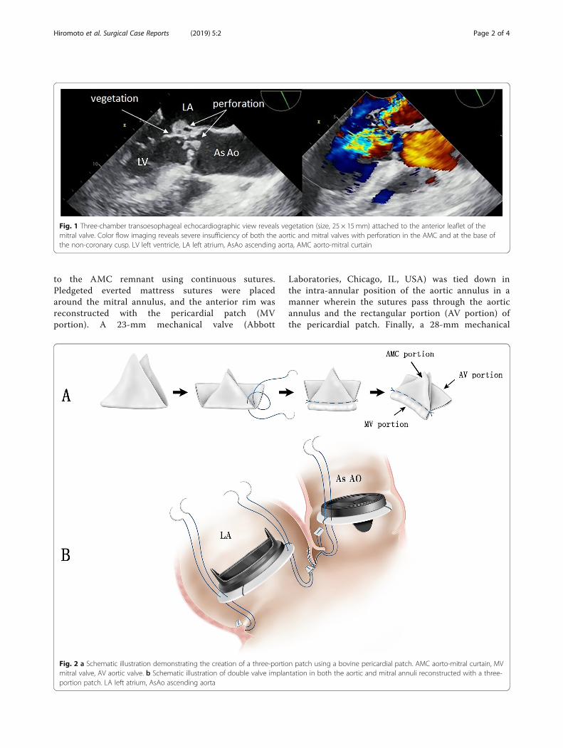
the anterior leaflet of the mitral valve. The vegetationexhibited oscillation and was connected to the thick-ened aortic valve. Color flow imaging showed severe in-sufficiency of both the aortic and mitral valves withperforation in the AMC (Fig. 1). Chest X-ray revealedbilateral lung congestion due to acute heart failure.Therefore, emergency surgery was indicated.The heart was approached via median full sternotomy.
An oblique incision was made in the ascending aortaunder conditions of cardiac arrest. The aortic valve wasbicuspid (type 1). Vegetation was observed at thenon-coronary cusp, extending to the AMC. The mitralvalve was exposed via the superior trans-septal ap-proach. The anterior leaflet was thickened and had at-tached vegetation. Debridement of the infected tissue ledto a defect in the middle portion of the anterior mitralannulus, AMC, and non-coronary cusp.For reconstructing the defective parts, a glutaraldehy-
de-treated bovine pericardial patch (Model 4700, Ed-wards Lifesciences, Irvine, CA, USA) was folded to makea three-portion patch (Fig. 1a). The triangular portion(AMC portion) of two pericardial patches was sutured
* Correspondence: [email protected] of Cardiovascular Surgery, Nippon Medical School, 1-1-5Sendagi, Bunkyo-ku, Tokyo 113-8603, Japan
© The Author(s). 2019 Open Access This article is distributed under the terms of the Creative Commons Attribution 4.0International License (http://creativecommons.org/licenses/by/4.0/), which permits unrestricted use, distribution, andreproduction in any medium, provided you give appropriate credit to the original author(s) and the source, provide a link tothe Creative Commons license, and indicate if changes were made.
Hiromoto et al. Surgical Case Reports (2019) 5:2 https://doi.org/10.1186/s40792-018-0558-5
to the AMC remnant using continuous sutures.Pledgeted everted mattress sutures were placedaround the mitral annulus, and the anterior rim wasreconstructed with the pericardial patch (MVportion). A 23-mm mechanical valve (Abbott
Laboratories, Chicago, IL, USA) was tied down inthe intra-annular position of the aortic annulus in amanner wherein the sutures pass through the aorticannulus and the rectangular portion (AV portion) ofthe pericardial patch. Finally, a 28-mm mechanical
Fig. 1 Three-chamber transoesophageal echocardiographic view reveals vegetation (size, 25 × 15mm) attached to the anterior leaflet of themitral valve. Color flow imaging reveals severe insufficiency of both the aortic and mitral valves with perforation in the AMC and at the base ofthe non-coronary cusp. LV left ventricle, LA left atrium, AsAo ascending aorta, AMC aorto-mitral curtain
Fig. 2 a Schematic illustration demonstrating the creation of a three-portion patch using a bovine pericardial patch. AMC aorto-mitral curtain, MVmitral valve, AV aortic valve. b Schematic illustration of double valve implantation in both the aortic and mitral annuli reconstructed with a three-portion patch. LA left atrium, AsAo ascending aorta
Hiromoto et al. Surgical Case Reports (2019) 5:2 Page 2 of 4
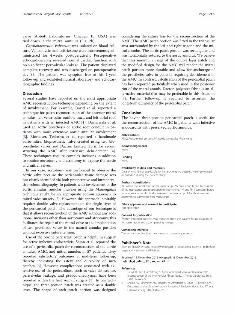
valve (Abbott Laboratories, Chicago, IL, USA) wastied down in the mitral annulus (Fig. 2b).Cardiobacterium valvarum was isolated on blood cul-
ture. Vancomycin and ceftriaxone were intravenously ad-ministered for 4 weeks postoperatively. Postoperativeechocardiography revealed normal cardiac function withno significant perivalvular leakage. The patient displayedcomplete recovery and was discharged on postoperativeday 33. The patient was symptom-free at his 1-yearfollow-up and exhibited normal laboratory and echocar-diographic findings.
DiscussionSeveral studies have reported on the most appropriateAMC reconstruction technique depending on the extentof involvement. For example, David et al. reported atechnique for patch reconstruction of the anterior mitralannulus, left ventricular outflow tract, and left atrial roofin patients with an infected AMC [1]. Davierwala et al.used an aortic prosthesis or aortic root conduit in pa-tients with more extensive aortic annular involvement[3]. Moreover, Tedoriya et al. reported a handmadeaorto-mitral bioprosthetic valve created using two bio-prosthetic valves and Dacron knitted fabric for recon-structing the AMC after extensive debridement [4].These techniques require complex incisions in additionto routine aortotomy and atriotomy to expose the aorticand mitral valves.In our case, aortotomy was performed to observe the
aortic valve because the periannular tissue damage wasnot clearly identified via both preoperative and intraopera-tive echocardiography. In patients with involvement of theaortic annulus, annular incision using the Manouguiantechnique might be an appropriate add-on approach tomitral valve surgery [5]. However, this approach inevitablyrequires double valve replacement on the single layer ofthe pericardial patch. The advantage of our technique isthat it allows reconstruction of the AMC without any add-itional incisions other than aortotomy and atriotomy; thisfacilitates the repair of the mitral valve or the implantationof two prosthetic valves in the natural annular positionwithout excessive suture tension.Use of the bovine pericardial patch is helpful in surgery
for active infective endocarditis. Shinn et al. reported theuse of a pericardial patch for reconstruction of the aorticannulus, AMC, and mitral annulus in 57 patients. Theyreported satisfactory outcomes at mid-term follow-up,thereby indicating the safety and durability of suchpatches [6]. However, complications associated with ex-tensive use of the pericardium, such as valve dehiscence,perivalvular leakage, and pseudo-aneurysms, have beenreported within the first year of surgery [3]. In our tech-nique, the three-portion patch was created as a doublelayer. The shape of each patch portion was designed
considering the suture line for the reconstruction of theAMC. The AMC patch portion was fitted in the triangulararea surrounded by the left and right trigone and the mi-tral annulus. The aortic patch portion was rectangular andwas horizontally sutured to the aortic annulus. We believethat this minimum usage of the double layer patch andthe modified design for the AMC will render the mitralpatch portion more durable and allow for anchorage ofthe prosthetic valve in patients requiring debridement ofthe AMC. In contrast, calcification of the pericardial patchhas been reported particularly when used in the posteriorrim of the mitral annuls. Dacron polyester fabric is an al-ternative material that may be preferable in this situation[7]. Further follow-up is required to ascertain thelong-term durability of the pericardial patch.
ConclusionThe bovine three-portion pericardial patch is useful forthe reconstruction of the AMC in patients with infectiveendocarditis with preserved aortic annulus.
AbbreviationsAMC: Aorto-mitral curtain; AV: Aortic valve; MV: Mitral valve
AcknowledgementsNone
FundingNone
Availability of data and materialsData sharing is not applicable to this article as no datasets were generatedor analyzed during the current study.
Authors’ contributionsAH wrote the initial draft of the manuscript. SS have contributed to revisionof the manuscript and preparation for submitting. YM and TN have contributedto interpretation and critically reviewed the manuscript. All authors read andapproved to submit the final manuscript.
Ethics approval and consent to participateNot applicable
Consent for publicationWritten informed consent was obtained from the patient for publication ofthis case report and accompanying images.
Competing interestsThe authors declare that they have no competing interests.
Publisher’s NoteSpringer Nature remains neutral with regard to jurisdictional claims in publishedmaps and institutional affiliations.
Received: 13 November 2018 Accepted: 18 December 2018
References1. David TE, Kuo J, Armstrong S. Aortic and mitral valve replacement with
reconstruction of the intervalvular fibrous body. J Thorac Cardiovasc Surg.1997;114:766–72.
2. Sheikh AM, Elhenawy AM, Maganti M, Armstrong S, David TE, Feindel CM.Outcomes of double valve surgery for active infective endocarditis. J ThoracCardiovasc Surg. 2009;138:69–75.
Hiromoto et al. Surgical Case Reports (2019) 5:2 Page 3 of 4
3. Davierwala PM, Binner C, Subramanian S, Luehr M, Pfannmueller B, Etz C,Dohmen P, Misfeld M, Borger MA, Mohr FW. Double valve replacement andreconstruction of the intervalvular fibrous body in patients with activeinfective endocarditis. Eur J Cardiothorac Surg. 2014;45:146–52.
4. Tedoriya T, Hirota M, Ishikawa N, Omoto T. Reconstruction of aorto-mitralcontinuity with a handmade aorto-mitral bioprosthetic valve for extensivebivalvular endocarditis. Interact Cardiovasc Thorac Surg. 2013;16:405–7.
5. Nishimura T, Ota T, Takayama H, Naka Y. Extended trans-septal and left atrialdome incision combined with Manouguian’s aortotomy for complexdouble valve surgery. Eur J Cardiothorac Surg. 2014;46:127–8.
6. Shinn SH, Sung K, Park PW, Lee YT, Kim WS, Jun TG, Lee SC, Park SW. Resultsof annular reconstruction with a pericardial patch in active infectiveendocarditis. J Heart Valve Dis. 2009;18:315–20.
7. De Oliveira NC, David TE, Armstrong S, Ivanov J. Aortic and mitral valvereplacement with reconstruction of the intervalvular fibrous body: ananalysis of clinical outcomes. J Thorac Cardiovasc Surg. 2005;129(2):286–90.
Hiromoto et al. Surgical Case Reports (2019) 5:2 Page 4 of 4