FA. FA guidelines/GuidelinesDocuments/guidelines-afib-FT.pdfFA.
http://repository.osakafu-u.ac.jp/dspace/
TitleEnzymatic synthesis of water-soluble ferulic acid derivatives and their im
proving effects for Alzheimer's disease.
Author(s) 菊川, 昌希
Editor(s)
Citation
Issue Date 2017
URL http://hdl.handle.net/10466/15683
Rights
大阪府立大学博士(応用生命科学)学位論文
Enzymatic synthesis of water-soluble ferulic acid derivatives and their improving effects for Alzheimer’s disease
水溶性フェルラ酸誘導体の酵素合成および
アルツハイマー病改善効果に関する研究
菊川 昌希
2017年

1
Contents
Contents ···························································································· 1
Abbreviations ····················································································· 2
Introduction ······················································································· 3
Chapter 1. Synthesis of water-soluble feruloyl diglycerols by esterification of an
Aspergillus niger feruloyl esterase.
Summary ········································································· 10
Materials and Methods ························································· 10
Results ··········································································· 14
Discussion ······································································· 29
Chapter 2. Ferulic acid and its water-soluble derivatives inhibit nitric oxide production
and inducible nitric oxide synthase expression in rat primary astrocytes.
Summary ········································································· 31
Materials and Methods ························································· 31
Results ··········································································· 34
Discussion ······································································· 40
Chapter 3. Water-soluble ferulic acid derivatives improve amyloid- induced neuronal
cell death and dysmnesia through inhibition of amyloid- aggregation.
Summary ········································································· 42
Materials and Methods ························································· 42
Results ··········································································· 46
Discussion ······································································· 55
Conclusion ························································································ 58
References ························································································ 60

2
Publications ······················································································ 68
Funding ··························································································· 68
Acknowledgements ············································································· 69
Abbreviations
A, amyloid-; AD, Alzheimer’s disease; BSA, bovine serum albumin; CA, caffeic acid;
CMC, carboxymethylcellulose; CNS, central nervous system; DG, diglycerol; DIV, days
in vitro; dmCA, 3,4-dimethoxycinnamic acid; DMEM, Dulbecco’s modified Eagle’s
medium; FA, ferulic acid; FA-DG1, 1-feruloyl diglycerol; FA-G1, 1-feruloyl glycerol;
FAE, ferulic acid esterase; FAE-PL, An Aspergillus niger FAE purified from Pectinase
PL “AMANO”; FBS, fetal bovine serum; IB-, inhibitor of NF-B- iNOS, inducible
NO synthase; LPS, lipopolysaccharide; MTT, 3-(4,5-dimethylthiazol-2-yl)-2,5-diphenyl
tetrazolium bromide; NF-B, nuclear factor (NF)-B; NO, nitric oxide; PBS, phosphate
buffered saline. pCA, p-coumaric acid; SA, sinapic acid; SDS-PAGE, sodium dodecyl
sulfate polyacrylamide gel electrophoresis; SE, standard error; TPI, triose-phosphate
isomerase.

3
Introduction
Location of ferulic acid (FA)
Ferulic acid (FA) (4-hydroxy-3-methoxycinnamic acid; Fig. 1) is a polyphenol
located in the cell walls of a wide variety of plants, such as artichokes, eggplants, wheat
bran, and maize bran (Smith and Hartley 1983; Saulnier et al. 1995). FA is esterified to
the C-5 of -L-arabinofuranosyl side chains of arabinoxylans in monocotyledons such as
maize bran (Saulnier et al. 1995) and wheat bran (Smith and Hartley 1983). In pectins
of dicotyledons such as sugar beet (Rombouts and Thibault 1986; Ralet et al. 1994;
Colquhoun et al. 1994) and spinach (Fry 1982; Ishii and Tobita 1993), FA mainly attaches
to the C-2 of -1,5-linked arabinofuranose residues or the C-6 of -1,4-linked
galactopyranose residues. FA is also linked to the C-4 position of -D-xylopyranosyl
residues in xyloglucans (Ishii et al. 1990). The amounts of FA and p-coumaric acid
(pCA) in various plants are summarized in Table 1. Some FAs occur in the form of
dehydrodimers which function to cross-link and strengthen the cell walls, and decrease
their digestibility by microorganisms (Eraso and Hartley 1990; Saulnier and Thibault
1999).
Table 1. The amounts of FA and pCA in various plants (Tsuchiyama 2007).
Class Source Hydroxy-
cinnamic acid
Amount (%)
in dry weight
Reference
Monocoty- Wheat bran FA 0.66 Smith et al. 1983
ledoneae pCA 0.014 Lequart et al. 1999
Wheat straw FA 0.28 Lequart et al. 1999
pCA 0.36 Lequart et al. 1999
Rice end sperm FA 0.9 Shibuya 1984
Maize bran FA 3.1 Saulnier et al. 1995
Barley grain FA 0.14 Nordkvist et al. 1984
Dicoty- Sugar beet pulp FA 0.8 Micard et al. 1994
ledoneae Chinese water
chestnut
FA 0.7 Parr et al. 1996
Fig. 1. Structure of FA
O
H3CO
HO
OH

4
Ferulic acid esterase (FAE)
Some microorganisms, to defeat this defensive strategy in plants, secrete FAEs (EC
3.1.1.73) as well as cellulases, pectinases, and hemicellulases. FAEs are enzymes that
catalyze the hydrolysis of ester bonds between FAs and sugars. FAE was first isolated
by Faulds and Williamson (1991). Since then, many microbial FAEs have been isolated
and classified based on their catalytic properties (Crepin et al. 2004).
Antioxidant action of FA FA is a cinnamic acid derivative with antioxidant properties (Castelluccio et al.
1995; Kikuzaki et al. 2002) and protects molecules that are essential to life (i.e., lipid,
protein, and DNA) from oxidative stress (Rice-Evans et al. 1996). Its antioxidant action
involves the up-regulation of heme-oxygenase-1, heat shock protein 70, and signal
transduction kinase 1/2, and protects cells from stress (Barone et al. 2009). Antioxidants
such as FA are commonly used to inhibit the oxidation of lipids in food, cosmetics, and
medicine.
Anti-inflammatory effect of FA FA has anti-inflammatory effects and can reduce the overexpression of
prostaglandin E2, tumor necrosis factor- (TNF-), and inducible NO synthase (iNOS)
induced by lipopolysaccharide (LPS) in cells (Ou et al. 2003; Tetsuka et al. 1996).
Administration of FA for one month counteracted the overexpression of endothelial NO
synthase induced by A in mouse hippocampal astrocytes (Cho et al. 2005). Moreover,
FA was shown to prevent iNOS induction in cerebral ischemia model rats (Koh 2012).
UV-absorbing action of FA FA is a strong UV absorber and can reduce UV-induced skin damage. FA is efficient
at absorbing the sunburn-inducing UVB (290–320 nm) and UVA (320–400 nm) radiation.
Indeed, FA has been proved to be an effective protectant from UVB-induced skin
erythema in healthy subjects (Saija et al. 2000). Importantly, FA prepared at an acidic
or neutral pH is readily absorbed by the skin.

5
Clinical and statistical data of Alzheimer's disease (AD) AD is a neurodegenerative disorder that mostly affects the elderly. Characteristics
of AD include memory deficits and delusions. AD is a significant problem for the super-
aged population in Japan, and many studies have aimed to produce drugs to treat AD.
Figure 2 illustrates the increase in the number of dementia patients over time. In Japan,
as of 2010, approximately 2 million people were considered senile, with some researchers
estimating that 10% of people 65 years of age or older (about 2.4 millions) having
dementia. Furthermore, the number of patients with dementia is expected to increase to
3.2 million by 2020, with the increasing number of people aged 65 years or above
(Ministry of Health, Labour and Welfare in Japan).
Fig. 2. Number of dementia patients
(Ministry of Health, Labour and Welfare in Japan 2010)
Treatment of AD in Japan Four drugs have been approved in Japan for the treatment of AD (Table 2).
Donepezil (Aricept) is used through it before, and binds and reversibly inactivates
cholinesterases, thus inhibiting acetylcholine hydrolysis. This results in an increased
acetylcholine concentration at cholinergic synapses. The three other drugs were given
approval in 2011, and this suggests that the Japanese people suffering from AD are
receiving increased attention.
0
5
10
15
0
50
100
150
200
250
300
1995年 2000年 2005年 2010年 2015年 2020年
Num
ber o
f pat
ient
s (th
ousa
nds)
3,000
2,500
2,000
1,5001,000
500
0
Num
ber o
f pat
ient
s (%
of t
he p
opul
atio
n ag
ed 6
5 an
d ov
er)
1995 2000 2005 2010 2015 2020

6
Table 2. Licensed palliative treatments for AD in Japan.
General name Action mechanisms Year of sale
(Japan)
Donepezil*1
(Aricept)
Acetylcholinesterase inhibitor 1999
Galantamine*2
Acetylcholinesterase inhibitor
Allosteric modulator of nicotinic receptors
2011
Memantine*3 Inhibitor of NMDA receptors 2011
Rivastigmine*4 Acetylcholinesterase inhibitor
Butylcholinesterase inhibitor
2011
*1 Eisai and Pfizer, *2Janssen Pharmaceutical K.K., *2 Takeda Pharmaceutical Co.,
Ltd, *3 Daiichi Sankyo Healthcare Co., Ltd., *4 Novartis Pharmaceuticals, *4 Ono
Pharmaceutical Co., Ltd.
Pathology of AD AD was first described in 1906 at a conference in Tubingen, Germany by Alois
Alzheimer (1906) as a loss of memory and cognitive dysfunction. The
clinicopathological features of AD are not completely understood. However, there are
three possible mechanisms: deposition of senile plaque, deposition of tau proteins in the
cytoplasm of neurons, and neuronal degeneration due to both of these components
(Masliah et al. 1993). Senile plaques are extracellular plaques in the brain consisting
mainly of amyloid- (A), a peptide thought to be a leading cause of neurotoxicity
(Masters et al. 1985). Overproduction and aberrant self-assembly of A into fibrillar
aggregates constitute the first steps of the so-called amyloid cascade hypothesis, which is
thought to trigger AD (Hardy and Higgins 1992). In fact, A aggregation is known to
cause neuronal cell death. Although it is not clear exactly how A causes neuronal loss
and tangle formation, the peptide disrupts calcium homeostasis and increases
intraneuronal calcium concentrations (Estus et al. 1997; Loo et al. 1993). Inhibition of
the aggregation of A and destabilization of aggregated A in the central nervous system
(CNS) would be an attractive therapeutic target for treatment of AD (Antonella et al.
2015).

7
Other hypotheses for AD include the cholinergic hypothesis, A oligomer
hypothesis, tau protein, and oxidative stress, among others (Terry and Buccafusco 2003;
Walsh et al. 2002). The cholinergic hypothesis is the first and most widely studied
approach describing AD pathophysiology and suggests that dysfunction of acetylcholine-
containing neurons in the brain contributes substantially to the cognitive decline observed
in AD. The cholinergic hypothesis is also the basis of most drug development
approaches (acetylcholinesterase inhibitors, allosteric cholinergic receptor potentiators,
N-methyl-D-aspartate (NMDA) receptor blockers) (Doggrell and Evans 2003).
Astrocytes and AD Astrocytes are the most abundant glial cells in the brain. They play important roles
in neuronal support and maintenance, such as providing structural support, delivery of
nutrients, maintaining the ionic environment inside the central CNS, and preserving the
blood-brain barrier (Sofroniew and Vinters 2010). In addition, astrocytes play a crucial
role in inflammatory and degenerative process in the CNS, conditions accompanied by a
surge in the expression of inducible nitric oxide synthase (iNOS), the enzyme that
synthesizes the inflammation mediator nitric oxide (NO) (Heneka et al. 2010; Hamby and
Sofroniew 2010; Moncada and Bolaños 2006).
FA and AD
FA has been shown to have beneficial effects in AD (Barone et al. 2009). FA
inhibits A aggregation, destabilizes preformed A fibrils in vitro (Ono et al. 2005) and
FA ethyl ester (EFA) protects cultured neuronal cells against A-induced cytotoxicity
(Sultana et al. 2005). Moreover, long term administration of FA protects mice against
A (1–42) peptide-induced learning and memory deficit by suppressing the activation of
astrocytes and microglia (Yan et al. 2001; Kim et al. 2004; Cho et al. 2005). A study by
Yan et al. (2013) used a transgenic mouse model of AD which overexpressed a mutant
form of amyloid precursor protein (APP) and a deletion mutant of presenilin-1 through
removal of exon 9. Their results suggested that administration of FA for six months
significantly reduces cortical levels of A and interleukin-1 (IL-1 Therefore, FA is
expected to be a key molecule for the development of therapeutics for AD.

8
Synthesis of FA derivatives using enzymes A problem is that FA is insoluble in water and oil, which limits its applications.
Esterification is one way of modifying the physical properties of FA. Many feruloylated
compounds are synthesized by lipases or FAEs to increase their hydrophobicity (Vafiadi
et al. 2008; Thörn et al. 2011; Sun et al. 2007b, 2009). One such study by Antonopoulou
et al. (2017) optimized synthesis of prenyl ferulate using an FAE from Myceliophthora
thermophile, however, very little work has been done to synthesize feruloylated
compounds with high solubility in water. The water solubility of FA can be increased
by linking it to hydrophilic compounds such as glycerol and sugars. Kelle et al. (2016)
synthesized feruloyl-saccharide esters using an FAE from Pleurotus sapidus. Esterases
such as lipases can catalyze esterifications (reverse reactions) and transesterifications in
non- or low-aqueous solvents. However, esterification reactions are more practical for
industrial applications than transesterification reactions, because FA can be directly used
as the donor in the reactions. Transesterification reactions are expensive and
complicated because of an additional required step of preparing FA esters, such as EFA,
to act as substrates.
We previously found that an FAE from A niger efficiently synthesized hydroxyl
cinnamic acid glycerol esters including 1-feruloyl glycerol (FA-G1), 1-sinapoyl glycerol,
and 1-p-coumaroyl glycerol through estesterification reaction for the first time
(Tsuchimaya et al. 2006, 2007). Since then, some researches of FA-Gs were reported
(Sun et al. 2007a, Compton et al. 2012). However, the water solubility of FA-G1 at
20 °C (1.20 mg/ml) was only slightly higher than that of FA (0.69 mg/ml).
Synthesis of derivatives for the purpose of water-soluble improvement Some compounds regardless of FA were reported about enzymatic synthesis for the
purpose of water solubility. For example, glucosyl rutin is more soluble 3,000 times
than free form rutin (Suzuki and Suzuki, 1991). Neohesperidin monoglucoside is more
soluble 1,500 times, and reduce bitter taste less than 10 times than free neohesperidin
(Kometani et al. 1996). Esterification is also possible that not only is souble but also
has other functions.

9
Aim of this study
As mentioned above, FA has multifunctions and can be applied in several industries
such as foods, cosmetics, and medicines. However, the solubility in both water and oil
is so low that the industrial applications have difficulties. The first aim of this study is
to develop FA derivatives with high water solubility to overcome this limitation. I also
evaluate the effects of the FA derivatives developed in improving AD. Because the
elderly population is increasing worldwide, AD is quickly becoming a major universal
healthcare problem (Ferri et al. 2005). Although FA has been shown to have beneficial
effects in AD, it is insoluble and it is difficult for elderly patients who have a decreased
ability to swallow to increase their FA intake. It would be easier to take FA by increasing
the water solubility.
In chapter 1, I describe the enzymatic synthesis of the diglycerol (DG) esters of FA,
one of which has a water solubility of at least two orders of magnitude higher than the
water solubility of FA and FA-G1. I also describe the optimal conditions for
esterification, the structures of the esters, and their seveal characteristics such as the
ability to scavenge 1,1-diphenyl-2-picrylhydrazyl (DPPH) radicals. Moreover, I
describe the biological activity of water-soluble FA derivatives against AD. In chapter
2, I examine whether FA and its derivatives inhibite NO production in primary astrocytes
stimulated with LPS. In chapter 3, I examine the effect of these compounds on A
aggregation, A-induced neurotoxicity in cortical neurons in vitro, and A-induced
learning and memory deficits in vivo.

10
Chapter 1. Synthesis of water-soluble feruloyl diglycerols
by esterification of an Aspergillus niger feruloyl
esterase.
Summary
As noted above, one problem with FA is its insolubility in both water and oil,
limiting its application. Here I describe the enzymatic synthesis of DG esters of FA with
the goal of increasing its water solubility. I synthesized water-soluble FA derivatives by
esterification of FA with DG using feruloyl esterase purified from a commercial enzyme
preparation produced by A niger. The major reaction product, FA-DG1, is a sticky
liquid whose water solubility (>980 mg/ml) is dramatically higher than that of FA (0.69
mg/ml). The structure of FA-DG1 was determined to be -feruloyl-,'-DG by
spectroscopic methods. Under suitable conditions, 95% of FA converted into feruloyl
DGs (FA-DG1, 2, and 3). I also developed a batch method which resulted in synthesis
of 729 mg of feruloyl DGs and 168 mg of diferuloyl DGs from 600 mg of FA and 1 g of
DG (corresponding to conversion of 69% of the FA to feruloyl DGs and 21% of the FA
to diferuloyl DGs). As an anti-oxidant, feruloyl DGs were essentially equal to FA and
butyl hydroxytoluene in scavenging 1,1-diphenyl-2-picrylhydrazyl radicals. In contrast,
the scavenging abilities of diferuloyl DGs were twice those of feruloyl DGs.
Materials and Methods
Chemicals and reagents
FA and EFA were kindly donated by Tsuno Food Industrial Co., Ltd. (Wakayama,
Japan). DG (Diglycerin S) was kindly gifted by Sakamoto Yakuhin Kogyo Co., Ltd.
(Osaka, Japan). Diglycerin S mainly contains DG isomers of ,′-DG (71%), ,′-DG
(24%), and ,′-DG (1%). Pectinase PL “AMANO” was from Amano Enzyme Inc.
(Nagoya, Japan). Sephadex LH-20 was from GE Healthcare UK Ltd. (Buckinghamshire,
UK). Mightysil RP-18 GP 250-4.6 (5 m) was from Kanto Chemical Co., Inc. (Tokyo,

11
Japan). Cosmosil C18-AR-2 (20×250 mm) and DPPH were purchased from Nacalai
Tesque, Inc. (Kyoto, Japan). All other chemicals were from Wako Pure Chemical
Industries, Ltd. (Osaka, Japan) unless otherwise stated and were of certified reagent grade.
Enzyme assay
An A.niger FAE, termed FAE-PL, was purified from Pectinase PL “AMANO” as
described previously (Tsuchiyama et al. 2006). Hydrolytic activity of FAE was assayed
by measuring the release of FA in a reaction mixture containing 200 l of 0.04% EFA in
100 mM acetate buffer (pH 5.0) and 5 l enzyme solution at 37°C for 5 min. After
inactivation of the enzyme by boiling for 5 min, the FA released was quantified by high-
performance liquid chromatography (HPLC). One unit was defined as the amount of
enzyme that releases 1 mol of FA in 1 min.
HPLC conditions
Reaction products were quantified by HPLC using Mightysil RP-18 GP 250-4.6.
Elution was carried out with a binary gradient of solvent A (0.1% acetic acid) and solvent
B (methanol containing 0.1% acetic acid) at 0.7 ml/min and 40°C. The elution sequence
consisted of 40% B for 10 min, then a linear gradient from 40% B to 70% B over 15 min,
followed by 100% B for 10 min. The effluent was monitored by measurement of
absorbance at 320 nm.
Isolation of feruloyl DGs synthesized from FA and DG using FAE-PL
The reaction mixture (50 ml) contained 42 ml of DG, 2.5 ml of 20% FA in
dimethylsulfoxide (DMSO), 2.5 ml of 1 M acetate buffer (pH 4.0), and 3 ml of FAE-PL
(10 U). The mixture was incubated at 50°C for 12 h followed by boiling for 10 min to
inactivate the enzyme. The mixture was added to an equal volume of water and
centrifuged at 15,000 rpm for 10 min to remove the precipitates. The supernatant was
loaded onto a Sephadex LH-20 column (4×50 cm) equilibrated with water. The
esterification products were eluted with water at a flow rate of 5 ml/min. Ten-milliliter
fractions were collected and the products in the fractions were analyzed by HPLC. The
fractions containing esterification products were pooled and concentrated to 5 ml under
reduced pressure. One milliliter of the concentrate was injected into a reversed-phase

12
preparative column of Cosmosil C18-AR-2 equilibrated with 35% methanol containing
0.1% acetic acid and chromatographed on a recycling preparative HPLC system
(Shimadzu LC-6 AD; Shimadzu Corp., Kyoto, Japan). Elution was performed with the
same solvent at a flow rate of 5 ml/min. The effluent was monitored by measurement
of absorbance at 320 nm.
Structure elucidation of esterification products 1H and 13C NMR spectra were recorded on a JNM-AL 400 spectrometer (JEOL,
Tokyo, Japan). Chemical shifts were referenced to the solvent peaks (DMSO, δH 2.49
and δC 39.5; methanol, δH 3.30 and δC 49.0) as an internal standard. Electrospray
ionization mass spectrometry (ESI–MS) experiments were achieved on a NanoFrontier
mass spectrometer (Hitachi High-Technologies, Tokyo, Japan) using semi-micro ESI ion
source.
Effect of reaction conditions for esterification of FAE-PL
The standard reaction mixture contained 100 mg of FA powder, 1 g (0.8 ml) of DG,
0.1 ml of 1 M phosphate buffer (pH 6.0) corresponding to 10% (vol/vol) phosphate buffer,
and 10 to 50 l of FAE-PL (2 U). All the components excluding enzyme were mixed
and heated in microwave oven to dissolve FA. After cooling the mixtures to 50°C, the
enzyme was added to them followed by incubation at 50°C at the times shown. Aliquots
(50 l) were taken at intervals, boiled for 10 min to inactivate the enzyme, and diluted
1,000 times with water followed by quantification of FA and the reaction products with
HPLC. In order to optimize reaction conditions for esterification of FAE-PL, several
factors in the above standard condition were varied. In examining the effect of pH on
esterification, acetate buffer (pH 4 and 5) and phosphate buffer (pH 6, 7, and 8) were used.
The conversion rate of FA was calculated based on the molar concentration by the
following formula: conversion rate of FA (%) = (FAbefore - FAafter ) / FAbefore × 100, where
FAbefore and FAafter are FA concentration (mM) before and after the enzyme reaction in
the mixture, respectively.

13
Enzymatic ester synthesis by a fed-batch method
FA was esterified in a 30-h reaction at 50°C under reduced pressure (to remove
water) as follows: a mixture containing 1 g of DG, 56 l of 2.5 M phosphate buffer (pH
7.0), and FAE-PL (12 U) was evaporated in a 50-ml evaporator flask, incubated for 9 h
with addition of 40 mg solid FA at the beginning of each hour, incubated for 4 h to
catalyze esterification reaction efficiently, incubated for 4 h with addition of 40 mg solid
FA at the beginning of each hour, incubated for 13 h to complete the esterification, and
boiled for 10 min to inactivate the enzyme.
Isolation of diferuloyl DGs synthesized by a fed-batch method
The reaction mixture obtained from a fed-batch method was mixed with 200 ml of
water and centrifuged at 15,000 rpm for 10 min. The supernatant was loaded onto a
Sephadex LH-20 column (4×50 cm) equilibrated with water. Stepwise elution was
carried out using water (1,000 ml), 10% ethanol (500 ml), 30% ethanol (500 ml), and
100% ethanol (500 ml). Ten-milliliter fractions were collected and the products in the
fractions were analyzed by HPLC. Diferuloyl DGs-containing fractions were pooled
and concentrated under reduced pressure. The concentrate was injected into a reversed-
phase preparative column of Cosmosil C18-AR-2 equilibrated with 40% methanol
containing 0.1% acetic acid. Diferuloyl DGs were eluted with the same solvent at a flow
rate of 5 ml/min. The effluent was monitored by measurement of absorbance at 320 nm.
Ability to scavenge DPPH radicals
The reaction mixture consisting of 100 l of 250 M DPPH in methanol and 100 l
of sample (0 to 200 M) in methanol was incubated at room temperature for 20 min.
Activity was expressed as the decrease in absorbance of the reaction mixture at 490 nm.

14
Results
Synthesis and isolation of feruloyl DGs
Direct esterification reaction was performed using FA and DG with FAE-PL at pH
4.0 and 50°C for 12 h. A major esterification product (FA-DG1) was detected by HPLC
together with a small amount of two other products (FA-DG2 and FA-DG3) as shown in
Fig. 1-1a. The feruloyl DG products were comprised of 82% FA-DG1, 11% FA-DG2,
and 7% FA-DG3. Although the chemical equilibrium of hydrolases favors the
hydrolysis rather than the synthesis of esters, approximately 70% of the initial FA was
converted to feruloyl DGs even in the presence of 11% water. A high esterification yield
seems to be obtained because high concentration of DG (84 %), which is an acceptor of
FA, is present in the reaction mixture. We previously obtained an 80% yield in the
synthesis of FA-Gs through a direct esterification reaction by PAE-PL in a 10% aqueous
solution (Tsuchiyama et al. 2006). To determine their structures, the three esterification
products were isolated from the above reaction mixture.
After inactivation of the enzyme, the esters were purified using two column
chromatographies. On the first column, a Sephadex LH-20 column, the three esters
(FA-DG1, 2, and 3) eluted together at around 600 ml with water. Hardly any residual
FA eluted from the column with water. DG did not adsorb to the column. The second
chromatography was performed using a preparative Cosmosil C18-AR-2 reversed phase
column on a recycling HPLC system, which was used to separate FA-DG1, 2, and 3.
Under reduced pressure at room temperature, the purified esters dried down to sticky
liquids. Like glycerol, they were miscible in all proportions with water. The water
solubility of FA-DG1 was more than 980 mg/ml at 20°C which is dramatically higher
than the solubilities of FA (0.69 mg/ml) and FA-G1 (1.20 mg/ml) at 20°C (Tsuchiyama
et al. 2006).

15
Fig. 1-1. HPLC chromatogram of compounds synthesized by treating FA and DG with
FAE-PL (a) and structures of FA-DG1, FA-DG2, and FA-DG3 (b).
The reaction products were analyzed by HPLC using a Mightysil RP-18 reversed-phase
column.
Structural determination of FA-DG1, 2, and 3
The structures of the isolated products were examined by spectroscopic methods.
ESI-MS of FA-DG1, 2, and 3 detected protonated molecules [M+H]+ at m/z 343,
indicating that their molecular masses are 342 and they are conjugated products of DG
with FA. The 1H, 13C NMR, 1H-1H COSY, and HMQC spectra of FA-DG1 confirmed the
presence of FA and ,′-DG units (Table 1-1). The HMBC correlation of γ-methylene
protons (δH 4.07–4.18 ppm) to carbonyl carbon at C-1 (δC 169.0 ppm) revealed that γ-
hydroxy group of ,′-DG was feruloylated (Fig. 1-1b). Thus, the structure of FA-DG1
was determined to be γ-feruloyl-,′-DG. Similarly, the structures of FA-DG2 and FA-
DG3 were identified as γ-feruloyl-,′-DG and -feruloyl-,′-DG (Fig. 1-1b),
respectively. Spectral data used for the structural determination of FA-DG2 and FA-
DG3 are presented in Tables 1-2 and 1-3, respectively.
0 5 10Retention time (min)
FA
FA-D
G1
FA-D
G2
FA-D
G3
a
O
CH3O
HOOHOH
OH
’
O O
’’1
98
7
65
43
2
(FA-DG1)
O
CH3O
HO OHOH
OH
O O
(FA-DG2)78
9
56 4
3 2 1’
’
’
7
8 9
56
4 32 1
’ ’’
O
OH
HO OH
O
O
HO
CH3O
(FA-DG3)
ba b

16
Table 1-1. Spectral data for FA-DG1.
Position C H (integral, mult, J Hz) HMBC (H to C)
73.6 3.38-3.51 (2H, m) ’
69.7 3.92 (1H, m)
66.6 4.07-4.18 (2H, m) , 1
’ 73.9 3.38-3.51 (2H, m) ’
’ 72.2 3.68 (1H, m) ’’
’ 64.2 3.38-3.51 (2H, m) ’’
1 169 -
2 115.1 6.29 (1H, d, 16.1) 1, 4
3 147.1 7.55 (1H, d, 16.1) 1, 2, 4, 5, 9
4 127.7 -
5 111.7 7.10 (1H, d, 1.7) 3, 6, 7, 9
6 149.4 -
7 150.6 -
8 116.5 6.71 (1H, d, 8.3) 4, 6, 7
9 124.1 6.98 (1H, dd, 8.3, 1.7) 3, 5, 7, 8
OMe 56.4 3.80 (3H, s) 6

17
Table 1-2. Spectral data for FA-DG2.
Position C H (integral, mult, J Hz) HMBC (H to C)
72.5 3.47-3.65 (2H, m) ’
70.1 3.93 (1H, m)
66.6 4.05-4.20 (2H, m) , 1
’ 62.6 3.47-3.65 (2H, m) ’,’
’ 83.4 3.35 (1H, m) ’, ’
’ 62.6 3.47-3.65 (2H, m) ’, ’
1 169.1 -
2 115.1 6.29 (1H, d, 16.1) 1, 4
3 147.1 7.55 (1H, d, 16.1) 1, 2, 4, 5, 9
4 127.7 -
5 111.7 7.10 (1H, d, 2.0) 3, 6, 7, 9
6 149.3 -
7 150.7 -
8 116.5 6.71 (1H, d, 8.3) 4, 6, 7
9 124.1 6.98 (1H, dd, 8.3, 2.0) 3, 5, 7, 8
OMe 56.4 3.80 (3H, s) 6

18
Table 1-3. Spectral data for FA-DG3.
Position C H (integral, mult, J Hz) HMBC (H to C)
69.6 3.50-3.62 (2H, m) ’
73.2 4.96 (1H, m) , 1
60.0 3.50-3.62 (2H, m)
’ 72.8 3.26-3.35 (1H, m) ’, ’
3.38-3.45 (1H, m)
’ 70.5 3.50-3.62 (1H, m) ’
’ 63.0 3.26-3.35 (2H, m) ’, ’
1 166.3 -
2 114.6 6.46 (1H, d, 15.6) 1, 4
3 145.0 7.54 (1H, d, 15.6) 1, 2, 4, 5, 9
4 125.5 -
5 111.1 7.31 (1H, d, 2.0) 3, 6, 7, 9
6 147.9 -
7 149.4 -
8 115.5 6.78 (1H, d, 8.4) 4, 6, 7
9 123.1 7.10 (1H, dd, 8.4, 2.0) 3, 5, 6
OMe 55.7 3.80 (3H, s) 6

19
Effects of substrate concentration, pH, temperature, and water concentration on
feruloyl DGs synthesis
Starting with 1 g DG, the yield of feruloyl DGs increased as the concentration of
FA increased from 11 to 142 mg/ml (Table 1-4). However, at a concentration of 142
mg/ml, the conversion rate of FA decreased, probably because some of the FA
precipitated. Therefore, I determined the optimal FA concentration for 1 g of DG to be
100 mg/ml. FAE-PL is stable from pH 4.0 to 9.5, and, in the presence of 0.02% FA, has
its highest esterification activity at pH 4.0 (Tsuchiyama et al. 2006). In the present study,
the conversion rate of FA (initial concentration 10%) varied little over the pH range 4-7,
but was slightly decreased at pH 8 (Fig. 1-2). FAE-PL had the highest esterification
activity at 50°C to 60°C and no activity at 70°C (Fig. 1-3a). After incubation of the
enzyme in the presence of 90% DG at 50°C for 4 days, more than 80% of the initial
activity remained (Fig. 1-3b). Feruloyl DG synthesis decreased with increasing
concentration of water (Fig. 1-4). Taking into account all the above results, I determined
the suitable conditions for esterification of FA with DG to be as follows: 100 mg of FA
in the presence of 1 g (0.8 ml) of DG and 0.1 ml of 1 M phosphate buffer (pH 6.0) at
50°C. When the reaction was conducted for 24 h with the optimized reaction mixture
under atmospheric pressure, the conversion rate of FA at 24 h was 70%. However, when
the reaction was conducted at reduced pressure in an evaporator flask, the conversion rate
increased to 95% (Fig. 1-5). About 168 mg of feruloyl DGs could be obtained from 100
mg of FA and 1 g of DG.
20
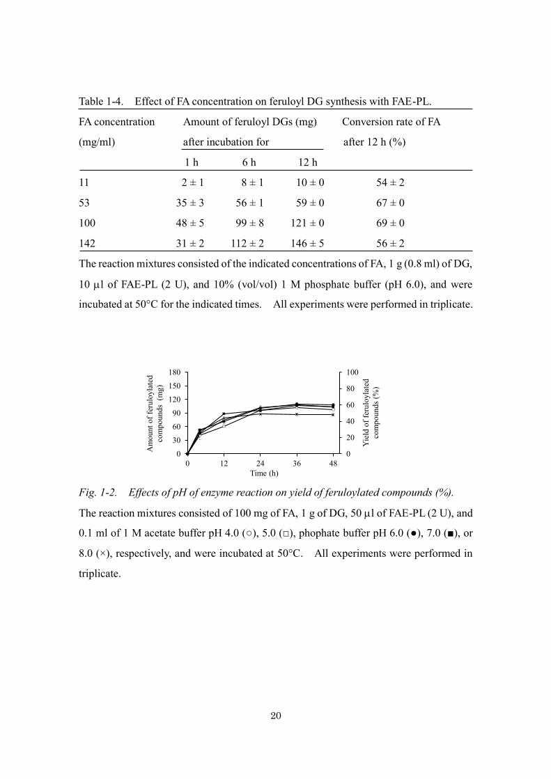
Table 1-4. Effect of FA concentration on feruloyl DG synthesis with FAE-PL.
FA concentration Amount of feruloyl DGs (mg) Conversion rate of FA
(mg/ml) after incubation for after 12 h (%)
1 h 6 h 12 h
11 2 ± 1 8 ± 1 10 ± 0 54 ± 2
53 35 ± 3 56 ± 1 59 ± 0 67 ± 0
100 48 ± 5 99 ± 8 121 ± 0 69 ± 0
142 31 ± 2 112 ± 2 146 ± 5 56 ± 2
The reaction mixtures consisted of the indicated concentrations of FA, 1 g (0.8 ml) of DG,
10 l of FAE-PL (2 U), and 10% (vol/vol) 1 M phosphate buffer (pH 6.0), and were
incubated at 50°C for the indicated times. All experiments were performed in triplicate.
Fig. 1-2. Effects of pH of enzyme reaction on yield of feruloylated compounds (%).
The reaction mixtures consisted of 100 mg of FA, 1 g of DG, 50 l of FAE-PL (2 U), and
0.1 ml of 1 M acetate buffer pH 4.0 (○), 5.0 (□), phophate buffer pH 6.0 (●), 7.0 (■), or
8.0 (×), respectively, and were incubated at 50°C. All experiments were performed in
triplicate.
Time (h)
Am
ount
of f
erul
oyla
ted
com
poun
ds (
mg)
Yie
ld o
f fer
uloy
late
dco
mpo
unds
(%)
0
20
40
60
80
100
0
30
60
90
120
150
180
0 12 24 36 48

21
Fig. 1-3. Effects of temperature on feruloyl DG synthesis (a) and the stability of FAE-
PL (b).
a Reaction mixtures consisted of 100 mg of FA, 1 g of DG, 50 l of FAE-PL (0.5 U), and
0.1 ml of 1 M phosphate buffer (pH 6.0), and incubated at various temperatures.
b Stability was evaluated by measuring the residual hydrolytic activity after 4 days pre-
incubation of the enzyme at temperatures between 30°C and 70°C in 100 mM phosphate
buffer (pH 6.0) containing 90% DG. All experiments were performed in triplicate.
Fig. 1-4. Effect of water concentration on feruloyl DG synthesis with FAE-PL.
The reaction mixtures consisted of 100 mg of FA, 1 g of DG, 50 l of FAE-PL (2 U), 0.1
ml of 1 M phosphate buffer (pH 6.0), and different volumes of water, and incubated at
50°C for 12 h. All experiments were performed in triplicate.
Reaction time (h)24 36120 48
0306090
150120
180
Am
ount
of f
erul
oylD
Gs (
mg)
Con
vers
ion
rate
of F
A (%
)
0
20
40
60
100
80a
Concentration of water (%)
Con
vers
ion
rate
of F
A (%
)
0
30
60
90
120
0 10 20 30 400
20
40
60
80
Am
ount
of f
erul
oyl D
Gs (
mg)
Temperature (°C)
60°C
50°C 40°C
30°C 70°C
20 40 605030 70 800
20
40
60
100
80
Rem
aini
ng a
ctiv
ity (%
)
b
a b

22
Fig. 1-5. Effects of enzyme reaction using vacuum-rotary evaporation on yield of
feruloylated compounds (%).
The reaction mixtures consisted of 100 mg of FA, 1 g of DG, 50 l of FAE-PL (2 U), and
0.1 ml of 1 M phosphate buffer (pH 6.0), and were incubated at 50°C under reduced
pressure (○) or atmospheric pressure (×). All experiments were performed in triplicate.
Feruloyl DG synthesis with FAE-PL by a fed-batch method
Under the optimized conditions, only 8% of the DG was converted to feruloyl DGs.
To obtain higher yields, I tried using a fed-batch method, in which 40 mg of FA was
added to the reaction mixture 15 times every hour. At first, the fed-batch reaction was
carried out in pH 6.0 phosphate buffer. However, feruloyl DG synthesis soon stopped,
probably because the addition of FA decreased the pH. Therefore, I performed the fed-
batch reaction in pH 7.0 phosphate buffer. The mixture was incubated in an evaporator
flask at 50°C under reduced pressure throughout the reaction. FA was added to the
mixture as a cloudy suspension, but as the reaction proceeded, the mixture became clear.
Three feruloyl DGs (FA-DG1, 2, and 3) and five other unidentified products, which were
subsequently identified as diferuloyl DGs by NMR and ESI-MS analyses, were detected
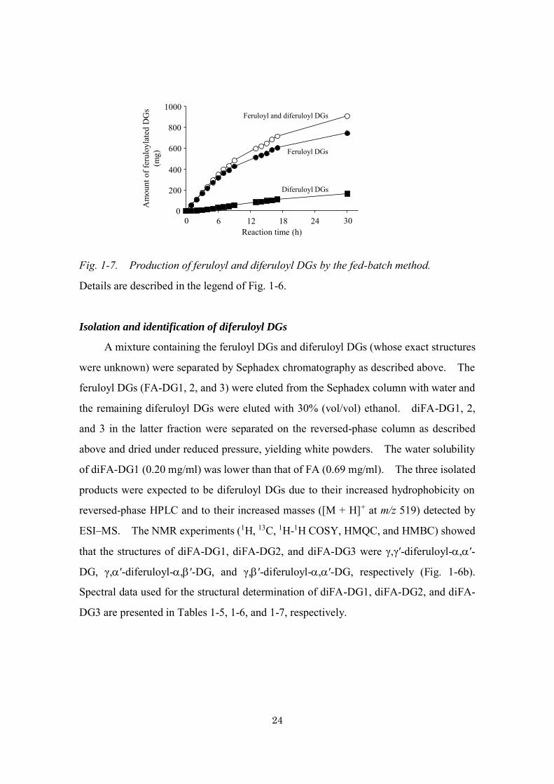
(Fig. 1-6a). The production of both feruloyl DGs and diferuloyl DGs increased with
time (Fig. 1-7). After the 30-h reaction, 729 mg of feruloyl DGs and 168 mg of
diferuloyl DGs were obtained from 600 mg of FA and 1 g of DG (corresponding to
conversion of 69% of the FA to feruloyl DGs and 21 % of the FA to diferuloyl DGs).
The conversion rates of FA and DG to the esters was 90% and 41% in the fed-batch
method, respectively.
Time (h)
Am
ount
of f
erul
oyla
ted
com
poun
ds(m
g)
Yie
ld o
f fer
uloy
late
dco
mpo
unds
(%)
0
20
40
60
80
100
0
30
60
90
120
150
180
0 3 6 9 12

23
Fig. 1-6. HPLC chromatogram of the enzymatic products obtained from FA and DG
using a fed-batch method (a) and structures of diFA-DG1, diFA-DG2, and diFA-DG3 (b).
Composition of the reaction mixture is described in the text. The mixture was incubated
at 50°C for 30 h followed by boiling for 10 min to inactivate the enzyme. The mixture
was diluted 5,000 times with water and analyzed by HPLC using a Mightysil RP-18
reversed-phase column. The upper figure shows the part from 20 to 29 min of the lower
figure.
20 25
FA-DG1
FA
0 5 10 15 20 25 30Retention time (min)
FA-DG3
FA-DG2
diFA-DG1
diFA-DG3
diFA-DG2
(diFA-DG2)
O
CH3O
HO OHOH
O O
O
OCH3
OH
O’
’’1
98
7
65
43
2
1’
9’8’
7’
6’5’
4’3’
2’
a
O
CH3O
HOOHOH
’
O O
’’1
98
7
65
43
2
O
OCH3
OH
O 1’
9’8’
7’
6’5’
4’3’
2’
(diFA-DG1)
b
O
CH3O
HO
OH
OH
O O
OH
OCH3
O
O
’
’’1
98
7
65
43
2
1’ 9’ 8’
7’6’5’
4’3’
2’
(diFA-DG3)
a
b
24
Fig. 1-7. Production of feruloyl and diferuloyl DGs by the fed-batch method.
Details are described in the legend of Fig. 1-6.
Isolation and identification of diferuloyl DGs
A mixture containing the feruloyl DGs and diferuloyl DGs (whose exact structures
were unknown) were separated by Sephadex chromatography as described above. The
feruloyl DGs (FA-DG1, 2, and 3) were eluted from the Sephadex column with water and
the remaining diferuloyl DGs were eluted with 30% (vol/vol) ethanol. diFA-DG1, 2,
and 3 in the latter fraction were separated on the reversed-phase column as described
above and dried under reduced pressure, yielding white powders. The water solubility
of diFA-DG1 (0.20 mg/ml) was lower than that of FA (0.69 mg/ml). The three isolated
products were expected to be diferuloyl DGs due to their increased hydrophobicity on
reversed-phase HPLC and to their increased masses ([M + H]+ at m/z 519) detected by
ESI–MS. The NMR experiments (1H, 13C, 1H-1H COSY, HMQC, and HMBC) showed
that the structures of diFA-DG1, diFA-DG2, and diFA-DG3 were γ,γ′-diferuloyl-,′-
DG, γ,′-diferuloyl-,′-DG, and γ,′-diferuloyl-,′-DG, respectively (Fig. 1-6b).
Spectral data used for the structural determination of diFA-DG1, diFA-DG2, and diFA-
DG3 are presented in Tables 1-5, 1-6, and 1-7, respectively.
0
400
200
600A
mou
nt o
f fer
uloy
late
dD
Gs
(mg)
1000
800
0 6 241812 30Reaction time (h)
Feruloyl DGs
Diferuloyl DGs
Feruloyl and diferuloyl DGs
25
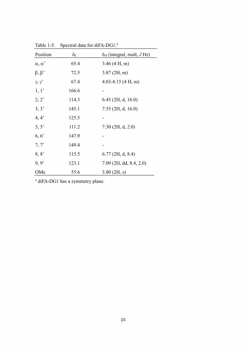
Table 1-5. Spectral data for diFA-DG1.a
Position C H (integral, mult, J Hz)
, ’ 65.4 3.46 (4 H, m)
, ’ 72.5 3.87 (2H, m)
, ’ 67.4 4.03-4.15 (4 H, m)
1, 1’ 166.6 -
2, 2’ 114.3 6.45 (2H, d, 16.0)
3, 3’ 145.1 7.55 (2H, d, 16.0)
4, 4’ 125.5 -
5, 5’ 111.2 7.30 (2H, d, 2.0)
6, 6’ 147.9 -
7, 7’ 149.4 -
8, 8’ 115.5 6.77 (2H, d, 8.4)
9, 9’ 123.1 7.09 (2H, dd, 8.4, 2.0)
OMe 55.6 3.80 (2H, s) a diFA-DG1 has a symmetry plane.
26
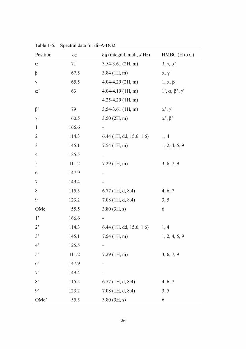
Table 1-6. Spectral data for diFA-DG2.
Position C H (integral, mult, J Hz) HMBC (H to C)
71 3.54-3.61 (2H, m) ’
67.5 3.84 (1H, m)
65.5 4.04-4.29 (2H, m) 1,
’ 63 4.04-4.19 (1H, m) 1’, ’, ’
4.25-4.29 (1H, m)
’ 79 3.54-3.61 (1H, m) ’, ’
’ 60.5 3.50 (2H, m) ’, ’
1 166.6 -
2 114.3 6.44 (1H, dd, 15.6, 1.6) 1, 4
3 145.1 7.54 (1H, m) 1, 2, 4, 5, 9
4 125.5 -
5 111.2 7.29 (1H, m) 3, 6, 7, 9
6 147.9 -
7 149.4 -
8 115.5 6.77 (1H, d, 8.4) 4, 6, 7
9 123.2 7.08 (1H, d, 8.4) 3, 5
OMe 55.5 3.80 (3H, s) 6
1’ 166.6 -
2’ 114.3 6.44 (1H, dd, 15.6, 1.6) 1, 4
3’ 145.1 7.54 (1H, m) 1, 2, 4, 5, 9
4’ 125.5 -
5’ 111.2 7.29 (1H, m) 3, 6, 7, 9
6’ 147.9 -
7’ 149.4 -
8’ 115.5 6.77 (1H, d, 8.4) 4, 6, 7
9’ 123.2 7.08 (1H, d, 8.4) 3, 5
OMe’ 55.5 3.80 (3H, s) 6

27
Table 1-7. Spectral data for diFA-DG3.
Position C H (integral, mult, J Hz) HMBC (H to C)
72.3 3.52-3.62 (2H, m) , , ’
67.4 3.83-3.86 (1H, m)
65.4 4.00-4.12 (2H, m) , , 1
’ 69.7 3.52-3.62 (2H, m) , ’
’ 73.1 4.98 (1H, m) ’, ’, 1’
’ 59.9 3.52-3.62 (2H, m) ’, ’
1 166.3-166.6 -
2 114.1-114.4 6.41-6.46 (1H, d, 16.0) 1, 4
3 145.1 7.53 (1H, d, 15.6) 1, 2, 4, 5, 9
4 125.3 -
5 111.1 7.28 (1H, m) 3, 6, 7, 9
6 148.0 -
7 148.0 -
8 115.5 6.77 (1H, d, 8.4) 4, 6, 7
9 123.1 7.07 (1H, d, 8.4) 3, 5
OMe 55.6 3.79 (3H, s) 6
1’ 166.3-166.6 -
2’ 114.1-114.4 6.41-6.46 (1H, d, 16.0) 1, 4
3’ 145.1 7.53 (1H, d, 15.6) 1, 2, 4, 5, 9
4’ 125.3 -
5’ 111.1 7.28 (1H, m) 3, 6, 7, 9
6’ 148.0 -
7’ 148.0 -
8’ 115.5 6.77 (1H, d, 8.4) 4, 6, 7
9’ 123.1 7.07 (1H, d, 8.4) 3, 5
OMe’ 55.6 3.79 (3H, s) 6

28
Anti-oxidant activity and UV properties of feruloylated DGs
The abilities of feruloyl DGs to scavenge DPPH radicals were similar to the
scavenging abilities of FA and butyl hydroxytoluene, a synthetic anti-oxidant. The
scavenging abilities of three diferuloyl DGs were twice those of feruloyl DGs (Table 1-
8). FA has an absorption peak at 321 nm. The UV spectra of feruloyl DGs and
diferuloyl DGs were very similar to the UV spectrum of FA (data not shown). The
molar absorption coefficients (ε) of the feruloyl DGs were very similar to the ε of FA, but
those of the diferuloyl DGs were twice the ε of FA (Table 1-8).
Table 1-8. DPPH scavenging ability and molar absorption coefficient of feruloylated
DGs.
Compound DPPH scavenging max (M-1cm-1)b ability (mmol)a (nm)
FA-DG1 0.76 ± 0.02 325 20,000 ± 300
FA-DG2 0.79 ± 0.00 325 20,000 ± 100
FA-DG3 0.78 ± 0.01 325 21,000 ± 900
diFA-DG1 0.40 ± 0.00 325 39,000 ± 1,500
diFA-DG2 0.41 ± 0.00 325 39,000 ± 1,700
diFA-DG3 0.40 ± 0.00 325 38,000 ± 400
FA 0.73 ± 0.01 321 19,000 ± 300
Butyl hydroxytoluene 0.79 ± 0.00 - - a Concentration that scavenges 1 mmol of DPPH radicals. The values are the means ±
standard errors of four experiments. b Molar absorption coefficients of the compounds
were determined at 320 nm. Experiments were performed in duplicate.

29
Discussion
Because FA has several beneficial functions, there have been many attempts in the
past decade to synthesize feruloylated compounds by esterification or transesterification
reactions with lipases or FAEs (reviewed by Figueroa-Espinoza and Villeneuve 2005;
Faulds 2010). The goal of many of these studies was to create more hydrophobic
feruloylated compounds such as butyl ferulate (Vafiadi et al. 2008; Thörn et al. 2011) and
feruloylated monoacyl- and diacyl-glycerol (Sun et al. 2007b, 2009). On the other hand,
Topakas et al. (2005) succeeded in synthesizing feruloylated L-arabinose with
Sporotrichum thermophile FAE (StFaeC), which is the first demonstration of enzymatic
feruloylation of hydrophilic compounds. Since then, several feruloyl polyols such as
sugars (Vafiadi et al. 2006, 2007) and glycerol (Tsuchiyama et al. 2006; Sun et al. 2007a;
Sun and Chen 2015) have been enzymatically synthesized. As described in the
Introduction, esterification reactions are more practical than transesterification reactions
for industrial applications. Tsuchiyama et al. (2006) first succeeded the esterification of
FA using an FAE. Since then, synthesis of FA derivatives through esterification
reactions have been reported (Ishihara et al 2010; Couto et al 2011; Zeng et al. 2014).
The goal of chapter 1 was to obtain feruloylated compounds that have high water
solubility and retain the beneficial functions of FA. We previously synthesized FA-G1,
whose water solubility was higher than that of FA. However, the solubility of the
product in water at 20°C was only 1.20 mg/ml (Tsuchiyama et al. 2006). Therefore, I
replaced glycerol with DG, which is liquid and has more hydroxyl groups than glycerol,
as the acceptor of the feruloylation. The water solubility of FA-DG1 (-feruloyl-,′-
DG; Fig. 1-1b), which is the major reaction product obtained from FA and DG by FAE-
PL, was more than 980 mg/ml (corresponding to an FA concentration of 556 mg/ml)
which was many times more than that of FA-G1. No other feruloylated compound has
been reported to have such a high water solubility.
Suitable conditions for esterification of FA with DG were 100 mg of FA in the
presence of 1 g of DG and 0.1 ml of 1 M phosphate buffer (pH 6.0) at 50°C under reduced
pressure. The conversion rate of FA was inversely related to water content in the

30
reaction mixture. Surprisingly, the conversion rate of FA was increased from 70% to
95% when the reaction was conducted under reduced pressure, indicating that continuous
removal of water from the reaction mixture using evaporation diminish the hydrolysis of
the synthesized esters by FAE-PL. The fed-batch reaction resulted in higher yields of
feruloylated DGs than the conventional reaction. This method made it possible to add
large amounts of FA to the reaction mixture, increasing the yield of feruloylated DGs by
more than five fold. In the fed-batch reaction, a considerable amount of the FA (21%)
was converted to diferuloyl DGs, probably because large amounts of feruloyl DGs
synthesized in the reaction mixture were used as acceptors of feruloylation. On the other
hand, diferuloyl DGs were not synthesized in the standard reaction, probably because the
amount of DG is much higher than that of feruloyl DGs.
Feruloylated DGs may have some applications. The DPPH scavenging abilities of
feruloyl DGs were comparable with those of FA and butyl hydroxytoluene, which is a
synthetic anti-oxidant. These results suggest that differences in the chemical structures
of feruloyl DGs did not affect their anti-oxidant activities. The scavenging abilities of
three diferuloyl DGs were twice those of feruloyl DGs, possibly because diferuloyl DGs
have two molecules of FA. Feruloyl and diferuloyl DGs also retained the UV absorption
properties of FA. Thus, the esterification of FA did not appear to alter its properties.
Based on these properties, feruloylated DGs might have applications in beverages, food,
and cosmetics.
31
Chapter 2. Ferulic acid and its water-soluble derivatives
inhibit nitric oxide production and inducible
nitric oxide synthase expression in rat primary
astrocytes.
Summary
In the previous chapter, I described the synthesis of 1-feruloyl diglycerol (FA-DG1),
which exhibited much higher solubility in water than FA. There is insufficient published
evidence regarding the properties of FA derivatives and the protective effects of FA itself
on activated astrocytes. Excessive iNOS expression in astrocytes is one of the factors
involved in the development of neurodegenerative diseases such as Parkinson’s disease
and AD. To evaluate the possible application of water-soluble FA derivatives as
neuroprotective agents, this chapter examines the ability of FA and FA derivatives to
suppress abnormal activation of astrocytes, an important event in the later stages of
disease progression and further neurodegeneration.
I investigated the effects of FA derivatives on NO production and iNOS expression
in rat primary astrocytes. The results showed that these compounds inhibited NO
production and iNOS expression in a concentration-dependent manner and that the
mechanism underlying these effects was the suppression of the nuclear factor-B (NF-
B) pathway. This evidence suggests that FA and its derivatives may be effective
neuroprotective agents.
Materials and Methods
Chemicals and reagents
FA was kindly gifted by Tsuno Food Industrial Co., Ltd. FA-DG1 and FA-G1 were
prepared as described in chapter 1 and previous paper (Tsuchiyama et al. 2006),

32
respectively. Dulbecco’s modified Eagle’s medium (DMEM) and penicillin-
streptomycin solutions were purchased from Wako Pure Chemical Industries. Sinapic
acid (SA), pCA, caffeic acid (CA), 3,4-dimethoxycinnamic acid (dmCA), LPS (from
Escherichia coli; L8274), protease inhibitor cocktail, deoxyribonuclease (DNase), and
anti-actin antibody were obtained from Sigma–Aldrich (St. Louis, Missouri, USA).
Fetal bovine serum (FBS) was purchased from Biowest (Nuaille, France). Mouse
monoclonal antibody against iNOS was prepared as previously described (Nakamura et
al. 2006). Anti-NF-B p65 antibody, anti-histone H2B antibody, and anti-NF-B
inhibitor (IB-) antibody were from Cell Signaling Technology (Danvers,
Massachusetts, USA). Anti-triose-phosphate isomerase (TPI) antibody was kindly
provided by Dr. R. Yamaji (Osaka prefecture University, Japan). Peroxidase-conjugated
anti-rabbit and anti-mouse secondary antibodies were from GE Healthcare Life Science
(Piscataway, New Jersey, USA). 3-(4,5-Dimethylthiazol-2-yl)-2,5-diphenyl tetrazolium
bromide (MTT) was from Dojindo Laboratories (Kumamoto, Japan). The rest of the
reagents were of analytical grade or higher and were obtained from standard sources.
Cell culture
Preparation of astrocytes was carried out in compliance with the Guidelines for
Animal Experimentation of Osaka Prefecture University. Primary astrocytes were
harvested from Wistar rats using a modification of a technique previously described
(Nakamura et al. 2006). Briefly, brain cortices from 20-day-old rat embryos were
deprived of their meninges, cut into blocks, and dissociated with 0.25% trypsin. An
equal volume of horse serum supplemented with 0.1 mg/ml DNase I was added to the
medium to inactivate the trypsin, and the tissues were centrifuged at 300 g for 5 min.
The tissue sediments were suspended in DMEM containing 10% FBS and 1% penicillin–
streptomycin. The cells were plated on 100-mm diameter polyethyleneimine-coated
plastic dishes at a density of 0.8–1.3 × 10⁵ cells/cm2. Cultures were maintained at 37°C
in 5% CO2 and 95% air, and the medium was changed every 3 days. After one week,
astrocytes were replated to remove the neurons by using a standard trypsin treatment
technique.

33
Cell treatment
To determine the effects of the tested compounds on NO production, iNOS protein
expression, NF-B translocation, and IB- degradation, the cells were pre-incubated in
culture medium containing the compounds for 30 min. After pre-incubation, the
astrocytes were treated with LPS (1 g/ml) and incubated at 37°C for 24 h.
Nitrite assay
NO synthesis was determined by quantification of nitrite, a stable product from the
reaction between NO and molecular oxygen, in the supernatant. A 100 l of culture
supernatant was dispensed into the wells of a 96-well plate and mixed with 50 l Griess
reagent A (1% sulfanilamide in 5% H3PO4) for 5 min before adding 50 l Griess reagent
B [0.1% N-(1-naphthyl)-ethylenediamine] to the mixture. After incubation for 5 min at
room temperature, absorbance was measured spectrophotometrically at 540 nm. Fresh
culture media served as the blank in all experiments. Nitrite concentration was
calculated by extrapolating from a standard curve obtained from the reaction of NaNO2
in the assay.
Cytotoxicity assay
Cytotoxicity of the compounds was estimated using the MTT assay (Mosmann
1983). MTT solution of DMEM (50 l, 0.5 mg/ml) was added to cells that had been
cultured for 24 h in the presence or absence of the compounds tested. After incubation
for 3 h at 37°C, the suspension was mixed with 50 l of an MTT stop solution (10%
sodium dodecyl sulfate (SDS) in 10 mM HCl) and further incubated for 24 h. The
optical density of the assay samples was measured at 540 nm.
Western blotting analysis
To obtain total proteins for immunoblot analysis of iNOS levels, astrocytes were
washed with PBS and then lysed with 1% Triton-X and protease inhibitor cocktail in PBS.
To prepare the nuclear fraction for an immunoblot analysis of NF-B p65 and the
cytosolic fraction for immunoblot analysis of IB-, the cell pellets were processed using
Nuclear Extraction Kit I (EpiQuik; Epigentek, New York, USA) according to the

34
manufacturer’s instructions. Soluble proteins from each sample were separated by SDS-
polyacrylamide gel electrophoresis (PAGE) and transferred onto a nitrocellulose
membrane (GE Healthcare). The membrane was blocked with Blocking One (Nacalai
Tesque) and incubated at 4°C overnight with the antibodies against the following
proteins: iNOS, -actin, NF-B p65, histone H2B, IB-, and TPI. After washing with
PBS, the membranes were incubated at room temperature for 1 h with peroxidase-
conjucated anti-rabbit or anti-mouse IgG secondary antibody. Immunoreactive bands
were detected using a chemiluminescence reagent (Millipore, Bedford, Massachusetts,
USA) with a luminescent image analyzer (LAS-1000: Fujifilm, Tokyo).
Protein concentrations
Protein concentrations were determined via the Bradford method with bovine serum
albumin (BSA) as the standard (Bradford 1976).
Statistical analysis
All the experiments were performed at least three times. Mean values for
individual experiments are presented as means ± standard error (SE) and were analyzed
using one-way analysis of variance (ANOVA) with the Newman-Keuls post-hoc test.
Statistical analysis was determined with a using the Prism program (GraphPad, San Diego,
CA, USA). A p value of less than 0.05 was considered significant.
Results
Inhibitory effects of cinnamic acid derivatives on LPS-induced NO production in
primary astrocytes
Excessive NO production plays a key role in neuropathological conditions such as
AD and Parkinson’s disease (Hamby and Sofroniew 2010). First, I evaluated the effects
of the following cinnamic acid derivatives: FA, SA, pCA, CA, and dmCA on LPS-induced
NO production in astrocytes. After pre-incubation with these compounds (1.5 mM), the
astrocytes were treated with LPS (1 g/ml) and incubated for 24 h. The production of

35
NO was determined by measuring the nitrite content of the culture medium. As shown
in Fig. 2-1, stimulation with LPS induced NO production in primary astrocyte cultures,
whereas pretreatment with FA and SA significantly inhibited NO production (p < 0.05).
However, pCA, CA, and dmCA did not significantly inhibit NO production.
According to the MTT assay results, none of the concentrations used during the
experiments were cytotoxic to the astrocytes (data not shown). These results suggest
that FA and SA may be useful as neuroprotective agents. Based on these findings, I
examined the effects of FA and its water-soluble derivatives: FA-G1 and FA-DG1.
Fig. 2-1. Inhibitory effects of various cinnamic acid derivatives on LPS-induced NO
production in primary astrocytes.
Astrocytes were pre-incubated in the presence of absence of FA, SA, pCA, CA, and
dmCA (1.5 mM) for 30 min, followed by treatment with LPS (1 g/ml) and incubated for
24 h. The cells were plated on polyethyleneimine-coated 96-well plate at a density of
1.3 × 10⁵ cells/cm2. The production of NO was determined by measuring the nitrite
content of the culture medium. Results are expressed as concentrations (M) of nitrite
content. Data are presented as the mean ± SE (n = 6). One-way ANOVA with the
Newman-Keuls post-hoc test was used for statistical analysis. * p < 0.05 vs. LPS (-).
** p < 0.05 vs. LPS (+).
Prod
uctio
n of
nitr
ite (
M)
02468
101214
**
*
**

36
Inhibitory effects of FA and its water-soluble derivatives on LPS-induced NO
production in primary astrocytes
Astrocytes were pre-incubated with FA and its water-soluble derivatives at several
concentrations (0.25, 0.5, 1.0, and 1.5 mM), treated with LPS (1 g/ml), and incubated
for 24 h. As shown in Fig. 2-2a, pretreatment with FA, FA-G1, and FA-DG1,
significantly inhibited NO production at concentration of 1.0 mM, 0.25 mM, and 0.25
mM or higher, respectively (p < 0.05). Cytotoxicity of the compounds at the above
concentrations to the astrocytes was not observed via the MTT assay (Fig. 2-2b),
indicating that FA, FA-G1, and FA-DG1 show potential as neuroprotective agents.

37
a
b
Fig. 2-2. Inhibitory effects of FA and its water-soluble derivatives on LPS-induced NO
production in primary astrocytes.
Astrocytes were prepared as described in the legend for Figure 2-1, except that they were
pre-incubated with different concentrations (from 0.25 to 1.5 mM) of FA or its water-
soluble derivatives before addition of LPS. The cells were plated on polyethyleneimine-
coated 96-well plate at a density of 1.3 × 10⁵ cells/cm2. (a) The production of NO was
determined by measuring the nitrite content of the culture medium. (b) Cell viability
was determined by MTT assay. Data are presented as the mean ± SE (n = 6). One-
way ANOVA with the Newman-Keuls post-hoc test was used for statistical analysis. *
p < 0.05 vs. control.
Cel
l via
bilit
y(%
of c
ontro
l)
20
40
60
80
100
Non FA FA-G1 FA-DG1
0.25 0.5 1.0 1.5 0.25 0.5 1.0 1.5 0.25 0.5 1.0 1.50
Prod
uctio
n of
NO
2-
(% o
f con
trol)
20
40
60
80
100
Non FA FA-G1 FA-DG1
0.25 0.5 1.0 1.5 0.25 0.5 1.0 1.5 0.25 0.5 1.0 1.5
*
*
*
** *
**0
* *

38
Inhibitory effect of FA and its water-soluble derivatives on LPS-induced iNOS
expression in primary astrocytes
LPS induces iNOS expression, and iNOS synthesizes NO in astrocytes (Hamby and
Sofroniew 2010). To determine whether the inhibition of NO production by FA and its
water-soluble derivatives was a result of the inhibition of iNOS protein expression, I
assessed the latter by immunoblotting using anti-iNOS antibodies. As shown in Fig. 2-
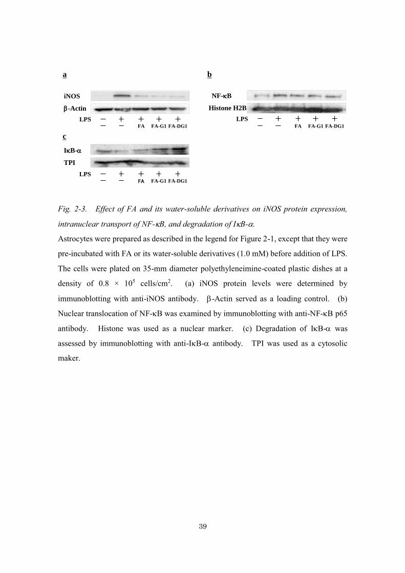
3a, LPS stimulation increased iNOS protein expression in primary astrocyte cultures,
whereas pretreatment with FA, FA-G1, and FA-DG1 (1.0 mM) significantly inhibited
such an increase in iNOS protein expression. These results suggest that FA, FA-G1, and
FA-DG1 repress NO production in astrocytes through inhibition of LPS-induced iNOS
expression.
Inhibitory effect of FA and its water-soluble derivatives on intranuclear transport of
NF-B and degradation of IB- in primary astrocytes
LPS-induced iNOS expression in astrocytes is mediated through NF-B signaling
(Nomura 2001; Saha and Pahan 2006). To determine whether inhibition of iNOS
expression by FA and its water-soluble derivatives is mediated through NF-B signaling,
I examined the effects of FA and its derivatives on LPS-induced NF-B nuclear
translocation and IB- degradation in astrocytes. As shown in Fig. 2-3b and Fig. 2-3c,
LPS stimulation increased NF-B nuclear translocation and IB- degradation in primary
astrocyte cultures. Pretreatment with FA, FA-G1, and FA-DG1 (1.0 mM) significantly
inhibited the increase in NF-B nuclear translocation and IB- degradation. These
results indicate that FA, FA-G1, and FA-DG1 inhibit LPS-induced activation of NF-B
signaling.
39
Fig. 2-3. Effect of FA and its water-soluble derivatives on iNOS protein expression,
intranuclear transport of NF-B, and degradation of IB-.
Astrocytes were prepared as described in the legend for Figure 2-1, except that they were
pre-incubated with FA or its water-soluble derivatives (1.0 mM) before addition of LPS.
The cells were plated on 35-mm diameter polyethyleneimine-coated plastic dishes at a
density of 0.8 × 10⁵ cells/cm2. (a) iNOS protein levels were determined by
immunoblotting with anti-iNOS antibody. -Actin served as a loading control. (b)
Nuclear translocation of NF-B was examined by immunoblotting with anti-NF-B p65
antibody. Histone was used as a nuclear marker. (c) Degradation of IB- was
assessed by immunoblotting with anti-IB- antibody. TPI was used as a cytosolic
maker.
-Actin
iNOS
a
Histone H2B
NF-B
b
FA-DG1FA FA-G1
LPSFA-DG1FA FA-G1
LPS
TPI
IB-
c
FA-DG1FA FA-G1
LPS

40
Discussion
In chapter 2, to expand the utility of FA, FA-G1, and FA-DG1, I examined the
effects of these compounds on NO production and iNOS expression in primary astrocytes.
Excessive iNOS expression in astrocytes is one of the factors involved in the development
of neurodegenerative diseases such as Parkinson’s disease and AD.
Prior to the experiments carried out with FA and its derivatives, I conducted
screening tests with various cinnamic acid derivatives. These experiments yielded FA
and SA as potential neuroprotective agents. Both FA and SA have a methoxy and a
hydroxy group in their benzene ring. The balance between hydrophilic and hydrophobic
groups makes possible for these compounds to bind to and stimulate cell membrane
proteins.
I found that FA inhibited NO production and iNOS expression in primary astrocytes.
Furthermore, inhibition of iNOS expression was mediated by a suppression of the NF-B
pathway. The water-soluble FA derivatives developed possess neuroprotective effects
similar to FA, suggesting that the neuroprotective effect is attributable to the phenol
groups rather than the ester groups.
In addition, in astrocytes, A induces expression of iNOS and other inflammatory
cytokines through the NF-B pathway (Kaltschmidt et al. 1997). Our results suggest
that treatment with FA, FA-G1, and FA-DG1 may improve AD through the inhibition of
A aggregation and the excessive activation of the NF-B pathway in astrocytes. The
NF-B pathway induces inflammatory cytokines that cause neuronal death (Nomura
2001). FA, FA-G1, and FA-DG1 may inhibit the overexpression of inflammatory
cytokines originating through the NF-B pathway and therefore exert a neuroprotective
effect.
Some polyphenols are known to exhibit neuroprotective properties, including a
therapeutic effect in AD (Granzotto and Zatta 2014; Malar and Devi 2014). Recent
research on plant-derived polyphenols has focused on their safe pharmacological profile
and their potential to positively affect human health. Water-soluble FA derivatives
showed antioxidant activity similar to that of FA, suggesting that they may be effective

41
as neuroprotective agents. The ability of certain compounds to inhibit the excessive
activation of astrocytes has been previously reported. For example,
polymethoxyflavone, extracted from young fruits of Citrus unshiu, was found to inhibit
NO production and iNOS expression in rat primary astrocytes (Ihara et al. 2012). In
addition, resveratrol was found to strongly prevent H2O2-induced increase in reactive
nitrogen species (RNS) production and iNOS expression in a C6 astrocyte cell line
(Quincozes-Santos et al. 2013). These compounds are considered to be neuroprotective
agents and the same could be true for FA, FA-G1, and FA-DG1, based on our latest results.

42
Chapter 3. Water-soluble ferulic acid derivatives improve
amyloid- induced neuronal cell death and
dysmnesia through inhibition of amyloid-
aggregation.
Summary
In the previous chapters, I describe the successful synthesis of water-soluble FA
derivatives (FA-DG1) and investigation of the inhibitory effects of these derivatives and
FA on LPS-induced NO production in primary astrocytes. The effects of FA derivatives
on A-induced neurodegeneration have not been reported previously. It is generally
accepted that aggregation of A is one of the crucial pathogenic events in AD.
Here in, I detail the neuroprotective effects of these water-soluble FA derivatives on
A-induced neurodegeneration in both in vitro and in vivo experiments. FA and water-
soluble FA derivatives inhibited A aggregation and destabilized pre-aggregated A to a
similar extent. Furthermore, water-soluble FA derivatives, as well as FA, inhibited A-
induced neuronal cell death in cultured neuronal cells. In in vivo experiments, oral
administration of water-soluble FA derivatives to mice improved A-induced dysmnesia
assessed by contextual fear conditioning test and protected hippocampal neurons against
A-induced neurotoxicity. This study provides useful evidence suggesting that water-
soluble FA derivatives are expected to be effective neuroprotective agents.
Materials and Methods
Chemicals and reagents
FA was kindly gifted by Tsuno Food Industrial Co., Ltd. FA-DG1 and FA-G1
were prepared as described in chapter 1 and previous paper (Tsuchiyama et al. 2006),
respectively. DMEM and penicillin-streptomycin solution were purchased from Wako

43
Pure Chemical Industries. FBS was from Biowest; horse serum, from Gibco (Carlsbad,
CA, USA); MTT, from Dojindo Laboratories; A (25–35), from AnaSpec (San Jose, CA,
USA); and A (1–40) and A (1–42), from Peptide Institute (Osaka, Japan). RPMI-
1640 medium, thioflavin-T, trypsin, DNase I and cytosine arabinofuranoside (Ara-C)
were bought from Sigma-Aldrich. PC12 cells and NeuroTrace 500/525 green-
fluorescent Nissl stain were from Invitrogen (Carlsbad, CA, USA). The rest of the
reagents were of analytical grade or higher and were obtained from standard sources.
Preparation of A
A (25–35) was dissolved in ultrapure water at a concentration of 1 mM and stored
at −80°C until use. A (1–40) and A (1–42) were dissolved in DMSO at a
concentration of 500 and 250 M, respectively, and stored at −80°C until use.
Thioflavin-T fluorescence assay
In order to determine the formation of aggregated A, reaction mixtures containing
fresh 200 M A (25–35), 25 M A (1–40), or 25 M A (1–42) with or without FA,
FA-G1, or FA-DG1 were incubated at 37°C for 7, 3, or 1 days, respectively. To
determine the destabilization of aggregated A, 400 M A (25–35), 100 M A (1–40),
or 50 M A (1–42) were incubated at 37°C for 3, 3, and 1 day, respectively. After the
aggregation reaction, the mixtures were incubated with or without FA, FA-G1, or FA-
DG1 at 37°C for 6 h. Ten microliters of the reaction mixtures were added to 700 l of
50 mM glycine–NaOH buffer (pH 8.0), containing 10 M thioflavin-T. Fluorescence
(excitation 450 nm and emission 485 nm) was monitored with a spectrofluorometer FP-
6200 (JASCO, Tokyo, Japan).
Cell culture
Primary cortical neurons were prepared from 20-day-old fetal Wistar rats as
described previously (Janssens and Lesage 2001) with minor modifications. Briefly,
brain cortices from embryos were cleaned of their meninges, cut into blocks, and
dissociated with 0.25% trypsin. An equal volume of horse serum supplemented with

44
0.1 mg/ml DNase I was added to the medium to inactivate the trypsin, and the tissues
were centrifuged at 300 × g for 5 min. The tissue sediments were resuspended in
DMEM containing 10% FBS and 1% penicillin-streptomycin. The cells were plated on
polyethyleneimine-coated 96 well plates at a density of 1.5 × 10⁶ cells/cm2. After
culturing for 2 to 4 days in vitro (DIV), the medium was replaced with fresh medium
containing 5 M Ara-C to inhibit proliferation of non-neuronal cells; thereafter, no further
medium change was carried out before the experiments. Cells were maintained at 37°C
in 5% CO2 and 95% air.
PC12 cells were plated on polyethyleneimine-coated 96 well plates at a density of
3.0 × 10⁷ cells/cm2 in RPMI-1640 containing 10% FBS and 1% penicillin-streptomycin.
Cells were maintained at 37°C in 5% CO2 and 95% air.
Cytotoxicity assay
Reaction mixtures containing fresh 2 mM A (25–35) with or without 2 mM FA,
FA-G1, or FA-DG1 were incubated at 37°C for 72 h. Primary cortical neurons (7 DIV)
or PC12 cells were treated with the reaction mixtures diluted in culture medium for 24 h
at a final concentration of 20 M A (25–35). Cell viability was evaluated by MTT
assay as described in chapter 2. Briefly, a 0.5 mg/mL solution of MTT in DMEM or
RPMI-1640 was added to the culture medium (10 l/well). Cultures were incubated at
37°C for 3 h. After incubation, the suspension was mixed with 110 l of MTT stop
solution (10% SDS in 10 mM HCl) and further incubated for 24 h. Absorbance was
measured at 540 nm.
Animals
Male C57BL/6J mice were obtained from Kiwa Laboratory Animal Co., Ltd. The
mice were allowed free access to food and water and maintained in a 12-hour dark-light
cycle for 1 week. Animal care and experimental procedures were carried out in
compliance with the Guide for Animal Experimentation at Osaka Prefecture University.

45
Intracerebroventricular injection of A
The mice were divided into five groups (n=6 each). FA, FA-G1, and FA-DG1
were dissolved in 0.5% carboxymethylcellulose (CMC) at a concentration of 10 mM.
After oral administration of FA, FA-G1, or FA-DG1 (0.1 mol/g/day) for 42 days, mice
were injected 4 nmol A (25–35) diluted in 2 l of ultrapure water by
intracerebroventricular injection, as described previously (Tsunekawa et al. 2008).
Briefly, using a 25-l Hamilton microsyringe fitted with a 27-gauge needle, each mouse
was injected at a point 0.8 mm lateral from bregma at a depth of 2.5 mm in the lateral
cerebral ventricle.
Contextual fear conditioning test
The contextual fear conditioning test was carried out on days 8–9 after A (25–35)
injection, according to a previous report (Tsunekawa et al. 2008) with a minor
modification. For training (conditioning phase), mice were placed in the conditioning
cage for 90 sec, and then a 15-sec tone (75 dB) was delivered as a conditioned stimulus.
During the last 2 sec of the tone stimulus, a foot shock of 0.15 mA (unconditioned
stimulus) was delivered through a shock generator (O’Hara & Co., LTD, Tokyo, Japan).
This procedure was repeated four times at 15-sec intervals. One day after fear
conditioning, mice were placed in the conditioning cage, and the freezing response for
tone stimulus was continuously measured for 2 min (retention session). The freezing
response was defined as no movement by any of the mice.
Nissl stain
After fear conditioning test, the mice were killed with a lethal dose of
pentobarbitone sodium (0.1 ml, 60 mg/ml) and perfused through the left ventricle with 60
ml of PBS, followed by 80 ml of PBS containing 4% paraformaldehyde. The brain was
removed and fixed in 4% paraformaldehyde for 2 h at 4°C, followed by cryoprotection in
30% sucrose overnight at 4°C. Ten-micrometer thick serial coronal sections were cut
using a Leica cryostat CM 1950 (Leica Microsystems, Wetzlar, Germany). Nissl
staining was performed using NeuroTrace 500/525 green-fluorescent Nissl stain,

46
according to the manufacturer's instructions. The sections were coverslipped and
observed under a fluorescence microscope (Nikon, Tokyo, Japany). Further image
processing and quantification were performed by using Adobe Photoshop v. 7.0 (Adobe
Systems, Waltham, MA, USA).
Statistical analysis
All results are represented as means ± S.E of three to five determinations and were
analyzed using one-way analysis of variance (ANOVA) with the Newman-Keuls post-
hoc test. Statistical analysis was determined with a using the Prism program (GraphPad,
San Diego, CA, USA). A p value of less than 0.05 was considered significant.
Results
Effects of water-soluble FA derivatives on A aggregation
FA has been shown to inhibit A aggregation in vitro (Ono et al. 2005). Therefore,
we examined whether water-soluble FA derivatives, FA-G1 and FA-DG1, showed similar
inhibitory effects on A aggregation. When fresh A (25–35), A (1–40), or A (1–
42) was incubated at 37°C for 5 days, thioflavin-T fluorescence was significantly
enhanced, indicating that aggregated A fibrils bound to thioflavin-T (data not shown).
As shown in Fig. 3-1, FA inhibited aggregation of A (25–35), A (1–42) in a
concentration of 10 M, 1 M or higher, respectively (p < 0.05), consistent with previous
data. FA-G1 and FA-DG1 also prevented A aggregation in a each concentration (Fig.
3-1).
These results show that, in vitro, FA-G1 and FA-DG1 as well as FA inhibit A
aggregation to a similar extent. Aggregabilities of As differ by differences in their
peptide lengths. The aggregability of A (1–42) is considerably high compared with
that of A (25–35). Therefore, the amounts of A or the amounts of FA derivatives in
the reaction mixtures were varied depending on the types of As used for the experiments.

47
Effects of water-soluble FA derivatives on destabilization of A aggregates
Since FA has been also shown to destabilize A aggregates in vitro (Ono et al. 2005),
we next tested whether the water-soluble FA derivatives also destabilize A aggregates.
As shown in Fig. 3-2, when the aggregated A (25–35), A (1–40), or A (1–42) were
incubated with FA, thioflavin-T fluorescence decreased, indicating that FA destabilized
the A aggregates. Similarly, FA-G1 and FA-DG1 also decreased the fluorescence
intensity of thioflavin-T; this reduction depended on FA-G1 and FA-DG1 concentration
(Fig. 3-2). These results indicate that water-soluble FA derivatives are able to
destabilize pre-aggregated A in vitro.

48
Fig. 3-1. Effects of FA and water-soluble FA derivatives on A aggregation.
The reaction mixtures containing 200 µM fresh A (25–35) (a), 25 µM A (1–40) (b), or
25 µM A (1–42) (c) were incubated at 37°C in the presence or absence of FA, FA-G1,
or FA-DG1. A aggregation was determined by thioflavin-T fluorescence assay, as
described in Materials and Methods. Data for all experiments are represented as means
± S.E. of values from three independent assays in triplicate experiments. One-way
ANOVA with the Newman-Kleus post-hoc test was used for statistical analysis. *P <
0.05 vs. values for control.
a bA(25-35) A(1-40)
* *
* *
control0.1 10 0.1 10 0.1 100
10
20
30
40
FA GFA Di-FA
(M)Th
io-T
flu
ore
scen
ce i
nte
nsit
y
(arb
itu
rary
un
it)
FA FA-G1 FA-DG1
control0.1 10 0.1 10 0.1 100
25
50
75
FA GFA Di-FA
(M)Th
io-T
flu
ore
scen
ce i
nte
nsit
y
(arb
itu
rary
un
it)
FA FA-G1 FA-DG1
Thio
-T fl
uore
scen
ce in
tens
ity
(arb
itura
ryun
it)
Non 0.1 10 0.1 10 0.1 10 (M)
FA FA-G1 FA-DG1
Thio
-T fl
uore
scen
ce in
tens
ity
(arb
itura
ryun
it)
Non 0.1 10 0.1 10 0.1 10 (M)
FA FA-G1 FA-DG1
c A(1-42)
* * * *
control 1 50 1 50 1 500
10
20
30
FA GFA Di-FA
(M)Th
io-T
flu
ore
scen
ce i
nte
nsit
y
(arb
itu
rary
un
it)
FA FA-G1 FA-DG1
Thio
-T fl
uore
scen
ce in
tens
ity
(arb
itura
ryun
it)
Non 1 50 1 50 1 50 (M)
FA FA-G1 FA-DG1
c
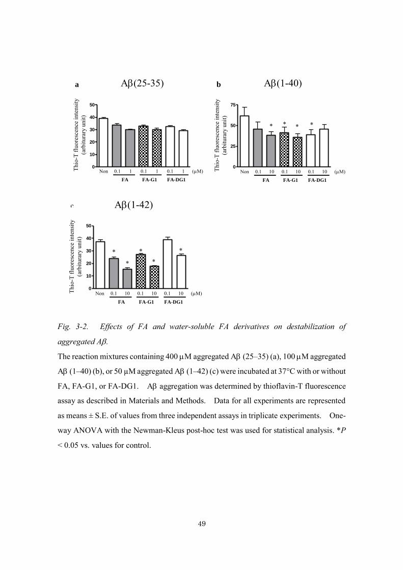
49
Fig. 3-2. Effects of FA and water-soluble FA derivatives on destabilization of
aggregated A.
The reaction mixtures containing 400 M aggregated A (25–35) (a), 100 M aggregated
A (1–40) (b), or 50 µM aggregated A (1–42) (c) were incubated at 37°C with or without
FA, FA-G1, or FA-DG1. A aggregation was determined by thioflavin-T fluorescence
assay as described in Materials and Methods. Data for all experiments are represented
as means ± S.E. of values from three independent assays in triplicate experiments. One-
way ANOVA with the Newman-Kleus post-hoc test was used for statistical analysis. *P
< 0.05 vs. values for control.
a bA(25-35) A(1-40)
* * * *
control0.1 1 0.1 1 0.1 1
0
10
20
30
40
50
FA GFA Di-FA
(M)
Th
io-T
flu
ore
scen
ce i
nte
nsit
y
(arb
itu
rary
un
it)
FA FA-G1 FA-DG1
control0.1 10 0.1 10 0.1 10
0
25
50
75
FA GFA Di-FA
(M)
Th
io-T
flu
ore
scen
ce i
nte
nsit
y
(arb
itu
rary
un
it)
FA FA-G1 FA-DG1
Thio
-T fl
uore
scen
ce in
tens
ity
(arb
itura
ryun
it)
Non 0.1 1 0.1 1 0.1 1 (M)
FA FA-G1 FA-DG1
Thio
-T fl
uore
scen
ce in
tens
ity
(arb
itura
ryun
it)
Non 0.1 10 0.1 10 0.1 10 (M)
FA FA-G1 FA-DG1
c A(1-42)
*
* *
* *
control0.1 10 0.1 10 0.1 10
0
10
20
30
40
50
FA GFA Di-FA
(M)
Th
io-T
flu
ore
scen
ce i
nte
nsit
y
(arb
itu
rary
un
it)
FA FA-G1 FA-DG1
Thio
-T fl
uore
scen
ce in
tens
ity
(arb
itura
ryun
it)
Non 0.1 10 0.1 10 0.1 10 (M)
FA FA-G1 FA-DG1
c

50
Effects of water-soluble FA derivatives on A-induced cell death in cultured neurons
or PC12 cells
Previous reports have indicated that EFA, whose structure is similar to FA, inhibited
A-induced cytotoxicity (Sultana et al 2005). Here, I examined whether water-soluble
FA derivatives have an inhibitory effects on A (25–35)-induced cytotoxicity in neuronal
cells. PC12 cells are rat pheochromocytoma cells that are a well-known model for
studying neuronal signaling pathways and other neurobiochemical events (Fujita et al.
1989). Many neurobiological investigations were studied using PC12 cells rather than
primary neurons. As shown in Fig. 3-3, aggregated-A (25–35) induced cytotoxicity in
primary cerebral cortical neuron cultures and PC12 cells, whereas pretreatment of A
(25–35) with FA significantly inhibited this cytotoxicity.
a Neurons b PC12 cells
Fig. 3-3. Inhibitory effects of FA and water-soluble FA derivatives on A-induced
neuronal cell death in cultured neurons or PC12 cells.
A (25–35) (2 mM) was aggregated in the presence or absence of 2 mM FA, FA-G1, or
FA-DG1 for 3 days. Primary cortical neurons (7 DIV) (a) or PC12 cells (b) were treated
with reaction mixtures for 24 h at a final concentration of 20 M A (25–35). Cell
viability was assessed by MTT assay as described in Materials and Methods. Data are
represented as means ± S. E. of values from triplicate experiments. One-way ANOVA
with the Newman-Kleus post-hoc test was used for statistical analysis. *P < 0.05 vs.
values for control, **P < 0.05 vs. values for A (25–35) alone.
* * *
*
- + + + +0
25
50
75
100
125
FA GFA Di-FA- -FA derivatives
A (25-35)
Cell
via
bil
ity (
%)
- - FA FA-G1 FA-DG1
*
* *
FA derivatives - - FA FA-G1 FA-DG1A(25-35) - + + + +
Cel
l via
bilit
y (%
)
* * *
*
- + + + +0
25
50
75
100
125
FA GFA Di-FA- -FA derivatives
A (25-35)
Cell
via
bil
ity (
%)
- - FA FA-G1 FA-DG1
*
* *
FA derivatives - - FA FA-G1 FA-DG1A(25-35) - + + + +
Cel
l via
bilit
y (%
)

51
Similarly, pre-incubation of A (25–35) with FA-G1 or FA-DG1 also inhibited A
(25–35)-induced neuronal cell death. These results indicate that water-soluble FA
derivatives, in addition FA, have a neuroprotective effect against A-induced neuronal
cell death.
Effects of water-soluble FA derivatives on A-induced dysmnesia
Previous in vivo studies have demonstrated that long-term administration of FA
protected mice against A-induced learning and memory deficits (Yan et al. 2001).
Here, I examined whether water-soluble FA derivatives improve A-induced dysmnesia
using a contextual fear conditioning test. No significant change in body-weight was
observed in FA, FA-G1, and FA-DG1-injected mice (Table 3-1). After administration
of FA, FA-G1, or FA-DG1 for 42 days, followed by injection of A (25–35), a fear-
conditioning test was performed (Fig. 3-4a).
Table 3-1. Effects of FA and water-soluble FA derivatives administration on the body
weight of mice.
Experimental group Initial (g) Final (g)
0.5% CMC + UPW 19.7±0.8 23.5±0.9
0.5% CMC + Ab(25–35) 16.3±0.9 23.6±0.8
FA + Ab(25–35) 19.5±0.7 25.1±0.7
FA-G1 + Ab(25–35) 16.7±0.9 24.6±0.6
FA-DG1 + Ab(25–35) 17.2±0.9 22.7±0.9
After orally administered FA or water-soluble FA derivatives (0.1 mol/g/day) for 42
days, mice were injected with A (25–35) as described in Fig. 3-4a. Body weight was
measured before the administration (initial) and after the injection (final). Data are
means ± S.E. (n = 6). UPW = ultra-pure water
Contextual-dependent tests were performed 24 h after conditioning. A (25–35)-
injected mice exhibited a lower contextual-dependent freezing response than distilled
water-injected mice, indicating an impairment of associative memory (Fig. 3-4b). FA-

52
G1 and FA-DG1 as well as FA attenuated the A (25–35)-induced impairment of
contextual freezing responses. However, there were no significicant differences
between FA and its derivatives. These results suggest that water-soluble FA derivatives,
in addition to FA, improve contextual freezing response impairment in mice injected with
A (25–35).
Fig. 3-4. Effects of FA and water-soluble FA derivatives on cognitive functions in A
(25–35)-injected mice.
(a) Behavioral experiments schedule. After orally administration of FA or water-soluble
FA derivatives (0.1 mol/g/day) for 42 days, mice were injected with A (25–35). Each
behavioral test was conducted on the indicated time point. (b) The freezing rate in fear
conditioning test was measured. Data for all experiments are represented as means ±
S.E. One-way ANOVA with the Newman-Kleus post-hoc test was used for statistical
analysis. *P < 0.05 vs. 0.5% CMC-administrated, vehicle-injected mice. **P < 0.05
vs. 0.5% CMC-administrated, A (25–35)-injected mice.
a
b
42nd
day
50th
day
51st
day
A (25-35)
injectionFear
conditioning
test
FA, FA-G1, FA-DG1
administration
0 day54th
day
* * * * *
* *
- + + + +
0
20
40
60
FA derivatives - - FA GFA Di-FA
A (25-35)
Fre
ezin
g r
ate
(%
)
- - FA FA-G1 FA-DG1
A(25-35) - + + + +
FA derivatives - - FA FA-G1 FA-DG1
Free
zing
rate
(%)
a
b

53
In vivo effects of water-soluble FA derivatives on A-induced neuronal dysfunction
In order to examine the protective effects of FA and water-soluble FA derivatives
on A-induced neuronal cell death in vivo, Nissl staining was performed in the
hippocampus of mice treated with A (25–35). As shown in Figs. 3-5a and b, the
number of Nissl-positive neuronal cells in the hippocampus decreased in mice treated
with A (25–35). Positive staining was recovered when mice were pretreated with FA.
In addition, FA-G1 and FA-DG1 dramatically suppressed A (25–35)-mediated neuronal
cell death. These results demonstrate that FA and water-soluble FA derivatives protect
neuronal cells against A (25–35)-induced neurotoxicity in vivo. The in vivo
neuroprotective effects were consistent with the results from in vitro experiments using
culture neuron (Fig. 3-3).
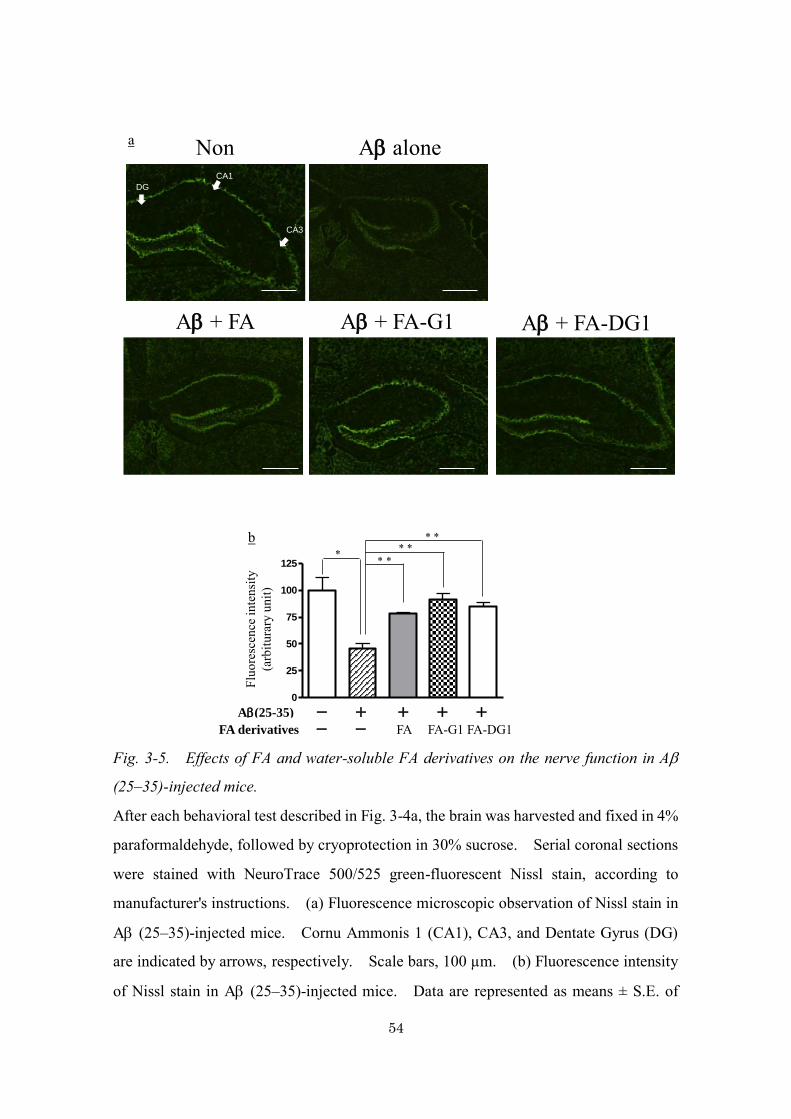
54
Fig. 3-5. Effects of FA and water-soluble FA derivatives on the nerve function in A
(25–35)-injected mice.
After each behavioral test described in Fig. 3-4a, the brain was harvested and fixed in 4%
paraformaldehyde, followed by cryoprotection in 30% sucrose. Serial coronal sections
were stained with NeuroTrace 500/525 green-fluorescent Nissl stain, according to
manufacturer's instructions. (a) Fluorescence microscopic observation of Nissl stain in
A (25–35)-injected mice. Cornu Ammonis 1 (CA1), CA3, and Dentate Gyrus (DG)
are indicated by arrows, respectively. Scale bars, 100 µm. (b) Fluorescence intensity
of Nissl stain in A (25–35)-injected mice. Data are represented as means ± S.E. of
A + FA A + FA-G1 A + FA-DG1
A aloneNona
CA1
CA3
DG
- + + + +
0
25
50
75
100
125
FA GFA Di-FA- -FA derivatives
A(25-35)
Flu
ore
scen
ce i
nte
nsit
y
(arb
itu
rary
un
it)
b
* * * * *
* *
FA-G1 FA-DG1
A(25-35) - + + + +
FA derivatives - - FA FA-G1 FA-DG1
Fluo
resc
ence
inte
nsity
(a
rbitu
rary
unit)
a
b

55
values from triplicate experiments. One-way ANOVA with the Newman-Kleus post-
hoc test was used for statistical analysis. *P < 0.05 vs. 0.5% CMC-administrated, vehicle-
injected mice. **P < 0.05 vs. 0.5% CMC-administrated, A (25–35)-injected mice.
Discussion
FA exerts neuroprotective effects against A-induced neurotoxicity in vitro
(Sultana et al. 2005) and in vivo (Yan et al. 2001; Kim et al. 2004). In chapter 3, I
compared the neuroprotective effects of two water-soluble FA derivatives, FA-G1 and
FA-DG1, with that of FA in vitro and in vivo.
A is a 40-42 amino acid peptide derived from the cleavage of the membrane
protein APP by- and -secretases. The overproduction and aberrant self-assembly of
A into fibrillar aggregates constitutes the first steps of the so-called amyloid cascade
hypothesis that is thought to trigger AD. In AD, neurodegeneration may be caused by
A aggregation in senile plaques in the brain, and this is generally regarded as a critical
pathogenic event in AD (Selkoe 2004; Lleo et al. 2006). A is also found in the brain
in healthy individuals; it is a secondary product of APP metabolism and may have some
physiological function. However, Ais generally degraded and discharged in healthy
individuals, resulting in little A deposition. Aaccumulation likely stems from an
abnormality in metabolism, possibly as the result of aging, where an imbalance occurs
between the production and degradation of A.
The mechanism by which Abecomes aggregated is complex and is composed of
several steps. These aggregates are characterized by having transitional aggregation
species as initial seeds, soluble small oligomers, protofibrils, and insoluble amyloid fibrils,
with a -sheet conformation. Ono et al. (2005) demonstrated that FA inhibited A
aggregation in vitro. Hence, I first confirmed whether water-soluble FA derivatives also
show this beneficial effect despite their structural modifications. Water-soluble FA
derivatives not only prevented A aggregation, but also destabilized aggregated A fibrils.
Therefore, differences in the chemical structures of these derivatives did not affect their

56
disaggregation activities (Fig. 3-1 and 3-2). This suggests that water-soluble FA
derivatives could be protective against A-induced neurotoxicity. Indeed, I show
evidence that the derivatives prevented A-induced cytotoxicity in cultured neuronal cells
(Fig. 3-3).
FA is also a well-known antioxidant. Since A-induced neurotoxicity is at least
partially due to oxidative stress, FA may exert its neuroprotective effect through its
antioxidant activity, besides its inhibitory effect on A aggregation. Sultana et al.
characterized the protective effect of EFA, which is more hydrophobic than FA, on A-
induced oxidative stress in rat primary neuronal cell cultures (Sultana et al. 2005).
Indeed, the water-soluble FA derivatives showed similar antioxidant activity compared
to FA, suggesting that the antioxidant activity of these derivatives may also be involved
in their neuroprotective effect. It is reported that FA binds A and induces cooperative
rupture of the backbone hydrogen bonds and of the Asp23-Lys28 salt bridge (Sgarbossa
et al. 2013). Cui et al. (2013) used thioflavin T fluorescence, CD spectroscopy, and
transmission electron microscopy and showed that FA inhibited the A aggregation
process by blocking the formation of -sheets, interacting with A by hydrogen bonding
with His14 and Glu22. Thus, it may be reasonable to speculate that water-soluble FA
derivatives could prevent AD development by directly inhibiting A aggregation in the
brain rather than scavenging reactive oxygen species. As shown previously,
intracerebroventricular administration of A increases A deposits, decreases choline
acetyltransferase activity, and causes a marked memory deficit in mice, whereas long-
term oral administration of FA significantly reverses neuroinflammation in mouse
hippocampus and improves memory loss (Yan et al. 2001; Kim et al. 2010). In this
study, I also evaluated the neuroprotective effects of water-soluble FA derivatives in vivo
by contextual fear conditioning test and measuring neuronal cell death in the
hippocampus. Our results showed that oral administration of FA and water-soluble FA
derivatives improved A-induced dysmnesia in parallel with exerting neuroprotective
effects in the hippocampus. Previous study shows that FA is cell-permeable and is
absorbed via stomach mucosal cells (Mori et al. 2013). FA is also reported to reach the
brain and protect neurons against A-induced oxidative stress and neurotoxicity (Sultana

57
et al. 2005; Qin et al. 2007). These observations suggest that water-soluble FA
derivatives could also reach the hippocampus, leading to neuroprotection against A-
induced cytotoxicity.
Polyphenols from green tea and grapes protected primary rat cortical neurons
against A-induced cytotoxicity and improved cognitive functions in a mouse model of
AD (Wang et al. 2012; Qin et al. 2012). The ability of plant-derived polyphenols to
reduce A toxicity suggests their therapeutic utility for age-related disorders such as AD
and dementia. In the present study, FA derivatives were orally administered to mice
with the dose level of 0.1 mol/g body weight/day, which is equivalent to about 1 g/day
in humans. It is difficult to ingest this quantity of FA or the derivatives from foods in
daily life. However, it is possible to take in large amounts of FA derivatives as
supplements, because they have high solubilities in water. It is worth noting that I did
not detect any adverse events including occurrence of atypical behavior, altered
food/water intake, or mortality associated with long-term administration in mice treated
with FA derivatives (data not shown). Moreover, no evidence of pathological changes
in body weight was observed (Table 3-1). These findings support the notion that the
dose of FA derivatives is safe, although toxicology analyses in humans should be
conducted. Collectively, our results provide insights on dietary polyphenolic
components that may benefit brain functions and clarify some mechanisms underlying
neurodegenerative disorders.

58
Conclusion
Because the elderly population is increasing worldwide, including in Japan, AD is
quickly becoming a major universal healthcare problem. Thus, I evaluated FA because
of its many biological activities such as its ability to improve AD symptoms. However,
its insolubility in water and oil limits its application in the food, health, cosmetic, and
pharmaceutical industries. The aim of this study was to develop FA derivatives with
high water solubility. In chapter 1, I succeeded in synthesizing highly water-soluble
feruloyl diglycerols by esterification using an A. niger feruloyl esterase. No other
feruloylated compound has been reported to have such high water solubility. Next, I
examined the neuroprotective effects of these water-soluble FA derivatives in vitro and
in vivo. In chapter 2, I found that FA and its derivatives inhibited NO production and
iNOS expression in primary astrocytes. Furthermore, inhibition of iNOS expression
was mediated by suppression of the NF-B pathway. In chapter 3, I found that FA and
its derivatives inhibited A aggregation, A-induced neurotoxicity in cortical neurons in
vitro, and A-induced learning and memory deficits in vivo. Figure 3 illustrates some
of the possible mechanisms by which water-soluble FA may improve the AD phenotype.
Although FA has been expected to be an effective and naturally occurring neuroprotective
agent, its use for a wide variety of applications is limited owing to its poor water solubility.
Fig. 3. Improvement mechanisms for AD of water-soluble FA derivatives
FA
Abnormal Activity
Nuron cellsAggregatted A-amyloid(A)
ROS
NO
Nuron
DeathAstrocytes
FA-G1 FA-DG1
Water-soluble ferulic acid derivatives

59
This study indicates that water-soluble FA derivatives not only overcome this
problem but also exhibit the same neuroprotective effects as observed with FA, and thus,
they may be more effective and useful molecules for development of AD therapeutics.
I thought that one of the ways to prevent AD was to consume phytochemical compounds
like FA, which occur naturally and are not expensive, as it is now which has only basic
remedy of AD. Additionally, the water-soluble FA derivatives might have novel
applications in the food, beverage, and cosmetics industries. I am presently attempting
to optimize the dose of these FA derivatives for their practical use.

60
References
Alzheimer A. Über einen eigenartigen schweren erkrankungsproze der hirnrincle.
Neurol Cent. 1906;25:1134.
Antonella S, Daniela G, Martadi C. Ferulic acid: A hope for Alzheimer’s disease therapy
from plants. Nutrients. 2015;7:5764-5782.
Antonopoulou I, Leonov L, Jütten P, Cerullo G, Faraco V, Papadopoulou A, Kletsas D,
Ralli M, Rova U, Christakopoulos P. Optimized synthesis of novel prenyl ferulate
performed by feruloyl esterases from Myceliophthora thermophila in
microemulsions. Appl Microbiol Biotechnol. 2017;101:3213-3226.
Barone E, Calabrese V, Mancuso C. Ferulic acid and its therapeutic potential as a
hormetin for age-related diseases. Biogerontology. 2009;10:97-108.
Bradford MM. A rapid and sensitive method for the quantitation of microgram quantities
of protein utilizing the principle of protein-dye binding. Anal Biochem.
1976;72:248-254.
Castelluccio C, Paganga G, Melikian N, Bolwell GP, Pridham J, Sampson J, Rice-Evans
C. Antioxidant potential of intermediates in phenylpropanoid metabolism in higher
plants. FEBS Lett. 1995;368:188-192.
Cho JY, Kim HS, Kim DH, Yan JJ, Suh HW, Song DK. Inhibitory effects of long-term
administration of ferulic acid on astrocyte activation induced by
intracerebroventricular injection of -amyloid peptide (1–42) in mice. Prog
Neuropsychopharmacol Biol Psychiatry. 2005;29:901-907.
Colquhoun IJ, Ralet MC, Thibault JF, Faulds CB, Williamson G. Structure identification
of feruloylated oligosaccharides from sugar-beet pulp by NMR spectroscopy.
Carbohydr Res. 1994;263:243-256.
Compton DL, Laszlo JA, Evans KO. Antioxidant properties of feruloyl glycerol
derivatives. Ind Crops Prod. 2012;36:217-221.
Couto J, St-Louis R, Karboune S. Optimization of feruloyl esterase-catalyzed synthesis
of feruloylated oligosaccharides by response surface methodology. J Mol Catal B:
Enzym. 2011;73:53-62.

61
Crepin VF, Faulds CB, Connerton IF. Functional classification of the microbial feruloyl
esterases. Appl Microbiol Biotechnol. 2004;63:647-652.
Cui L, Zhang Y, Cao H, Wang Y, Teng T, Ma G, Li Y, Li K, Zhang Y. Ferulic acid
inhibits the transition of amyloid-42 monomers to oligomers but accelerates the
transition from oligomers to fibrils. J Alzheimers Dis. 2013;37:19-28.
Doggrell SA, Evans S. Treatment of dementia with neurotransmission modulation. Expert
Opin Investig Drugs. 2003;12:1633-1654.
Eraso F, Hartley RD. Monomeric and dimeric phenolic constituents of plant cell walls
possible factors influencing wall biodegradability. J Sci Food Agric. 1990;51:163-
170.
Estus S, Tucker HM, van Rooyen C, Wright S, Brigham EF, Wogulis M, Rydel RE.
Aggregated amyloid-beta protein induces cortical neuronal apoptosis and
concomitant "apoptotic" pattern of gene induction. J Neurosci. 1997;17:7736-7745.
Faulds CB. What can feruloyl esterases do for us? Phytochem Rev. 2010;9:121-132.
Faulds CB, Williamson G. The purification and characterization of 4-hydroxy-3-
methoxycinnamic (ferulic) acid esterase from Streptomyces olivochromogenes. J
Gen Microbiol. 1991;137:2339-2345.
Ferri CP. Prince M, Brayne C, Brodaty H, Fratiglioni L, Ganguli M, Hall K, Hasegawa
K, Hendrie H, Huang Y. Global prevalence of dementia: A Delphi consensus study.
Lancet. 2005;366:2112-2117.
Figueroa-Espinoza MC, Villeneuve P. Phenolic acids enzymatic lipophilization. J Agric
Food Chem.2005;53:2779-2787.
Fry SC. Phenolic components of the primary cell wall. Feruloylated disaccharides of D-
galactose and L-arabinose from spinach polysaccharide. Biochem J. 1982;203:493-
504.
Fujita K, Lazarovici P, Guroff G. Regulation of the differentiation of PC12
pheochromocytoma cells. Environ Health Perspect. 1989;80:127-142.
Granzotto A, Zatta P. Resveratrol and Alzheimer's disease: message in a bottle on red
wine and cognition. Front Aging Neurosci. 2014;6:95.
Hamby ME, Sofroniew MV. Reactive astrocytes as therapeutic targets for CNS disorders.
62
Neurotherapeutics. 2010;7:494-506.
Hardy JA, Higgins GA. Alzheimer's disease: the amyloid cascade hypothesis. Science.
1992;256:184-185.
Heneka MT, Rodríguez JJ, Verkhratsky A. Neuroglia in neurodegeneration. Brain Res
Rev. 2010;63:189-211.
Ihara H, Yamamoto H, Ida T, Tsutsuki H, Sakamoto T, Fujita T, Okada T, Kozaki S.
Inhibition of nitric oxide production and inducible nitric oxide synthase expression
by a polymethoxyflavone from young fruits of Citrus unshiu in rat primary
astrocytes. Biosci Biotechnol Biochem. 2012;76:1843-1848.
Ishihara K, Katsube Y, Kumazawa N, Kuratani M, Masuoka N, Nakajima N. Enzymatic
preparation of arbutin derivatives: lipase-catalyzed direct acylation without the need
of vinyl ester as an acyl donor. J Biosci Bioeng. 2010;109:554-556.
Ishii T, Hiroi T, Thomas JR. Feruloylated xyloglucan and p-coumaroyl arabinoxylan
oligosaccharides from bamboo shoot cell-walls. Phytochemistry. 1990;29:1999-
2003.
Ishii T, Tobita T. Structural characterization of feruloyl oligosaccharides from spinach-
leaf cell walls. Carbohydr Res. 1993;248:179-190.
Janssens N, Lesage AS. Glutamate receptor subunit expression in primary neuronal and
secondary glial cultures. J Neurochem. 2001;77:1457-1474.
Kaltschmidt B, Uherek M, Volk B, Baeuerle PA, Kaltschmidt C. Transcription factor NF-
B is activated in primary neurons by amyloid peptides and in neurons
surrounding early plaques from patients with Alzheimer disease. Proc Natl Acad Sci
USA. 1997;94:2642-2647.
Kelle S, Nieter A, Krings U, Zelena K, Linke D, Berger RG. Heterologous production of
a feruloyl esterase from Pleurotus sapidus synthesizing feruloyl-saccharide esters.
Biotechnol Appl Biochem. 2016;63:852-862.
Kikuzaki H, Hisamoto M, Hirose K, Akiyama K, Taniguchi H. Antioxidant properties of
ferulic acid and its related compounds. J Agric Food Chem. 2002;50:2161-2168.
Kim EA, Hahn HG, Kim TU, Choi SY, Cho SW. Attenuation of -amyloid-induced
neuroinflammation by KHG21834 in vivo. BMB Rep. 2010;43:413-418.

63
Kim HS, Cho JY, Kim DH, Yan JJ, Lee HK, Suh HW, Song DK. Inhibitory effects of
long-term administration of ferulic acid on microglial activation induced by
intracerebroventricular injection of -amyloid peptide (1–42) in mice. Biol Pharm
Bull. 2004;27:120-121.
Koh PO. Ferulic acid modulates nitric oxide synthase expression in focal cerebral
ischemia. Lab Anim Res. 2012;28:273-278.
Kometani T, Nishimura T, Nakae T, Takii H, Okada S. Synthesis of neohesperidin
glycosides and naringin glycosides by cyclodextrin glucanotransferase from an
alkalophilic Bacillus species. Biosci Biotechnol Biochem. 1996;60:645-649.
Lequart C, Nuzillard JM, Kurek B, Debeire P. Hydrolysis of wheat bran and straw by an
endoxylanase: production and structural characterization of cinnamoyl-
oligosaccharides. Carbohydr Res. 1999;319:102-111.
Lleo A, Greenberg SM, Growdon JH. Current pharmacotherapy for Alzheimer's disease.
Annu Rev Med. 2006;57:513-533.
Loo DT, Copani A, Pike CJ, Whittemore ER, Walencewicz AJ, Cotman CW. Apoptosis
is induced by beta-amyloid in cultured central nervous system neurons. Proc Natl
Acad Sci U S A. 1993;90:7951-7955.
Malar DS, Devi KP. Dietary polyphenols for treatment of Alzheimer's disease-future
research and development. Curr Pharm Biotechnol. 2014;15:330-342.
Masliah E, Mallory M, Deerinck T, DeTeresa R, Lamont S, Miller A. Re-evaluation of
the structural organization of neuritic plaques in Alzheimer’s disease. J Neuropathol
Exp Neurol. 1993;52:619-632.
Masters CL, Simms G, Weinman NA, Multhaup G, McDonald BL, Beyreuther K.
Amyloid plaque core protein in Alzheimer disease and Down syndrome. Proc Natl
Acad Sci USA. 1985;82:4245-4249.
Micard V, Renard CMGC, Thibault JF. Studies on enzymatic release of ferulic acid from
sugar beet pulp. Lebensm Wiss Technol. 1994;22:59-66.
Moncada S, Bolaños JP. Nitric oxide, cell bioenergetics and neurodegeneration. J
Neurochem. 2006;97:1676-1689.
Mori T, Koyama N, Guillot-Sestier MV, Tan J, Town T. Ferulic acid is a nutraceutical -
64
secretase modulator that improves behavioral impairment and alzheimer-like
pathology in transgenic mice. PLoS One. 2013;8:e55774.
Mosmann T. Rapid colorimetric assay for cellular growth and survival: application to
proliferation and cytotoxicity assays. J Immunol Methods. 1983;65:55-63.
Nakamura Y, Kitagawa T, Ihara H, Kozaki S, Moriyama M, Kannan Y. Potentiation by
high potassium of lipopolysaccharide-induced nitric oxide production from cultured
astrocytes. Neurochem Int. 2006;48:43-49.
Nomura Y. NF-B activation and IB dynamism involved in iNOS and chemokine
induction in astroglial cells. Life Sci. 2001;68:1695-1701.
Nordkvist E, Salomonsson AC, Amar P. Distribution of insoluble bound phenolic acids
in barley grain. J Sci Food Agric. 1984;35:657-661.
Ono K, Hirohata M, Yamada M. Ferulic acid destabilizes preformed -amyloid fibrils in
vitro. Biochem Biophys Res Commun. 2005;336:444-449.
Ou L, Kong LY, Zhang XM, Niwa M. Oxidation of ferulic acid by Momordica charantia
peroxidase and related anti-inflammation activity changes. Biol Pharm Bull
2003;26:1511-1516.
Parr AJ, Waldron KW, Ng A, Parker ML. The Wall-Bound Phenolics of Chinese Water
Chestnut (Eleocharisdulcis). J Sci Food Agric. 1996;71:501-507.
Qin J, Chen D, Hu H, Qiao M, Zhao X, Chen B. Body distributioin of RGD-mediated
liposome in brain-targeting drug delivery. Yakugaku Zasshi 2007;127:1497-1501.
Qin XY, Cheng Y, Yu LC. Potential protection of green tea polyphenols against
intracellular amyloid beta-induced toxicity on primary cultured prefrontal cortical
neurons of rats. Neurosci Lett. 2012;513:170-173.
Quincozes-Santos A, Bobermin LD, Latini A, Wajner M, Souza DO, Gonçalves CA,
Gottfried C. Resveratrol protects C6 astrocyte cell line against hydrogen peroxide-
induced oxidative stress through heme oxygenase 1. PLoS One. 2013;8:e64372.
Ralet MC, Thibault JF, Faulds CB, Williamson G. Isolation and purification of
feruloylated oligosaccharides from cell walls of sugar-beet pulp. Carbohydr Res.
1994;263:227-241.
Rice-Evans CA, Miller NJ, Paganga G. Structure-antioxidant activity relationships of

65
flavonoids and phenolic acids. Free Radic Biol Med. 1996;20:933-956.
Rombouts FM, Thibault JF. Feruloylated pectic substances from sugar-beet pulp.
Carbohydr Res. 1986;154:177-187.
Saha RN, Pahan K. Regulation of inducible nitric oxide synthase gene in glial cells.
Antioxid. Redox Signal. 2006;8:929-947.
Saija A, Tomaino A, Trombetta D, De Pasquale A, Uccella N, Barbuzzi T, Paolino D,
Bonina F. In vitro and in vivo evaluation of caffeic and ferulic acids as topical
photoprotective agents. Int J Pharm. 2000;199:39-47.
Saulnier L, Thibault JF. Ferulic acid and diferulic acids as components of sugar-beet
pectins and maize bran heteroxylans. J Sci Food Agric. 1999;79:396-402.
Saulnier L, Vigouroux J, Thibault JF. Isolation and partial characterization of feruloylated
oligosaccharides from maize bran. Carbohydr Res. 1995;272:241-253.
Selkoe DJ. Cell biology of protein misfolding: the examples of Alzheimer's and
Parkinson's diseases. Nat Cell Biol. 2004;6:1054-1061.
Sgarbossa A, Monti S, Lenci F, Bramanti E, Bizzarri R, Barone V. The effects of ferulic
acid on -amyloid fibrillar structures investigated through experimental and
computational techniques. Biochim Biophys Acta. 2013;1830:2924-37.
Shibuya N. Phenolic acids and their carbohydrate esters in rice endosperm cell walls.
Phytochem Lett. 1984;23:2233-2237.
Smith MM, Hartley RD. Occurrence and nature of ferulic acid substitution of cell-wall
polysaccharides in graminaceous plants. Carbohydr Res. 1983;118:65-80.
Sofroniew MV, Vinters HV. Astrocytes: biology and pathology. Acta Neuropathol.
2010;119:7-35.
Sultana R, Ravagna A, Mohmmad-Abdul H, Calabrese V, Butterfield DA. Ferulic acid
ethyl ester protects neurons against amyloid beta- peptide (1–42) -induced oxidative
stress and neurotoxicity: relationship to antioxidant activity. J Neurochem.
2005;92:749-758.
Sun S, Chen X. Kinetics of enzymatic synthesis of monoferuloyl glycerol and diferuloyl
glycerol by transesterification in PF6. Bioengineered. 2015;97:25-31.
Sun S, Shan L, Jin Q, Liu Y, Wang X. Solvent-free synthesis of glyceryl ferulate using a

66
commercial microbial lipase. Biotechnol Lett. 2007a;29:945-949.
Sun S, Shan L, Liu Y, Jin Q, Song Y, Wang X. Solvent-free enzymatic synthesis of
feruloylated diacylglycerols and kinetic study. J Mol Catal B: Enzym. 2009;57:104-
108.
Sun S, Shan L, Liu Y, Jin Q, Wang X, Wang Z. A novel, two consecutive enzyme synthesis
of feruloylated monoacyl- and diacyl-glycerols in a solvent-free system. Biotechnol
Lett. 2007b;29:1947-1950.
Suzuki Y, Suzuki K. Enzymatic formation of 4G--D-glucopyranosyl-rutin. Agric Biol
Chem. 1991;55:181-187.
Terry AV, Buccafusco JJ. The cholinergic hypothesis of age and Alzheimer's disease-
related cognitive deficits: recent challenges and their implications for novel drug
development. J Pharmacol Exp Ther. 2003;306:821-827.
Tetsuka T, Baier LD, Morrison AR. Antioxidants inhibit interleukin-1-induced
cyclooxygenase and nitric-oxide synthase expression in rat mesangial cells.
Evidence for post-transcriptional regulation. J Biol Chem. 1996;271:11689-11693.
Thörn C, Gustafsson H, Olsson L. Immobilization of feruloyl esterases in mesoporous
materials leads to improved transesterification yield. J Mol Catal B: Enzym.
2011;72:57-64.
Topakas E, Vafiadi C, Stamatis H, Christakopoulos P. Sporotrichum thermophile type C
feruloyl esterase (StFaeC): purification, characterization, and its use for phenolic
acid (sugar) ester synthesis. Enzyme Microb Technol. 2005;36:729-736.
Tsuchiyama M, Sakamoto T, Fujita T, Murata S, Kawasaki H. Esterification of ferulic
acid with polyols using a ferulic acid esterase from Aspergillus niger. Biochim
Biophys Acta. 2006;1760:1071-1079.
Tsuchiyama M, Sakamoto T, Tanimori S, Murata S, Kawasaki H. Enzymatic synthesis of
hydroxycinnamic acid glycerol esters using type A feruloyl esterase from
Aspergillus niger. Biosci Biotechnol Biochem. 2007;71:2606-2609.
Tsunekawa H, Noda Y, Mouri A, Yoneda F, Nabeshima T. Synergistic effects of
selegiline and donepezil on cognitive impairment induced by amyloid beta (25-35).
Behav Brain Res. 2008;190:224-232.

67
Vafiadi C, Topakas E, Alderwick LJ, Besra GS, Christakopoulos P. Chemoenzymatic
synthesis of feruloyl D-arabinose as a potential anti-mycobacterial agent. Biotechnol
Lett. 2007;29:1771-1774.
Vafiadi C, Topakas E, Christakopoulos P. Preparation of multipurpose cross-linked
enzyme aggregates and their application to production of alkyl ferulates. J Mol Catal
B: Enzym. 2008;54:35-41.
Vafiadi C, Topakas E, Christakopoulos P. Regioselective esterase-catalyzed feruloylation
of L-arabinobiose. Carbohydr Res. 2006;341:1992-1997.
Walsh DM, Klyubint I, Fadeeva JV, Rowan MJ, Selkoe DJ. Amyloid- oligomers: their
production, toxicity and therapeutic inhibition. Biochemical Society Transactions.
2002;30:552-557.
Wang J, Ferruzzi MG, Ho L, Blount J, Janle EM, Gong B, Pan Y, Gowda GA, Raftery D,
Arrieta-Cruz I, Sharma V, Cooper B, Lobo J, Simon JE, Zhang C, Cheng A, Qian
X, Ono K, Teplow DB, Pavlides C, Dixon RA, Pasinetti GM. Brain-targeted
proanthocyanidin metabolites for Alzheimer's disease treatment. J Neurosci.
2012;32:5144-5150.
Yan JJ, Cho JY, Kim HS, Kim KL, Jung JS, Huh SO, Suh HW, Kim YH, Song DK.
Protection against -amyloid peptide toxicity in vivo with long-term administration
of ferulic acid. Br J Pharmacol. 2001;133:89-96.
Yan JJ, Jung JS, Kim TK, Hasan A, Hong CW, Nam JS, Song DK. Protective effects of
ferulic acid in amyloid precursor protein plus presenilin-1 transgenic mouse model
of Alzheimer disease. Biol Pharm Bull. 2013;36:140-143.
Zeng Y, Yin X, Wu MC, Yu T, Feng F, Zhu TD, Pang QF. Expression of a novel feruloyl
esterase from Aspergillus oryzae in Pichia pastoris with esterification activity. J
Mol Catal B Enzym. 2014;110:140-146.
土山 守安. 桂皮酸類の水溶性エステル誘導体の酵素合成法に関する研究 .
(2007) 大阪府立大学大学院博士 (農学) 学位論文

68
Publications
Kikugawa M, Tsuchiyama M, Kai K, Sakamoto T. Synthesis of highly water-soluble
feruloyl diglycerols by esterification of an Aspergillus niger feruloyl esterase. Appl
Microbiol Biotechnol. 2012;95:615-622.
Kikugawa M, Tsutsuki H, Ida T, Nakajima H, Ihara H, Sakamoto T. Water-soluble ferulic
acid derivatives improve amyloid- induced neuronal cell death and dysmnesia
through inhibition of amyloid- aggregation. Biosci Biotechnol Biochem.
2016;80:547-553.
Kikugawa M, Ida T, Ihara H, Sakamoto T. Ferulic acid and its water-soluble derivatives
inhibit nitric oxide production and inducible nitric oxide synthase expression in rat
primary astrocytes. Biosci Biotechnol Biochem. 2017;81:1607-1611.
Funding
This work was supported by Joint Project for Advanced Research in Science and
Technology of Osaka Prefecture University and in part by Glovia Co., ltd.

69
Acknowledgements
I wish to thank Professor Tatsuji Sakamoto of the University of Osaka Prefecture
for his kind advice and encouragement throughout the course of these studies.
I am indebted to Professor Takashi Inui and Ryoichi Yamaji, both of the University
Osaka Prefecture, for their valuable advice and encouragement. Thanks are due to
Associate Professor Hideshi Ihara, Associate Professor Hidemitsu Nakajima, and
Assistant Professor Kenji Kai at the University Osaka Prefecture.
Thanks are due to Assistant Professor Hiroyasu Tsutsuki of the University of
Kumamoto and Tomoaki Ida of the University of Tohoku.
Finally, I wish to express my thanks to my parents for their encouragement
throughout the course of these studies.