Anti-Protein C Monoclonal Antibody Induces Thrombus in Mice · Anti-Protein C Monoclonal Antibody...
9
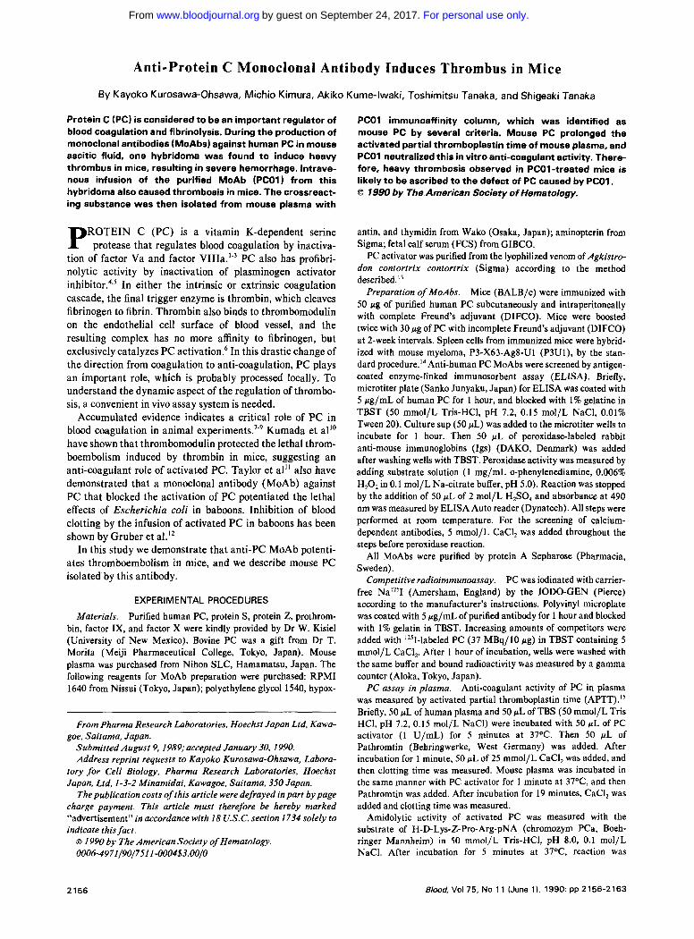
Anti-Protein C Monoclonal Antibody Induces Thrombus in Mice By Kayoko Kurosawa-Ohsawa, Michio Kimura, Akiko Kume-lwaki, Toshimitsu Tanaka, and Shigeaki Tanaka Protein C (PC) is consideredto be an important regulator of blood coagulationand fibrinolysis. During the production of monoclonal antibodies (MoAbs) against human PC in mouse ascitic fluid, one hybridoma was found to induce heavy thrombus in mice, resulting in severe hemorrhage. Intrave- nous infusion of the purified MoAb (PCO1) from this hybridomaalso causedthrombosis in mice. The crossreact- ing substance was then isolated from mouse plasma with ROTEIN C (PC) is a vitamin K-dependent serine P protease that regulates blood coagulation by inactiva- tion of factor Va and factor VIIIa.’” PC also has profibri- nolytic activity by inactivation of plasminogen activator inhibit~r.~.~ In either the intrinsic or extrinsic coagulation cascade, the final trigger enzyme is thrombin, which cleaves fibrinogen to fibrin. Thrombin also binds to thrombomodulin on the endothelial cell surface of blood vessel, and the resulting complex has no more affinity to fibrinogen, but exclusively catalyzes PC activation.6 In this drastic change of the direction from coagulation to anti-coagulation, PC plays an important role, which is probably processed locally. To understand the dynamic aspect of the regulation of thrombo- sis, a convenient in vivo assay system is needed. Accumulated evidence indicates a critical role of PC in blood coagulation in animal experiment^.'.^ Kumada et all0 have shown that thrombomodulin protected the lethal throm- boembolism induced by thrombin in mice, suggesting an anti-coagulant role of activated PC. Taylor et all’ also have demonstrated that a monoclonal antibody (MoAb) against PC that blocked the activation of PC potentiated the lethal effects of Escherichia coli in baboons. Inhibition of blood clotting by the infusion of activated PC in baboons has been shown by Gruber et a1.I’ In this study we demonstrate that anti-PC MoAb potenti- ates thromboembolism in mice, and we describe mouse PC isolated by this antibody. EXPERIMENTAL PROCEDURES Purified human PC, protein S, protein Z, prothrom- bin, factor IX, and factor X were kindly provided by Dr W. Kisiel (University of New Mexico). Bovine PC was a gift from Dr T. Morita (Meiji Pharmaceutical College, Tokyo, Japan). Mouse plasma was purchased from Nihon SLC, Hamamatsu, Japan. The following reagents for MoAb preparation were purchased: RPMI 1640 from Nissui (Tokyo, Japan); polyethylene glycol 1540, hypox- Materials. From Pharma Research Laboratories, Hoechst Japan Ltd, Kawa- Submitted August 9, 1989; accepted January 30. 1990. Address reprint requests to Kayoko Kurosawa-Ohsawa, Labora- tory for Cell Biology. Pharma Research Laboratories, Hoechst Japan, Ltd. 1-3-2 Minamidai, Kawagoe, Saitama, 350 Japan. The publication costs of this article were defrayed in part by page charge payment. This article must therefore be hereby marked “advertisement” in accordance with 18 U.S.C. section 1734 solely to indicate this fact. goe. Saitama, Japan. 0 1990 by The American Society of Hematology. 0006~4971/90/7511-0004$3.00/0 PCOl immunoaffinity column, which was identified as mouse PC by several criteria. Mouse PC prolonged the activated partial thromboplastin time of mouse plasma, and PCOl neutralizedthis in vitro anti-coagulantactivity. There- fore, heavy thrombosis observed in PCOl -treated mice is likely to be ascribed to the defect of PC caused by PCOl . 0 1990 by The Americen Society Of Hem8tOlOgy. antin, and thymidin from Wako (Osaka, Japan); aminopterin from Sigma; fetal calf serum (FCS) from GIBCO. PC activator was purified from the lyophilized venom of Agkistro- don contortrix contortrix (Sigma) according to the method described.” Preparation of MoAbs. Mice (BALB/c) were immunized with 50 pg of purified human PC subcutaneously and intraperitoneally with complete Freund’s adjuvant (DIFCO). Mice were boosted twice with 30 pg of PC with incomplete Freund’s adjuvant (DIFCO) at 2-week intervals. Spleen cells from immunized mice were hybrid- ized with mouse myeloma, P3-X63-Ag8-U1 (P3U1), by the stan- dard pr~cedure.’~ Anti-human PC MoAbs were screened by antigen- coated enzyme-linked immunosorbent assay (ELISA). Briefly, microtiter plate (Sanko Junyaku, Japan) for ELISA was coated with 5 pg/mL of human PC for 1 hour, and blocked with 1% gelatine in TBST (50 mmol/L Tris-HC1, pH 7.2, 0.15 mol/L NaC1, 0.01% Tween 20). Culture sup (50 pL) was added to the microtiter wells to incubate for 1 hour. Then 50 pL of peroxidase-labeled rabbit anti-mouse immunoglobins (Igs) (DAKO, Denmark) was added after washing wells with TBST. Peroxidase activity was measured by adding substrate solution (1 mg/mL o-phenylenediamine, 0.006% H,O, in 0.1 mol/L Na-citrate buffer, pH 5.0). Reaction was stopped by the addition of 50 pL of 2 mol/L H,S04 and absorbance at 490 nm was measured by ELISA Auto reader (Dynatech). All steps were performed at room temperature. For the screening of calcium- dependent antibodies, 5 mmol/L CaCI, was added throughout the steps before peroxidase reaction. All MoAbs were purified by protein A Sepharose (Pharmacia, Sweden). Competitive radioimmunoassay. PC was iodinated with carrier- free Na”’I (Amersham, England) by the IODO-GEN (Pierce) according to the manufacturer’s instructions. Polyvinyl microplate was coated with 5 pg/mL of purified antibody for 1 hour and blocked with 1% gelatin in TBST. Increasing amounts of competitors were added with I2’I-labeled PC (37 MBq/10 pg) in TBST containing 5 mmol/L CaC1,. After 1 hour of incubation, wells were washed with the same buffer and bound radioactivity was measured by a gamma counter (Aloka, Tokyo, Japan). Anti-coagulant activity of PC in plasma was measured by activated partial thromboplastin time (APTT).’’ Briefly, 50 pL of human plasma and 50 pL of TBS (50 mmol/L Tris HC1, pH 7.2, 0.15 mol/L NaCI) were incubated with 50 pL of PC activator (1 U/mL) for 5 minutes at 37OC. Then 50 pL of Pathromtin (Behringwerke, West Germany) was added. After incubation for 1 minute, 50 pL of 25 mmol/L CaCI, was added, and then clotting time was measured. Mouse plasma was incubated in the same manner with PC activator for 1 minute at 37”C, and then Pathromtin was added. After incubation for 19 minutes, CaCI, was added and clotting time was measured. Amidolytic activity of activated PC was measured with the substrate of H-D-Lys-Z-Pro-Arg-pNA (chromozym PCa, Boeh- ringer Mannheim) in 50 mmol/L Tris-HC1, pH 8.0, 0.1 mol/L NaC1. After incubation for 5 minutes at 37OC. reaction was PC assay in plasma. 2156 Blood, Vol 75, No 11 (June 1). 1990: pp 2156-2163 For personal use only. on September 24, 2017. by guest www.bloodjournal.org From
Transcript of Anti-Protein C Monoclonal Antibody Induces Thrombus in Mice · Anti-Protein C Monoclonal Antibody...
Anti-Protein C Monoclonal Antibody Induces Thrombus in Mice
By Kayoko Kurosawa-Ohsawa, Michio Kimura, Akiko Kume-lwaki, Toshimitsu Tanaka, and Shigeaki Tanaka
Protein C (PC) is considered to be an important regulator of blood coagulation and fibrinolysis. During the production of monoclonal antibodies (MoAbs) against human PC in mouse ascitic fluid, one hybridoma was found to induce heavy thrombus in mice, resulting in severe hemorrhage. Intrave- nous infusion of the purified MoAb (PCO1) from this hybridoma also caused thrombosis in mice. The crossreact- ing substance was then isolated from mouse plasma with
ROTEIN C (PC) is a vitamin K-dependent serine P protease that regulates blood coagulation by inactiva- tion of factor Va and factor VIIIa.’” PC also has profibri- nolytic activity by inactivation of plasminogen activator i n h i b i t ~ r . ~ . ~ In either the intrinsic or extrinsic coagulation cascade, the final trigger enzyme is thrombin, which cleaves fibrinogen to fibrin. Thrombin also binds to thrombomodulin on the endothelial cell surface of blood vessel, and the resulting complex has no more affinity to fibrinogen, but exclusively catalyzes PC activation.6 In this drastic change of the direction from coagulation to anti-coagulation, PC plays an important role, which is probably processed locally. To understand the dynamic aspect of the regulation of thrombo- sis, a convenient in vivo assay system is needed.
Accumulated evidence indicates a critical role of P C in blood coagulation in animal experiment^.'.^ Kumada et all0 have shown that thrombomodulin protected the lethal throm- boembolism induced by thrombin in mice, suggesting an anti-coagulant role of activated PC. Taylor et all’ also have demonstrated that a monoclonal antibody (MoAb) against PC that blocked the activation of PC potentiated the lethal effects of Escherichia coli in baboons. Inhibition of blood clotting by the infusion of activated PC in baboons has been shown by Gruber et a1.I’
In this study we demonstrate that anti-PC MoAb potenti- ates thromboembolism in mice, and we describe mouse PC isolated by this antibody.
EXPERIMENTAL PROCEDURES
Purified human PC, protein S, protein Z, prothrom- bin, factor IX, and factor X were kindly provided by Dr W. Kisiel (University of New Mexico). Bovine PC was a gift from Dr T. Morita (Meiji Pharmaceutical College, Tokyo, Japan). Mouse plasma was purchased from Nihon SLC, Hamamatsu, Japan. The following reagents for MoAb preparation were purchased: RPMI 1640 from Nissui (Tokyo, Japan); polyethylene glycol 1540, hypox-
Materials.
From Pharma Research Laboratories, Hoechst Japan Ltd, Kawa-
Submitted August 9, 1989; accepted January 30. 1990. Address reprint requests to Kayoko Kurosawa-Ohsawa, Labora-
tory for Cell Biology. Pharma Research Laboratories, Hoechst Japan, Ltd. 1-3-2 Minamidai, Kawagoe, Saitama, 350 Japan.
The publication costs of this article were defrayed in part by page charge payment. This article must therefore be hereby marked “advertisement” in accordance with 18 U.S.C. section 1734 solely to indicate this fact.
goe. Saitama, Japan.
0 1990 by The American Society of Hematology. 0006~4971/90/7511-0004$3.00/0
PCOl immunoaffinity column, which was identified as mouse PC by several criteria. Mouse PC prolonged the activated partial thromboplastin time of mouse plasma, and PCOl neutralized this in vitro anti-coagulant activity. There- fore, heavy thrombosis observed in PCOl -treated mice is likely to be ascribed to the defect of PC caused by PCOl . 0 1990 by The Americen Society O f Hem8tOlOgy.
antin, and thymidin from Wako (Osaka, Japan); aminopterin from Sigma; fetal calf serum (FCS) from GIBCO.
PC activator was purified from the lyophilized venom of Agkistro- don contortrix contortrix (Sigma) according to the method described.”
Preparation of MoAbs. Mice (BALB/c) were immunized with 50 pg of purified human PC subcutaneously and intraperitoneally with complete Freund’s adjuvant (DIFCO). Mice were boosted twice with 30 pg of PC with incomplete Freund’s adjuvant (DIFCO) at 2-week intervals. Spleen cells from immunized mice were hybrid- ized with mouse myeloma, P3-X63-Ag8-U1 (P3U1), by the stan- dard pr~cedure . ’~ Anti-human PC MoAbs were screened by antigen- coated enzyme-linked immunosorbent assay (ELISA). Briefly, microtiter plate (Sanko Junyaku, Japan) for ELISA was coated with 5 pg/mL of human PC for 1 hour, and blocked with 1% gelatine in TBST (50 mmol/L Tris-HC1, pH 7.2, 0.15 mol/L NaC1, 0.01% Tween 20). Culture sup (50 pL) was added to the microtiter wells to incubate for 1 hour. Then 50 pL of peroxidase-labeled rabbit anti-mouse immunoglobins (Igs) (DAKO, Denmark) was added after washing wells with TBST. Peroxidase activity was measured by adding substrate solution (1 mg/mL o-phenylenediamine, 0.006% H,O, in 0.1 mol/L Na-citrate buffer, pH 5.0). Reaction was stopped by the addition of 50 pL of 2 mol/L H,S04 and absorbance at 490 nm was measured by ELISA Auto reader (Dynatech). All steps were performed at room temperature. For the screening of calcium- dependent antibodies, 5 mmol/L CaCI, was added throughout the steps before peroxidase reaction.
All MoAbs were purified by protein A Sepharose (Pharmacia, Sweden).
Competitive radioimmunoassay. PC was iodinated with carrier- free Na”’I (Amersham, England) by the IODO-GEN (Pierce) according to the manufacturer’s instructions. Polyvinyl microplate was coated with 5 pg/mL of purified antibody for 1 hour and blocked with 1% gelatin in TBST. Increasing amounts of competitors were added with I2’I-labeled PC (37 MBq/10 pg) in TBST containing 5 mmol/L CaC1,. After 1 hour of incubation, wells were washed with the same buffer and bound radioactivity was measured by a gamma counter (Aloka, Tokyo, Japan).
Anti-coagulant activity of PC in plasma was measured by activated partial thromboplastin time (APTT).’’ Briefly, 50 pL of human plasma and 50 pL of TBS (50 mmol/L Tris HC1, pH 7.2, 0.15 mol/L NaCI) were incubated with 50 pL of PC activator (1 U/mL) for 5 minutes at 37OC. Then 50 pL of Pathromtin (Behringwerke, West Germany) was added. After incubation for 1 minute, 50 pL of 25 mmol/L CaCI, was added, and then clotting time was measured. Mouse plasma was incubated in the same manner with PC activator for 1 minute at 37”C, and then Pathromtin was added. After incubation for 19 minutes, CaCI, was added and clotting time was measured.
Amidolytic activity of activated PC was measured with the substrate of H-D-Lys-Z-Pro-Arg-pNA (chromozym PCa, Boeh- ringer Mannheim) in 50 mmol/L Tris-HC1, pH 8.0, 0.1 mol/L NaC1. After incubation for 5 minutes at 37OC. reaction was
PC assay in plasma.
2156 Blood, Vol 75, No 11 (June 1). 1990: pp 2156-2163
For personal use only.on September 24, 2017. by guest www.bloodjournal.orgFrom
ANTI-PROTEIN C ANTIBODY INDUCES THROMBUS 2157
CaC12 (mM)
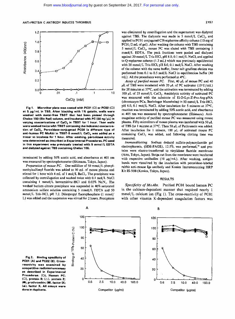
Fig 1. Microtiter plate was coated with PCOl (0) or PC02 (0 ) at 5 pg/mL in TBS. After blocking with 1% gelatin, wells were washed with metal-free TBST that had been passed through Chelex 100 (Bio Rad) column, and incubated with PC (50 nglpL) in varying concentrations of CaCI, in TBST for 1 hour. Then wells were washed twice with TBST containing the indicated concentra- tion of CaCI,. Peroxidase-conjugated PCO8 (a different type of anti-human PC MoAb) in TBST-5 mmol/L CaCI, was added as a tracer to incubate for 1 hour. After washing, peroxidase activity wes determined as described in Experimental Procedures. PC used in this experiment was previously treated with 6 mmol/L EDTA and dialyzed against TBS containing Chelex 100.
terminated by adding 50% acetic acid, and absorbance at 405 nm was measured by spectrophotometer (Shimazu, Tokyo, Japan).
One milliliter of 50 mmol/L phenyl- methylsulfonyl fluoride was added to 50 mL of mouse plasma and stirred for 1 hour with 4 mL of 1 mol/L BaCI,. The precipitate was collected by centrifugation and washed twice with 0.1 mol/L NaCl containing 1 mmol/L benzamidine-HC1 and 0.02% NaN,. The washed barium citrate precipitate was suspended in 40% saturated ammonium sulfate solution containing 5 mmol/L EDTA and 50 mmol/L Tris-HC1, pH 7.2. Diisopropyl fluorophosphate (1 mmol/ L) was added and the suspension was stirred for 2 hours. Precipitate
Preparation of mouse PC.
Fig 2. Binding specificity of PCOl (A) and PC02 (B). Cross- reactivity was examined by competitive radioimmunoassay as described in Experimental Procedures. (0). Human PC; (0). protein S; (A), protein 2; (01, prothrombin; (DL factor IX; (A), factor X. All assays were done in duplicate.
A
120
100
80
60
40
20
0
was eliminated by centrifugation and the supernatant was dialyzed against TBS. The dialyzate was made in 5 mmol/L CaCl,, and applied to PCOl conjugated CH-sepharose affinity column (10 mg of PCO1/2 mL of gel). After washing the column with TBS containing 5 mmol/L CaCI,, mouse PC was eluted with TBS containing 5 mmol/L EDTA. The peak fractions were pooled and dialyzed against 20 mmol/L Tris-HCI, pH 8.0,O.l mol/L NaCI, and applied to Q-sepharose column (1.2 mL) which was previously equilibrated with 20 mmol/L Tris HCI, pH 8.0.0.1 mol/L NaCl. After washing of the column with the same buffer, linear salt gradient elution was performed from 0.1 to 0.5 mol/L NaCl in equilibration buffer (40 mL). All the procedures were performed at 4OC.
First, 40 pL of mouse PC and 40 pL of TBS were incubated with 20 pL of PC activator (10 U/mL) for 20 minutes at 37OC, and the activation was terminated by adding 100 pL of 25 mmol/L CaCI,. Amidolytic activity of activated PC was measured with the substrate of H-D-Lys-Z-Pro-Arg-pNA (chromozym PCa, Boehringer Mannheim) in 50 mmol/L Tris-HCI, pH 8.0, 0.1 mol/L NaCI. After incubation for 5 minutes at 37OC, reaction was terminated by adding 50% acetic acid, and absorbance at 405 nm was measured by spectrophotometer (Shimazu). Anti- coagulant activity of purified mouse PC was measured using mouse plasma. Fifty microliters of mouse plasma was incubated with 50 pL of TBS for 1 minute at 37OC. Then 50 pL of Pathromtin was added. After incubation for 1 minute, 100 pL of activated mouse PC containing CaCI, was added, and following clotting time was measured.
Immunoblotting. Sodium dodecyl sulfate-polyacrylamide gel electrophoresis, (SDS-PAGE), 12.5%, was performed,I6 and pro- teins were electro-transferred to vinylidene fluoride membrane (Atto, Tokyo, Japan). Strips cut from the membrane were incubated with respective antibodies (10 pg/mL). After washing, antigen bands were visualized by the incubation with peroxidase-labeled rabbit anti-mouse Ips antibody and Konica Immunostaining HRP Kit IS-SOB (Konica, Tokyo, Japan).
Assay of purified mouse PC.
RESULTS
Specificity of MoAbs. Purified PCOl bound human PC in the calcium-dependent manner that required nearly 1 mmol/L calcium ion (Fig 1). The cross-reactivity of PCOl with other vitamin K-dependent coagulation factors was
B
120 I
Competitor (vg/ml) Competitor (pg/ml)
For personal use only.on September 24, 2017. by guest www.bloodjournal.orgFrom
2168 KUROSAWA-OHSAWA ET Al
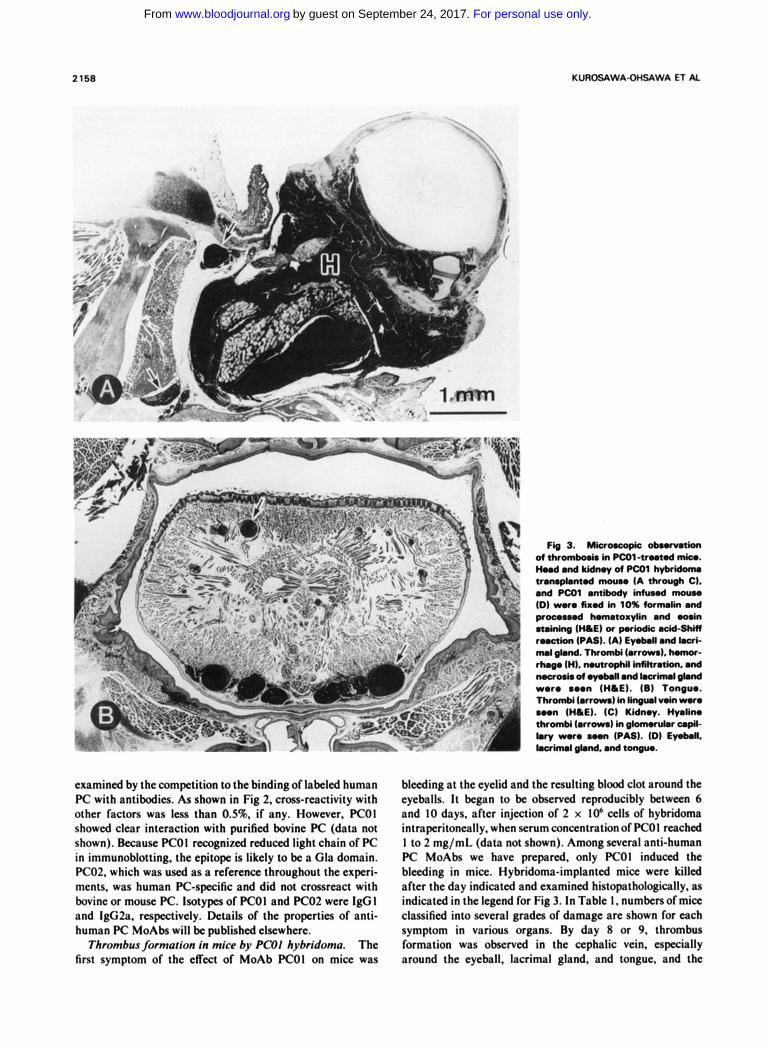
Fig 3. WCVOW@C okmnkn d thranbd. &I Pco1-tmt.d Ink.. HoodmdkidrnydPCOlhyktdorru transplontod mouu (A through C). and Pcol antibody infuaod mouu ID) woro f ir4 in 10% fornulin and praco.ud homotoxylin and -sin staining ( H I E ) or poriodk ocid-Shiif ruetion (PAS). (A) E y o l u l and L.aC MI gbnd. ThromM (arrows). homor- rhogo (H). noutraphil infiltration. ond
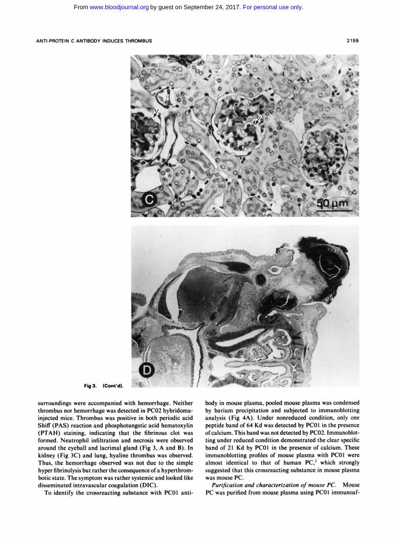
woro ooon ( H I E ) . ( 6 ) Tonguo. Thrombi (arrows) In lingurl voin wore soon (H IE) . IC) Kidnoy. Hyalin. t h r d (arrows) in glom.n*.r upil-
McIo./. of .y.bll and loc3inuI gland
I.V WWO ~ . n (PAS). ID) Eyolun. locriwul gbnd, and tongcH.
examined by the competition to the binding of labeled human PC with antibodies. As shown in Fig 2. cross-reactivity with other factors was less than 0.5%. if any. However. K O 1 showed clear interaction with purified bovine PC (data not shown). Because PCOl recognixd reduced light chain of PC in immunoblotting. the epitope is likely to be a Gla domain. PC02. which was used as a reference throughout the experi- ments. was human PC-specific and did not crossreact with bovine or mouse PC. lsotypes of PCOl and K O 2 were IgG I and IgG2a. respectively. Details of the properties of anti- human PC MoAbs will be published elsewhere.
Thrombus formation in mice by PCOl hybridoma. The first symptom of the eKcct of MoAb PCOl on mice was
bleeding at the eyelid and the resulting blood clot around the eyeballs. It began to be observed reproducibly between 6 and IO days, after injection of 2 x 10⁶ cells of hybridoma intraperitoneally. when serum concentration of PCOl reached I to 2 mg/mL (data not shown). Among several anti-human PC MoAbs we have prepared, only PCOl induced the bleeding in mice. Hybridoma-implanted mice were killed after the day indicated and examined histopathologically. as indicatcd in the legend for Fig 3. In Table I , numbers of mice classified into several grades of damage arc shown for each symptom in various organs. By day 8 or 9. thrombus formation was observed in the cephalic vein, especially around the eyeball, lacrimal gland, and tongue. and the
For personal use only.on September 24, 2017. by guest www.bloodjournal.orgFrom
ANTIPROTEIN C ANTIBODY INDUCES THROMBUS 2169
.*, - ma.-. . a 0 - - I * -
Fig3. (Cont'd).
surroundings were accompanied with hemorrhage. Neither thrombus nor hemorrhage was detected in PC02 hybridoma- injected mice. Thrombus was positive in both periodic acid Shiff (PAS) reaction and phosphotungstic acid hematoxylin (PTAH) staining, indicating that the fibrinous clot was formed. Neutrophil infiltration and nectosis were observed around the eyeball and lacrimal gland (Fig 3, A and B). In kidney (Fig 3C) and lung, hyaline thrombus was observed. Thus. the hemorrhage observed was not due to the simple hyper fibrinolysis but rather theconsequenceofa hyperthrom- botic state. The symptom was rather systemic and looked like disseminated intravascular coagulation (DIC).
To identify the crossreacting substance with PCOl anti-
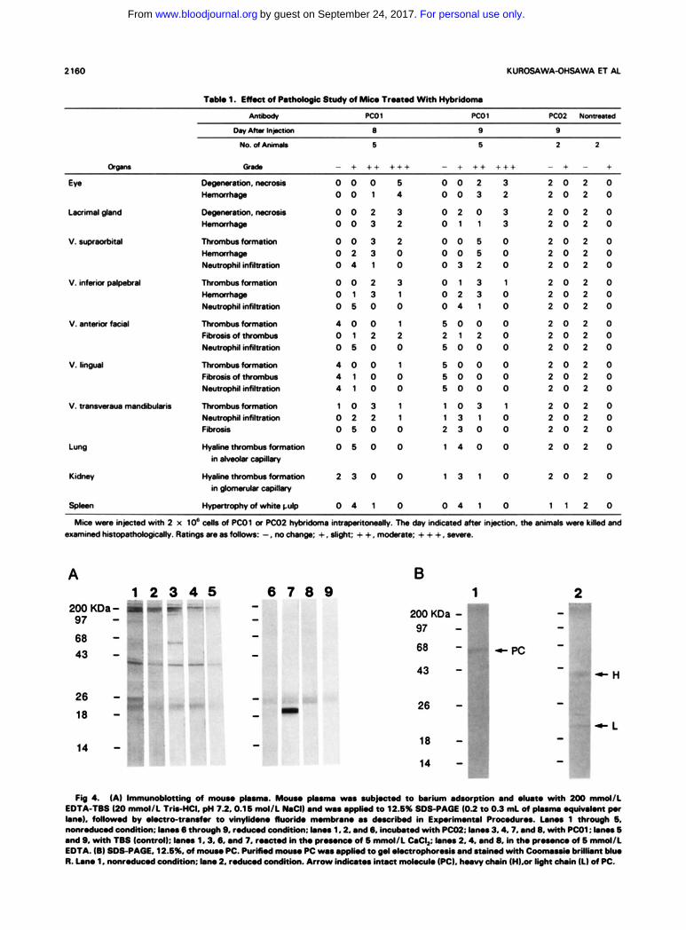
body in mouse plasma, pooled mouse plasma was condensed by barium precipitation and subjected to immunoblotting analysis (Fig 4A). Under nonrcduced condition. only one peptide band of 64 Kd was detected by PCOl in the presence ofcalcium. This band was not detected by PC02. Immunoblot- ting under reduced condition demonstrated the clear specific band of 21 Kd by P C O I in the presence of calcium. These immunoblotting profiles of mouse plasma with PCOl were almost identical to that of human PC,' which strongly suggested that this crossreacting substance in mouse plasma was mouse PC.
Mouse PC was purified from mouse plasma using PCOl immunoaf-
Purification and characterization of mouse PC.
For personal use only.on September 24, 2017. by guest www.bloodjournal.orgFrom
2160 KUROSAWA-OHSAWA E l AI.
Bd. - + ++ +++ - + + + +++ - + - +
v. supmc&w
DeQ”h.naorir 0 0 0 6 0 0 2 3 2 0 2 0 - 0 0 1 4 0 0 3 2 2 0 2 0
DeQ”h,m 0 0 2 3 0 2 0 3 2 0 2 0 - 0 0 3 2 0 1 1 3 2 0 2 0
Tluunbmtamnkn 0 0 3 2 0 0 6 0 2 0 2 0 - 0 2 3 0 0 0 6 0 2 0 2 0 Neutmphilinfihth 0 4 1 0 0 3 2 0 2 0 2 0
Thranburfamnbn 0 0 2 3 0 1 3 1 2 0 2 0 - 0 1 3 1 0 2 3 0 2 0 2 0 Namophi lMR” 0 6 0 0 0 4 1 0 2 0 2 0
Thromkaia” 4 0 0 1 6 0 0 0 2 0 2 0 Fikor i rdthrank* 0 1 2 2 2 1 2 0 2 0 2 0 Nartrophi(mmion 0 6 0 0 6 0 0 0 2 0 2 0
”bV8- 4 0 0 1 6 0 0 0 2 0 2 0 F&airo(thrank* 4 1 0 0 5 0 0 0 2 0 2 0 Nwtrophil infiltrmion 4 1 0 0 5 0 0 0 2 0 2 0
v. emwawto - Thranburtamnia, 1 0 3 1 1 0 3 1 2 0 2 0 Namophi(infiltraion 0 2 2 1 1 3 1 0 2 0 2 0 Fibrosis 0 6 0 0 2 3 0 0 2 0 2 0
Hydin*rankr- 0 6 0 0 1 4 0 0 2 0 2 0
HvJin-- 2 3 0 0 1 3 1 0 2 0 2 0
indrnobuoilby
in-crpilbry
A 1 2 3 4 5
200 KDa- 7” 97 - 68 - 43 -
26 - 18 - 14 -
B 1 2
43 - 26 -
- 18 - - 14 -
Fig 4. (A) d moun ph”. Mouse pbanm rm rub- to krkm 8doo@on end .(ut. with 200 “ d l L
).no). foitowd by . k c n o - n o ~ to vlnylidono Iluorido momkono os doocribod in Exp.rimontol Proaduroa. Lonos 1 thr- 6, nonroducod condition: ).nos 6 through 9, roducod aonditkn: ).nos 1,2. ond 6. incubotod with P a ; ).nos 3.4.7. ond 8. with PCO1: hnm 6 and 9, with TBS (control): lonos 1.3,O. and 7. r..ctod in tho pr...nc* of 6 mmd/L CoCI,: ).no# 2.4. ond 8. In tho pr...nc* of 6 m d / L EDTA. (8) SDS-PAQE. 12.6%. of “o PC. PurHHd mocl.. PC was oppbd to 0.1 oloctrophoroais ond otoinod with coonU..k brilllont Mrw R. lono 1. nonrodwod eonditkn: ).no 2. rod& condition. Arrow Indiwtoa i m m md.culo (PC). h..vy d i n (H1.w light thoin (1) of PC.
EDTA-TBS (20 &/I. T W C I . pH 7.2.0.16 m d l L b C I ) ~ n d w.. 0pgR.d to 12.6% (IDS-PAGE (0.2 to 0.3 ml d pl.mu .quM.m P.r
For personal use only.on September 24, 2017. by guest www.bloodjournal.orgFrom
ANTI-PROTEIN C ANTIBODY INDUCES THROMBUS
8o I 0
2161
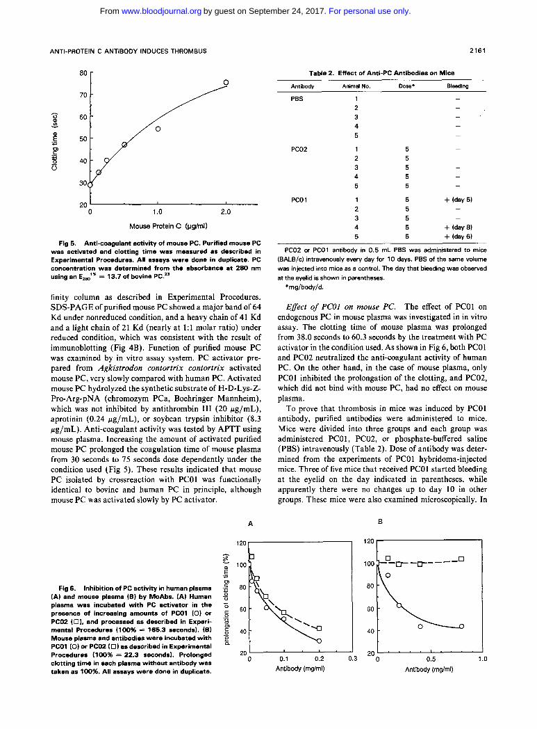
Table 2. Effect of Anti-PC Antibodies on Mice
Antiboh, Animal No. Dose. Bleeding
20 0 1 .o 2.0
Mouse Protein C (vs/ml)
Fig 5. Anti-coagulant activity of mouse PC. Purified mouse PC was activated and clotting time was measured as described in Experimental Procedures. All assays were done in duplicate. PC concentration was determined from the absorbance at 280 nm using an E,," = 13.7 of bovine PC.2'
finity column as described in Experimental Procedures. SDS-PAGE of purified mouse PC showed a major band of 64 Kd under nonreduced condition, and a heavy chain of 41 Kd and a light chain of 21 Kd (nearly a t 1:l molar ratio) under reduced condition, which was consistent with the result of immunoblotting (Fig 4B). Function of purified mouse PC was examined by in vitro assay system. PC activator pre- pared from Agkistrodon contortrix contortrix activated mouse PC, very slowly compared with human PC. Activated mouse PC hydrolyzed the synthetic substrate of H-D-Lys-Z- Pro-Arg-pNA (chromozym PCa, Boehringer Mannheim), which was not inhibited by antithrombin I11 (20 pg/mL), aprotinin (0.24 pg/mL), or soybean trypsin inhibitor (8.3 pg/mL). Anti-coagulant activity was tested by APTT using mouse plasma. Increasing the amount of activated purified mouse PC prolonged the coagulation time of mouse plasma from 30 seconds to 15 seconds dose dependently under the condition used (Fig 5). These results indicated that mouse PC isolated by crossreaction with PCOl was functionally identical to bovine and human PC in principle, although mouse PC was activated slowly by PC activator.
Fig 6. Inhibition of PC activity in human plasma (A) and mouse plasma (6) by MoAbs. (A) Human plasma was incubated with PC activator in the presence of increasing amounts of PCOl (0) or PC02 (0). and processed as described in Experi- mental Procedures (100% = 166.3 seconds). (6) Mouse plasma and antibodies were incubated with PCOl (0) or PC02 (0) as described in Experimental Procedures (1 00% = 22.3 seconds). Prolonged clotting time in each plasma without antibody was taken as 100%. All assays were done in duplicate.
A
PBS
PC02
PCO 1
1 2 3 4 5
1 2 3 4 5
1 2 3 4 5
5 5 5 5 5
5 5 5 5 5
PC02 or PCOl antibody in 0.5 mL PBS was administered to mice (BALB/c) intravenously every day for 10 days. PBS of the same volume was injected into mice as a control. The day that bleeding was observed at the eyelid is shown in parentheses.
*mg/body/d.
Effect of PCOl on mouse PC. The effect of PCOl on endogenous PC in mouse plasma was investigated in in vitro assay. The clotting time of mouse plasma was prolonged from 38.0 seconds to 60.3 seconds by the treatment with PC activator in the condition used. As shown in Fig 6, both PCOl and PC02 neutralized the anti-coagulant activity of human PC. On the other hand, in the case of mouse plasma, only PCOl inhibited the prolongation of the clotting, and PC02, which did not bind with mouse PC, had no effect on mouse plasma.
To prove that thrombosis in mice was induced by PCOl antibody, purified antibodies were administered to mice. Mice were divided into three groups and each group was administered PCOl, PC02, or phosphate-buffered saline (PBS) intravenously (Table 2). Dose of antibody was deter- mined from the experiments of PCOl hybridoma-injected mice. Three of five mice that received PCOl started bleeding at the eyelid on the day indicated in parentheses, while apparently there were no changes up to day 10 in other groups. These mice were also examined microscopically. In
B
100
80
60
40
20 ' ' ' ' 20 0 0.1 0.2 0.3 0 0.5 1 .o
Antibody (mglml) Antibody (mg/ml)
For personal use only.on September 24, 2017. by guest www.bloodjournal.orgFrom
2162 KUROSAWA-OHSAWA ET AL
all mice treated with PCO1, the observed thrombi were similar to those of the mice that were injected with PCOl hybridoma (Fig 3D). The thrombi were slightly observed in mice in which bleeding had not yet been detected. The mice that were administered PC02 or PBS showed no symptoms of thrombosis or hemorrhage. Clear contrast between the ef- fects of PCOl and PC02 on mice suggests that this thrombo- sis is very likely to be induced by the blockage of mouse PC function by PCOl .
DISCUSSION
Hybridoma technology has provided new probes for the research in biology and medicine, and several types of MoAbs already contributed to the characterization of human
In addition, our findings of thrombus induction in mice by anti-human PC MoAb gave another view of MoAb application.
We have shown that PCOl bound mouse PC. Mouse PC was isolated for the first time from mouse plasma, which showed similar activities to bovine and human PC. The structure and molecular size of mouse PC were also similar to those in bovine and human PC. PCOl crossreacted with bovine, rat, and rabbit PC, suggesting that PCOl recognizes the conserved structure in the Gla-domain of PC. Details of other species' PC will be published elsewhere. PCOl neutral- ized mouse PC activity in vitro. We could not show the decrease of PC activity level in PCO1-treated mice, since mouse PC-PCOl complex was dissociated in citrate plasma. However, it may be reasonable to explain these severe thromboses by the decrease of functional PC in plasma. Platelet number and APTT of mouse plasma were not affected by the treatment with either PCOl and PC02 (data not shown), suggesting that PCOl directly affected neither
pc. 17-20
platelet activation nor coagulation cascade. Relatively high doses of antibody required for in vivo experiments may be due to the relatively low affinity of PCOl to mouse PC in plasma, which was suggested by a preliminary competition experiment. It is not well-understood whether PC in 'the blood stream forms a complex with calcium ion that can be recognized by PCO1. In this regard, PCO1, a specific reagent to PC, could be a useful probe to understand the behavior of PC in in vivo animal models. In our mouse model, large excess amounts of PCOl may be necessary to keep the hyperthrombotic state for a certain period before coagulation cascade starts. Deficiency of PC alone is not likely to be sufficient to start coagulation cascade, but some other induc- tion(s) may be required. The reason thrombi were observed mainly in the cephalic vein is not known. Because thrombosis is not localized in the cephalic vein in the case of human PC-deficient patients, this localized effect may be specific for mice or may be the result of unidentified interaction of PCOl in mouse, which remains to be solved.
It has been known that the deficiency of PC causes thrombotic disease." Typical clinical features in homozy- gous PC-deficient patients are reported to be skin necrosis, microthrombi, and DIC.22 It should be noted that PCO1- injected mice, either with hybridoma or antibodies, shared some symptoms with homozygous PC-deficient patients. This mouse disease model will be useful for in vivo PC research for hereditary and acquired PC deficiency. Our study also suggests possible application of functional MoAbs to produce a relevant factor-deficient animal model in general.
ACKNOWLEDGMENT
We thank Drs E. Konz, H.B. Maruyama, and T. Matsuishi for their support and encouragement during this work.
REFERENCES
1. Walker FJ, Sexton PW, Esmon CT: The inhibition of blood coagulation by activated protein C through the selective inactivation of activated factor V. Biochem Biophys Acta 571:333,1979
2. Vehar GA, Davie EW: Preparation and properties of bovine factor VI11 (antihemophilic factor). Biochemistry 19:401, 1980
3. Esmon CT: The regulation of natural anticoagulant pathways. Science 235:1348, 1987
4. Stenflo J: A new vitamin K-dependent protein. J Biol Chem 251:355, 1976
5. Kisiel W: Human plasma protein C. J Clin Invest 64:761, 1979 6. Esmon CT, Owen WG: Identification of an endothelial cell
cofactor for thrombin-catalyzed activation of protein C. Proc Natl Acad Sci USA 78:2249,1981
7. Comp PC, Esmon CT: Generation of fibrinolytic activity by infusion of activated protein C into dogs. J Clin Invest 68:1221, 1981
8. Burdick MD, Schaub RG: Human protein C induces anticoag- ulation and increased fibrinolytic activity in the cat. Thromb Res 45:413, 1987
9. Colucci M, Stassen JM, Collen D: Influence of protein C activation on blood coagulation and fibrinolysis in squirrel monkeys. J Clin Invest 74:200, 1984
10. Kumada T, Dittman WA, Majerus PW: A role for thrombo- modulin in the pathogenesis of thrombin-induced thromboembolism in mice. Blood 71:728, 1987
1 1 . Taylor FB, Chang A, Esmon CT, DAngelo A, Vigano- DAngelo S, Blick KE: Protein C prevents the coagulopathic and letal effects of Escherichia coli infusion in the baboon. J Clin Invest 79:918, 1987
12. Gruber A, Griffin JH, Harker LA, Hanson SR Inhibition of platelet-dependent thrombus formation by human activated protein C in a primate model. Blood 73:639, 1989
13. Kisiel W, Choi E, Kondo S: Isolation of protein C activator from southern copperhead venom. Biochem Biophys Res Commun 143:917, 1987
14. Oi VT, Herzenberg LA: Immunoglobulin-producing hybrid cell lines, in Mishell BB, Shiigi SM (eds): Selected Methods in Cellular Immunology. San Francisco, CA, Freeman, 1980, p 351
15. Lobermann H, Kolde H-J, Deubel R, Peter R, Tourte E, Becker U: Detection of protein C in plasma. Behring Inst Mitt 79:112, 1986
16. Laemmli UK Cleavage of structural proteins during the assembly of the head of bacteriophage T4. Nature 227:680, 1970
17. Suzuki K, Matsuda Y, Kusumoto H, Nishioka J, Terada M, Yamashita T, Hashimoto S: Monoclonal antibodies to human protein C: Effects on the biological activity of activated protein C and the thrombin-catalyzed activation of protein C. J Biochem (Tokyo) 97:127,1985
18. Wakabayashi K, Sakata Y, Aoki N: Conformation-specific monoclonal antibodies to the calcium-induced structure of protein C. J Biol Chem 261:11097,1986
For personal use only.on September 24, 2017. by guest www.bloodjournal.orgFrom

ANTI-PROTEIN C ANTIBODY INDUCES THROMBUS 2163
19. Ohlin A-K, Stenflo J: Calcium-dependent interaction be- tween the epidermal growth factor precursor-like region of human protein C and a monoclonal antibody. J Biol Chem 262:13798, 1987
20. Stearns DJ, Kurosawa S, Sims PJ, Esmon NL, Esmon C T The interaction of Ca2+-dependent monoclonal antibody with the protein C activation peptide region. J Biol Chem 263:826, 1988
21. Griffin JH, Evatt B, Zimmerman TS, Kleiss AJ, Wideman C: Deficiency of protein C in congenital thrombotic disease. J Clin Invest 68:1370, 1981
22. Pabinger I: Clinical relevance of protein C. Blut 53:63, 1986 23. Kisiel W, Ericsson LH, Davie EW: Proteolytic activation of
protein C from bovine plasma. Biochemistry 15:4893, 1976
For personal use only.on September 24, 2017. by guest www.bloodjournal.orgFrom
1990 75: 2156-2163
K Kurosawa-Ohsawa, M Kimura, A Kume-Iwaki, T Tanaka and S Tanaka Anti-protein C monoclonal antibody induces thrombus in mice
http://www.bloodjournal.org/content/75/11/2156.full.htmlUpdated information and services can be found at:
Articles on similar topics can be found in the following Blood collections
http://www.bloodjournal.org/site/misc/rights.xhtml#repub_requestsInformation about reproducing this article in parts or in its entirety may be found online at:
http://www.bloodjournal.org/site/misc/rights.xhtml#reprintsInformation about ordering reprints may be found online at:
http://www.bloodjournal.org/site/subscriptions/index.xhtmlInformation about subscriptions and ASH membership may be found online at:
Copyright 2011 by The American Society of Hematology; all rights reserved.Society of Hematology, 2021 L St, NW, Suite 900, Washington DC 20036.Blood (print ISSN 0006-4971, online ISSN 1528-0020), is published weekly by the American
For personal use only.on September 24, 2017. by guest www.bloodjournal.orgFrom