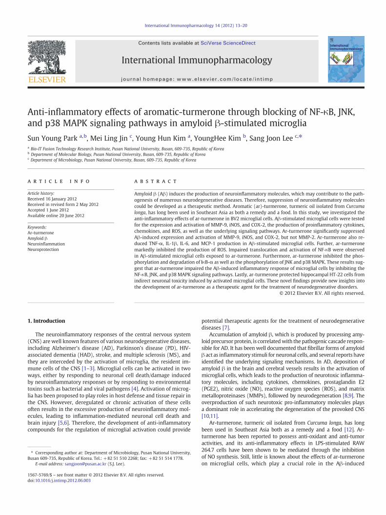
Anti-inflammatory effects of aromatic-turmerone through blocking of NF-κB, JNK, and p38 MAPK...
8
Anti-inflammatory effects of aromatic-turmerone through blocking of NF-κB, JNK, and p38 MAPK signaling pathways in amyloid β-stimulated microglia Sun Young Park a, b , Mei Ling Jin c , Young Hun Kim a , YoungHee Kim b , Sang Joon Lee c, ⁎ a Bio-IT Fusion Technology Research Institute, Pusan National University, Busan, 609‐735, Republic of Korea b Department of Molecular Biology, Pusan National University, Busan, 609‐735, Republic of Korea c Department of Microbiology, Pusan National University, Busan, 609‐735, Republic of Korea abstract article info Article history: Received 16 January 2012 Received in revised form 2 May 2012 Accepted 1 June 2012 Available online 20 June 2012 Keywords: Ar-turmerone Amyloid β Neuroinflammation Neuroprotection Amyloid β (Aβ) induces the production of neuroinflammatory molecules, which may contribute to the path- ogenesis of numerous neurodegenerative diseases. Therefore, suppression of neuroinflammatory molecules could be developed as a therapeutic method. Aromatic (ar)-turmerone, turmeric oil isolated from Curcuma longa, has long been used in Southeast Asia as both a remedy and a food. In this study, we investigated the anti-inflammatory effects of ar-turmerone in BV2 microglial cells. Aβ-stimulated microglial cells were tested for the expression and activation of MMP-9, iNOS, and COX-2, the production of proinflammatory cytokines, chemokines, and ROS, as well as the underlying signaling pathways. Ar-turmerone significantly suppressed Aβ-induced expression and activation of MMP-9, iNOS, and COX-2, but not MMP-2. Ar-turmerone also re- duced TNF-α, IL-1β, IL-6, and MCP-1 production in Aβ-stimulated microglial cells. Further, ar-turmerone markedly inhibited the production of ROS. Impaired translocation and activation of NF-κB were observed in Aβ-stimulated microglial cells exposed to ar-turmerone. Furthermore, ar-turmerone inhibited the phos- phorylation and degradation of IκB-α as well as the phosphorylation of JNK and p38 MAPK. These results sug- gest that ar-turmerone impaired the Aβ-induced inflammatory response of microglial cells by inhibiting the NF-κB, JNK, and p38 MAPK signaling pathways. Lastly, ar-turmerone protected hippocampal HT-22 cells from indirect neuronal toxicity induced by activated microglial cells. These novel findings provide new insights into the development of ar-turmerone as a therapeutic agent for the treatment of neurodegenerative disorders. © 2012 Elsevier B.V. All rights reserved. 1. Introduction The neuroinflammatory responses of the central nervous system (CNS) are well known features of various neurodegenerative diseases, including Alzheimer's disease (AD), Parkinson's disease (PD), HIV- associated dementia (HAD), stroke, and multiple sclerosis (MS), and they are interceded by the activation of microglia, the resident im- mune cells of the CNS [1–3]. Microglial cells can be activated in two ways, either by responding to neuronal cell death/damage induced by neuroinflammatory responses or by responding to environmental toxins such as bacterial and viral pathogens [4]. Activation of microg- lia has been proposed to play roles in host defense and tissue repair in the CNS. However, deregulated or chronic activation of these cells often results in the excessive production of neuroinflammatory mol- ecules, leading to inflammation-mediated neuronal cell death and brain injury [5,6]. Therefore, the development of anti-inflammatory compounds for the regulation of microglial activation could provide potential therapeutic agents for the treatment of neurodegenerative diseases [7]. Accumulation of amyloid β, which is produced by processing amy- loid precursor protein, is correlated with the pathogenic cascade respon- sible for AD. It has been well documented that fibrillar forms of amyloid β act as inflammatory stimuli for neuronal cells, and several reports have identified the underlying signaling mechanisms. In AD, deposition of amyloid β in the brain and cerebral vessels results in the activation of microglial cells, which leads to the production of neurotoxic inflamma- tory molecules, including cytokines, chemokines, prostaglandin E2 (PGE2), nitric oxide (NO), reactive oxygen species (ROS), and matrix metalloproteinases (MMPs), followed by neurodegeneration [8,9]. The overproduction of such neurotoxic pro-inflammatory molecules plays a dominant role in accelerating the degeneration of the provoked CNS [10,11]. Ar-turmerone, turmeric oil isolated from Curcuma longa, has long been used in Southeast Asia both as a remedy and a food [12]. Ar- turmerone has been reported to possess anti-oxidant and anti-tumor activities, and its anti-inflammatory effects in LPS-stimulated RAW 264.7 cells have been shown to be mediated through the inhibition of NO synthesis. Still, little is known about the effects of ar-turmerone on microglial cells, which play a crucial role in the Aβ-induced International Immunopharmacology 14 (2012) 13–20 ⁎ Corresponding author at: Department of Microbiology, Pusan National University, Busan 609‐735, Republic of Korea. Tel.: +82 51 510 2268; fax: +82 51 514 1778. E-mail address: [email protected] (S.J. Lee). 1567-5769/$ – see front matter © 2012 Elsevier B.V. All rights reserved. doi:10.1016/j.intimp.2012.06.003 Contents lists available at SciVerse ScienceDirect International Immunopharmacology journal homepage: www.elsevier.com/locate/intimp
-
Upload
sun-young-park -
Category
Documents
-
view
214 -
download
2
Transcript of Anti-inflammatory effects of aromatic-turmerone through blocking of NF-κB, JNK, and p38 MAPK...
International Immunopharmacology 14 (2012) 13–20
Contents lists available at SciVerse ScienceDirect
International Immunopharmacology
j ourna l homepage: www.e lsev ie r .com/ locate / in t imp
Anti-inflammatory effects of aromatic-turmerone through blocking of NF-κB, JNK,and p38 MAPK signaling pathways in amyloid β-stimulated microglia
Sun Young Park a,b, Mei Ling Jin c, Young Hun Kim a, YoungHee Kim b, Sang Joon Lee c,⁎a Bio-IT Fusion Technology Research Institute, Pusan National University, Busan, 609‐735, Republic of Koreab Department of Molecular Biology, Pusan National University, Busan, 609‐735, Republic of Koreac Department of Microbiology, Pusan National University, Busan, 609‐735, Republic of Korea
⁎ Corresponding author at: Department of MicrobioloBusan 609‐735, Republic of Korea. Tel.: +82 51 510 22
E-mail address: [email protected] (S.J. Lee).
1567-5769/$ – see front matter © 2012 Elsevier B.V. Alldoi:10.1016/j.intimp.2012.06.003
a b s t r a c t
a r t i c l e i n f oArticle history:Received 16 January 2012Received in revised form 2 May 2012Accepted 1 June 2012Available online 20 June 2012
Keywords:Ar-turmeroneAmyloid βNeuroinflammationNeuroprotection
Amyloid β (Aβ) induces the production of neuroinflammatory molecules, which may contribute to the path-ogenesis of numerous neurodegenerative diseases. Therefore, suppression of neuroinflammatory moleculescould be developed as a therapeutic method. Aromatic (ar)-turmerone, turmeric oil isolated from Curcumalonga, has long been used in Southeast Asia as both a remedy and a food. In this study, we investigated theanti-inflammatory effects of ar-turmerone in BV2 microglial cells. Aβ-stimulated microglial cells were testedfor the expression and activation of MMP-9, iNOS, and COX-2, the production of proinflammatory cytokines,chemokines, and ROS, as well as the underlying signaling pathways. Ar-turmerone significantly suppressedAβ-induced expression and activation of MMP-9, iNOS, and COX-2, but not MMP-2. Ar-turmerone also re-duced TNF-α, IL-1β, IL-6, and MCP-1 production in Aβ-stimulated microglial cells. Further, ar-turmeronemarkedly inhibited the production of ROS. Impaired translocation and activation of NF-κB were observedin Aβ-stimulated microglial cells exposed to ar-turmerone. Furthermore, ar-turmerone inhibited the phos-phorylation and degradation of IκB-α as well as the phosphorylation of JNK and p38 MAPK. These results sug-gest that ar-turmerone impaired the Aβ-induced inflammatory response of microglial cells by inhibiting theNF-κB, JNK, and p38 MAPK signaling pathways. Lastly, ar-turmerone protected hippocampal HT-22 cells fromindirect neuronal toxicity induced by activated microglial cells. These novel findings provide new insights intothe development of ar-turmerone as a therapeutic agent for the treatment of neurodegenerative disorders.
© 2012 Elsevier B.V. All rights reserved.
1. Introduction
The neuroinflammatory responses of the central nervous system(CNS) are well known features of various neurodegenerative diseases,including Alzheimer's disease (AD), Parkinson's disease (PD), HIV-associated dementia (HAD), stroke, and multiple sclerosis (MS), andthey are interceded by the activation of microglia, the resident im-mune cells of the CNS [1–3]. Microglial cells can be activated in twoways, either by responding to neuronal cell death/damage inducedby neuroinflammatory responses or by responding to environmentaltoxins such as bacterial and viral pathogens [4]. Activation of microg-lia has been proposed to play roles in host defense and tissue repair inthe CNS. However, deregulated or chronic activation of these cellsoften results in the excessive production of neuroinflammatory mol-ecules, leading to inflammation-mediated neuronal cell death andbrain injury [5,6]. Therefore, the development of anti-inflammatorycompounds for the regulation of microglial activation could provide
gy, Pusan National University,68; fax: +82 51 514 1778.
rights reserved.
potential therapeutic agents for the treatment of neurodegenerativediseases [7].
Accumulation of amyloid β, which is produced by processing amy-loid precursor protein, is correlatedwith the pathogenic cascade respon-sible for AD. It has been well documented that fibrillar forms of amyloidβ act as inflammatory stimuli for neuronal cells, and several reports haveidentified the underlying signaling mechanisms. In AD, deposition ofamyloid β in the brain and cerebral vessels results in the activation ofmicroglial cells, which leads to the production of neurotoxic inflamma-tory molecules, including cytokines, chemokines, prostaglandin E2(PGE2), nitric oxide (NO), reactive oxygen species (ROS), and matrixmetalloproteinases (MMPs), followed by neurodegeneration [8,9]. Theoverproduction of such neurotoxic pro-inflammatory molecules playsa dominant role in accelerating the degeneration of the provoked CNS[10,11].
Ar-turmerone, turmeric oil isolated from Curcuma longa, has longbeen used in Southeast Asia both as a remedy and a food [12]. Ar-turmerone has been reported to possess anti-oxidant and anti-tumoractivities, and its anti-inflammatory effects in LPS-stimulated RAW264.7 cells have been shown to be mediated through the inhibitionof NO synthesis. Still, little is known about the effects of ar-turmeroneon microglial cells, which play a crucial role in the Aβ-induced

14 S.Y. Park et al. / International Immunopharmacology 14 (2012) 13–20
inflammatory response. In this study, we investigated the effects ofar-turmerone on the neuroinflammatory response of Aβ-stimulatedmicroglial cells. Here, we were able to show that ar-turmerone at-tenuated the expression and activation of MMP-9, iNOS, and COX-2 in Aβ-stimulated microglial cells. Further, we observed that ar-turmerone inhibited the production of pro-inflammatory cytokines,chemokines, and ROS. We observed that ar-turmerone inhibitedMAPK and NF-κB signaling pathways, which leads to anti-inflammatory effect. Lastly, we found that ar-turmerone protectedthe HT-22 hippocampal cells by inhibiting Aβ-induced microglialactivation. Together, these findings provide novel insights into theanti-neuroinflammatory and neuroprotective mechanisms of ar-turmerone. More broadly, these findings may be useful in develop-ing novel therapeutic strategies for inhibiting Aβ-mediated neu-rodegeneration in CNS diseases.
2. Materials and methods
2.1. Materials
Ar-turmerone and other reagents were purchased from Sigma(St. Louis, MO, USA). Protoporphyrin IX (SnPP) and antibodiesagainst iNOS, COX-2, NF-κB, IκBα, TBP, and α-tubulin were purchasedfrom Santa Cruz Biotechnology (Santa Cruz, CA, USA). Antibodiesagainst phosphorylated p38 (p-p38), p-JNK, p‐ERK, p-IkB α, andMMP-9 were purchased from Cell Signaling Technology (Beverly, MA,USA). Amyloidβ (1–42)was obtained fromBachemBioscience. Cell cul-ture medium, DMEM, and fetal bovine serum (FBS) were purchasedfrom Gibco BRL (now Invitrogen Corporation, Carlsbad, CA, USA). TheFuGENE 6 transfection reagent and X-treme GENE siRNA TransfectionReagent were purchased from Roche (Indianapolis, IN, USA).
2.2. Isolation of mouse primary microglia and cell culture
The protocols for animal experiments were approved by the AnimalExperiment Committee of Pusan National University. Isolated primarymicroglial cultures were prepared as described previously [13]. Briefly,primarymixed glial cell cultures fromwhole brains of ICRmouse at post-natal day 2–5were prepared in culture flasks andmaintained in DMEM/F12 containing 10% FBS, 0.1 mM None essential nonessential aminoacids, 1 mM Sodium pyruvate, 2 mM L-glutamine and 50 mg/mL Penicil-lin/streptomycin at 37 °C in 5% CO2. After 2 weeks, the culture flaskswere shaken on an orbital shaker at 180 rpm at 37 °C for 5 h and theme-diumwas harvested. The attracted cells were removed by trypsinizationand seeded onto new plates for further studies. To monitor purity, cellswere immunostained with CD11b antibody; more than 90% of cellswere stained positively. Mouse BV2 microglial cells and mouse HT-22hippocampal cells were a generous gift from Prof. Youn-Chul Kim atWonkwang University (Iksan, Korea). Cells were cultured in DMEMme-dium supplemented with 5% heat-inactivated FBS and 0.1% penicillin–streptomycin (BioSource International, Camarillo, CA, USA) at 37 °C in ahumidified atmosphere of 5% CO2 and 95% air.
2.3. Cell viability assay
The cytotoxicity of ar-turmerone was assessed using a microcul-ture (3-(4,5-dimethylthiazol-2-yl)-2,5-diphenyltetrazolium bromide(MTT)-based colorimetric assay. Cells were incubated in 24-wellplates at a density of 5×10⁵ cells per well, after which MTT solution(5 μL of 5 mg/mL) was added to each well (final concentration62.5 μg/mL). After incubation for 3 h at 37 °C in 5% CO2, the superna-tant was removed and the formazan crystals produced in viable cellswere solubilized with 150 μL of dimethylsulfoxide (DMSO). The ab-sorbance of each well was then read at 570 nm using a microplatereader (Wallac 1420 Boston, MA USA).
2.4. Gelatin zymography assay
The activity of MMP-9 in conditioned medium was determined bygelatin zymography protease assay. Briefly, 2×10⁵ cells were seededin 6-well plates and allowed to grow to 80% confluency. The cellswere then maintained in serum-free medium for 12 h prior to treat-ment with ar-turmerone, and then amyloid β for 16 h. Conditionedmedia were collected, cleared by centrifugation, mixed with 2× sodi-um dodecyl sulfate (SDS) sample buffer (Invitrogen Corporation), andelectrophoresed in a polyacrylamide gel containing 0.1% (w/v) gela-tin. Following electrophoresis, the gels were incubated in renaturingbuffer (2.5% Triton X-100) with gentle agitation to remove SDS,followed by incubation in developing buffer (50 mM Tris–HCl buffer,pH 7.4, and 10 mM CaCl2) overnight at 37 °C to digest the gelatin.Gels were then stained with SimplyBlue SafeStain (Invitrogen Corpo-ration) until clear bands suggestive of gelatin digestion appeared.
2.5. Measurement of TNF-α, IL-1β, IL-6, MCP-1, and PGE2 concentrations
Cells were incubated first with various concentrations of ar-turmerone for 1 h, followed by amyloid β for 16 h. Following 24-h in-cubation, TNF-α, IL-1β, IL-6, MCP-1, and PGE2 levels were quantifiedin the culture media using an enzyme-linked immunosorbent assay(ELISA) kit (R&D Systems, Minneapolis, MN) according to the manu-facturer's instructions.
2.6. Measurement of ROS
To evaluate the levels of intracellular ROS, cells were treated withCM-H2DCFDA, an indicator of general oxidative stress, for 1 h at 37 °Cunder 5% CO2. The cells were then harvested and washed three timeswith PBS. The fluorescence intensity was then measured by confocalmicroscopy, microplate flurometry, and flow cytometry at an excita-tion wavelength of 488 nm and an emission wavelength of 525 nm.Data analyses were performed using CXP software 2.0 (BeckmanCoulter).
2.7. Transient transfection and dual luciferase assay
Cells were transfected with a κB-luc reporter plasmid consisting of3-kB concatemers from the Igγ chain and firefly luciferase gene aswell as an ARE-reporter plasmid (Stratagene, Grand Island, NY)using FuGENE-HD reagent (Roche Applied Science) according to themanufacturer's instructions. Then, Renilla luciferase control plasmid,pRL-CMV (Promega), was co-transfected as an internal control forthe verification of transfection efficiency. Twenty-four hours aftertransfection, the cells were incubated with the ar-turmerone for 1 hand then treated with amyloid β for 16 h. Luciferase activity wasassayed using a dual luciferase assay kit (Promega) according to themanufacturer's instructions. Luminescence was measured using amicroplate luminometer (Wallac 1420).
2.8. Western blot analysis
Cells were harvested in ice-cold lysis buffer consisting of 1% TritonX-100, 1% deoxycholate, and 0.1% SDS. The protein content of the celllysates was then determined using Bradford reagent (Bio-Rad,Hercules, CA, USA). Total proteins in each sample (50 μg) were re-solved by 7.5% SDS-polyacrylamide gel electrophoresis (SDS-PAGE),transferred to a polyvinylidene difluoride (PVDF) membrane, andincubated with the appropriate antibodies. The proteins were thenvisualized using an enhanced chemiluminescence detection system(Amersham Biosciences, Piscataway, NJ, USA) with horseradishperoxidase-conjugated anti-rabbit or anti-mouse secondary antibody.Images were acquired using an ImageQuant 350 analyzer (AmershamBiosciences).
15S.Y. Park et al. / International Immunopharmacology 14 (2012) 13–20
2.9. Neurotoxicity of microglial conditioned medium
The BV2 microglial cells were treated with amyloid-β and/or ar-turmerone for 16 h and then centrifuged to obtain a cell-free super-natant. The HT-22 hippocampal cells were serum-starved for 4 hand then treated with 50% BV2 microglial cell-conditioned mediumand 50% fresh DMEM. The viability of the HT-22 hippocampal cellswas assessed by MTT assay and TdT-mediated dUTP-biotin nickend-labeling (TUNEL) assay after 24 h of incubation.
2.10. TUNEL assay
The cells were evaluated by TUNEL assay using APO-BrdU™TUNEL Assay kit (Invitrogen, San Diego, CA, USA) according to themanufacturer's instructions. The results were analyzed by flowcytometry (fluorescent isothiocyanate, excitation at 488 nm andemission at 520 nm). Data analyses were performed with CXP soft-ware 2.0 (Beckman Coulter).
2.11. Statistical analysis
Data are expressed as the mean (standard deviation, S.D.). Eachexperiment was repeated at least three times. Statistical analysiswas performed using the statistical package for the Social Sciences(SPSS) software (version 16.0) to determine significant differences.We used either 1-way or 2-way analysis of variance (ANOVA)followed by Dunn's post-hoc tests for analyses. P-valuesb0.05 wereconsidered statistically significant.
3. Results
3.1. Ar-turmerone inhibits Aβ-induced production of neuroinflammatorymolecules in mouse primary microglia and BV2 microglial cells
There is a close resemblance between BV2 microglial cells and pri-mary microglia in terms of the inflammatory signaling pathwaysstimulated by Aβ. Thus, BV-2 microglial cells can be used as an appro-priate model for the activation of microglia in vitro [14]. Upon activa-tion, BV2 microglial cells are known to increase their production ofneuroinflammatory molecules (such as MMP, iNOS cytokine, ROS).Here, we observed that Aβ-induced activation resulted in the in-creased expression and activation of MMP-9, iNOS, and COX-2 inmouse primary microglia and BV2 microglial cells. On the otherhand, ar-turmerone inhibited the Aβ-induced expression and activa-tion of MMP-9, iNOS and COX-2. Interestingly, there was no signifi-cant change in MMP-2 expression or activation (Fig. 1A and B). Wenext examined the effect of ar-turmerone on the expression of vari-ous inflammatory cytokines and chemokines. Stimulation of mouseprimary microglia and BV2 microglial cells with Aβ increased theexpression of pro-inflammatory cytokines and chemokines, includingTNF-α, IL-1β, IL-6, and MCP-1. However, treatment with ar-turmerone significantly attenuated the Aβ-induced production ofthese pro-inflammatory cytokines and chemokines (Fig. 1C and D).To confirm that the anti-inflammatory activity of ar-turmerone wasnot due to a direct cytotoxic effect on mouse primary microglia andBV2 microglial cells, we performed MMP-based cell viability assays.Mouse primary microglia and BV2 microglial cells were pre-treatedwith various concentrations of ar-turmerone for 1 h, followed bystimulation with Aβ for 24 h. As shown in Fig. 2, cell viability wasnot reduced by ar-turmerone. Conversely, ar-turmerone protectedmouse primary microglia and BV2 microglial cells from Aβ cytotoxic-ity in a dose-dependent manner. Ar-turmerone treatment alone hadno effect on the viability of mouse primary microglia and BV2 micro-glial cells. These results suggest that ar-turmerone impaired the neu-roinflammatory response of Aβ-induced mouse primary microglia
and BV2 microglial cells by suppressing the activation of various neu-roinflammatory molecules.
3.2. Ar-turmerone inhibits Aβ-induced ROS production in BV2 microglialcells
Recent studies have shown that Aβ-induced ROS production inmicroglia in the brain is involved in oxidative damage and neu-rodegeneration in neurological diseases [2,5]. Therefore, we exam-ined whether the anti-neuroinflammatory effects of ar-turmeroneare a result of reduced ROS production or not. To test this, we mea-sured ROS production using CM-H2DCFDA and by confocal microsco-py and flow cytometry. BV2 microglial cells were pre-incubated withar-turmerone for 1 h at various doses, followed by Aβ treatment for16 h. Consistent with previous studies, treatment of BV2 microglialcells with Aβ induced a significant increase in ROS production. Mean-while, pre-treatment with ar-turmerone suppressed ROS productionin Aβ-stimulated BV-2 microglial cells (Fig. 3). At a concentration of20 μM, ar-turmerone did not significantly affect basal ROS levels(data not shown). We used NAC, an antioxidant agent, as a positivecontrol. These results suggest that ar-turmerone mediated its anti-neuroinflammatory effects by restricting ROS production in Aβ-stimulated microglial cells.
3.3. Ar-turmerone inhibits Aβ-induced activation of NF-κB, JNK, and p38MAPKs in BV-2 microglial cells
Since NF-κB is known to play a critical role in the inflammatoryresponse, we investigated whether ar-turmerone inhibits NF-κBtranslocation and activation or not. We examined the effect of ar-turmerone on Aβ-induced NF-κB translocation by Western blotting.Indeed, treatment with ar-turmerone inhibited Aβ-induced translo-cation of NF-κB p65 subunit from the cytosol into the nucleus. Fur-ther, we examined the effect of ar-turmerone on Aβ-stimulatedphosphorylation and degradation of IκBα. The results show that ar-turmerone inhibited Aβ-induced IκB phosphorylation and degrada-tion (Fig. 4A). Next, we transiently transfected BV-2 microglial cellswith luciferase reporter genes driven by NF-κB binding consensusconcatamers (κB-luc), after which we examined the effect of ar-turmerone on reporter gene expression. As shown in Fig. 4B, ar-turmerone inhibited Aβ-induced transactivation of NF-κB in a dose-dependent manner. As there is crosstalk between MAPK and NF-κB,and crosstalk is important for maximal activation of inflammatory-related genes, we examined the effect of ar-turmerone on MAPK ac-tivity, which regulates the function of NF-κB. For this, BV-2 microglialcells were pre-treated with different concentrations of ar-turmeronefor 1 h, followed by stimulation with Aβ for 1 h. As shown in Fig. 4C,ar-turmerone inhibited Aβ-induced JNK and p38 activation. However,up to 20 μM ar-turmerone had no effect on Aβ-induced ERKphosphorylation. Further, ar-turmerone treatment alone had no ef-fect on phosphorylation (data not shown). The levels of non-phosphorylated ERK, JNK, and P38 were unaffected by either Aβ orar-turmerone treatment. These data suggest that the inhibitory effectof ar-turmerone on the Aβ-induced inflammatory response occurredthrough the suppressed activation of NF-κB, JNK, and P38 MAPKs inBV-2 microglial cells.
3.4. Ar-turmerone protects the HT-22 hippocampal cells from indirecttoxicity
A number of studies have demonstrated that the neuro-inflammatory molecules released by activated microglial cells haveindirect neuronal toxic effects [4,6]. Such a process could contributeto neurodegenerative disorders. Since ar-turmerone inhibits Aβ-induced neuroinflammatory and toxic molecules, we investigatedwhether ar-turmerone induces indirect toxicity in the HT-22

0
200
400
0
200
400
0
200
400
0
200
400
0
200
400
600
0
10
20
ar-turmerone
(μM) 5 10 20
Aβ 1-42 (5 μM)
− − 20
MMP-9
MMP-9,2(Zym)
α-tubulin
iNOS
COX-2
- + +- - +
- + +- - +
- + +- - +
- + +- - +
- + +- - +
TN
F-α
(pg/
ml)
IL-6
(pg
/ml)
NO
(μM
)
PGE
2 (p
g/m
l)
MC
P-1
(pg/
ml)
- + +- - +
B
D
** **
** **
** **
MMP-2
A
C
5 10 20− − 20
MMP-9
MMP-9,2(Zym)
α-tubulin
iNOS
COX-2
MMP-2
Aβ 1-42 (5 µM)ar-turmerone
(μM)
Primary microglia BV-2 Macroglial
0
20
40
0
200
400
0
100
200
0
200
400
0
200
400
0
200
400
Aβ 1-42ar-T
Aβ 1-42ar-T
Aβ 1-42ar-T
Aβ 1-42ar-T
Aβ 1-42ar-T
Aβ 1-42ar-T
Aβ 1-42ar-T
Aβ 1-42ar-T
Aβ 1-42ar-T
Aβ 1-42ar-T
Aβ 1-42ar-T
Aβ 1-42ar-T
- + +- - +
- + +- - +
- + +- - +
- + +- - +
- + +- - +
TN
F-α
(pg/
ml)
IL-1
β (p
g/m
l)
IL-1
β (p
g/m
l)
IL-6
(pg
/ml)
NO
(μM
)
MC
P-1
(pg/
ml)
- + +- - +
****
** **
****
PGE
2 (p
g/m
l)
Fig. 1. Ar-turmerone inhibits production of neuroinflammatory molecules in Aβ-stimulated mouse primary microglia and BV2 microglial cells. (A and B) Mouse primary microgliaand BV2 microglial cells were treated with different concentrations of ar-turmerone (ar-T: 5, 10 and 20 μM) for 1 h, followed by amyloid β (Aβ: 5 μM) under serum-free conditions.After 16 h of stimulation, MMP-9 enzymatic activity was analyzed by gelatin zymography (Zym). Protein expression levels of MMP-9, iNOS, COX-2 and tubulin were detected byWestern blotting using specific antibodies. (C and D) Nitrite content was measured using the Griess reaction. The concentrations of PGE2, TNF-α, IL-1β, IL-6, and MCP-1 in the cul-ture media were measured using a commercial ELISA kit. Each bar represents the mean (S.D.) from three independent experiments per group. *Pb0.05 vs. the amyloid β-treatedgroup.
16 S.Y. Park et al. / International Immunopharmacology 14 (2012) 13–20
hippocampal cells or not. First, we performed MTT assay to assess cellviability. When the HT-22 hippocampal cells were treated withconditioned media from Αβ-stimulated BV2 microglial cells, a signifi-cant decrease in cell viability was observed. However, when cellswere treated with conditioned media from Αβ- and ar-turmerone-stimulated BV-2 microglial cells, increased cell viability was observed.These results were further corroborated by TUNEL assay, which mea-sures the number of apoptotic cells. Consistent with the MTT assay
results, treatment with conditioned media from Αβ-treated BV-2microglial cells resulted in an increased number of TUNEL-positivecells. Similar to above, when HT-22 cells were treated with condi-tioned media from Αβ- and ar-turmerone-stimulated BV-2 microglialcells, there was no increase in the number of TUNEL-positive cells.Furthermore, we observed that stimulation of HT-22 cells with Aβsignificantly increased the number of TUNEL-positive cells. Mean-while, ar-turmerone had no effect on Αβ-induced TUNEL-positive

0
40
80
120
A
ar-turmerone (μM)
5 10 20
ar-T+Aβ 1-42
−
ar-T
Cel
l Via
bilit
y (%
)
Primary microglia BV-2 Macroglial
0
40
80
120
ar-turmerone (μM)
5 10 20
ar-T+Aβ 1-42
−
ar-T
Cel
l Via
bilit
y (%
)
B
Fig. 2. Effect of ar-turmerone on cell viability in mouse primary microglia and BV2 microglial cells. (A and B) Cell viability was assessed by MTT assay, and the results are presentedas the percentage of surviving cells compared to control cells. Each bar represents the mean (S.D.) from three independent experiments per group. *Pb0.05 vs. the amyloid β-treated group.
17S.Y. Park et al. / International Immunopharmacology 14 (2012) 13–20
cells (Fig. 5). Therefore, the anti-inflammatory effect of ar-turmeronein activated microglia effectively increased the viability of neighbor-ing neurons.
4. Discussion
Recently, there has beenmuch research into themechanism of actionof phytochemicals, which are natural substances extracted from plants.C. longa is considered to be an antioxidant, anti-tumor, and anti-inflammatory agent used for the treatment of a variety of diseases, in-cluding rheumatism, ischemic heart disease, hepatitis, and gastric ulcers[15–17]. Curcumin, demethoxycurcumin, bisdemethoxycurcumin, ar-
Control Aβ1-42A
CON ar-T+Aβ
B
100 101 102 103
Fluorescence intensity
Cou
nts
Aβ
Fig. 3. Effect of ar-turmerone on Aβ-induced reactive oxygen species production in BV-2 mturmerone (ar-T: 20 μM) for 1 h, followed by amyloid β (Aβ: 5 μM). After 16 h of stimulatof ROS were then determined by confocal microscopy (A) and flow cytometry (B). The vaper group. *Pb0.05 vs. the amyloid β-treated group.
turmerone,α-turmerone, andβ-turmerone are themajor bioactive com-pounds found in C. longa. Particularly, curcumin has been shown to haveanti-neuroinflammatory, neuroprotective, chemopreventive, immuno-modulatory, and potentially chemotherapeutic properties [18–20]. How-ever, the effects of ar-turmerone from C. longa on neuroinflammationhave not been investigated.
Regulation of the signaling pathways in activated microglia isimportant in maintaining CNS homeostasis, as deregulated neuro-inflammatory responses can induce neurodegeneration through the re-lease of pro-inflammatory molecules such as cytokines, chemokines,NO, and ROS. Microglia cells undergo an increase in the production ofthese neuroinflammatory molecules following exposure to amyloid β
ar-T +Aβ NAC+Aβ
0
100
200
300
- + +- - +
Rel
ativ
e R
OS
Aβ 1-42ar-T
**
icroglial cells. BV-2 microglial cells were treated with different concentrations of ar-ion, cells were incubated with CM-H2DCFDA for an additional 1 h. Intracellular levelslues represent mean fluorescence intensity±S.D. from four independent experiments

18 S.Y. Park et al. / International Immunopharmacology 14 (2012) 13–20
[21]. Furthermore, production of neuroinflammatory molecules likelyharms neighboring healthy cells, including neurons. Thus, controllingmicroglia-meditated neuroinflammation and neurotoxicity has beensuggested to be an important therapeutic approach to the treatmentof neurodegenerative diseases [22,23]. Although many studies have in-vestigated the biochemistry driving the beneficial functions of ar-turmerone, only a few have reported the anti-inflammatory properties
p65
TBP
p-IkB α
IkB α
5 10 20− −
tubulin
p65
ar-turmerone (μM)
Aβ 1-42 (5 μM)A
0
2
4
6
NF-
κBR
elat
ive
Luc
ifer
ase
activ
ity
ar-turmerone(μM)
5 10 20
Aβ 1-42
− −
B
*
5 10 20− −ar-turmerone
(μM)
Aβ 1-42 (5 μM)C
pho-ERK
ERK
pho-JNK
JNK
pho-p38
p38
of ar-turmerone. Furthermore, the mechanisms responsible for theanti-inflammatory effects of ar-turmerone have not yet been elucidat-ed. Therefore, in the current study, we tried to investigate the potentialof ar-turmerone as a therapeutic agent against inflammation and, inparticular, against Aβ-induced neuroinflammation. The results of thisstudy indicate that ar-turmerone exerts its anti-inflammatory effectsthrough the inhibition of various neuroinflammatory molecules(MMP-9, iNOS, COX-2, inflammatory cytokines, chemokines, and ROS).
MMP-9 is an important regulatory molecule in normal physiolog-ical processes and neuronal development. Recent studies have shownthat abnormal MMP-9 expression may play a key role in neurodegen-erative disorders. For example, in animal models of cerebral ischemia,MMP-9 is upregulated in microglia cells. Accordingly, amyloid β maystimulate microglial cells by inducing the release of MMP-9 as an im-portant intermediary of neuroinflammatory diseases. In this regard,inhibition of MMP-9 could be an effective therapeutic approach forthe treatment of neurodegenerative diseases. In fact, many recent re-ports have demonstrated that natural products can inhibit MMP-9and are thus potential therapeutic agents for alleviating neurodegen-erative disorders [24]. MMP-9 (also known as gelatinase B and 92-kDa type IV collagenase) and MMP-2 (also known as gelatinase Aand 72-kDa type IV collagenase) are the most important enzymes inthe degradation of the basement membrane due to their ability to de-grade type IV collagen. In this study, conditioned medium was col-lected, and the enzyme activity of MMP-9 was analyzed by gelatinzymography [25,26]. The results establish that ar-turmerone dose-dependently inhibited Aβ-induced enzyme activity of MMP-9 in mi-croglia cells. Furthermore, ar-turmerone decreased Aβ-stimulated in-tracellular expression of MMP-9 in a dose-dependent manner.
ROS accumulation is strongly associated with microglial neu-roinflammation in neurodegenerative diseases. Accordingly, natural anti-oxidant defense mechanisms in microglial cells maintain low levels ofintracellular ROS. However, during chronic neuroinflammation, ROSamplify neuroinflammatory signals in microglia through activation ofkinases such as JNK and p38 MAPK, transcription factors such as NF-κB,and over-expression of neuroinflammatorymolecules [27]. In the presentstudy, we observed that ar-turmerone inhibited Aβ-stimulated ROSproduction in BV2 microglial cells. Regarding NF-κB, it can be activatedby various stimuli associated with stress and injury, including amyloidβ. Several studies have reported that NF-κB is a major signalingmoleculeinvolved in inflammation. In terms ofmechanism,NF-κB is bound to IκBαin the cytosol in inactive form. However, in response to stress, phosphor-ylated IκBα is degraded through selective ubiquitination, resulting in theactivation of NF-κB. Activated NF-κB then translocates into the nucleusand binds to the promoter regions of pro-inflammatorymolecules, there-by upregulating gene expression. Our data show that ar-turmeroneinhibited both NF-κB transactivity and translocation from the cytosolinto the nucleus. We observed that both IκBα phosphorylation and deg-radation decreased in BV2microglial cells upon ar-turmerone treatment.MAPKs have been shown to be involved in the Aβ-mediated induction of
Fig. 4. Inhibitory effects of ar-turmerone on Aβ-induced activation of NF-κB, JNK, andp38 MAPKs. (A) BV-2 microglial cells were treated with ar-turmerone, followed byamyloid β (Aβ: 5 μM) treatment for 0.5 h. Nuclear translocation of NF-κB p65 wasconfirmed by Western blotting. Cytosolic extracts were analyzed by Western blottingwith anti-IκB-α and anti-p-IκB-α antibodies. For Western blot detection of TBP, α-tubulin was used as a protein-loading control for each lane. (B) Cells were co-transfected with the κB-luc reporter and the control Renilla luciferase plasmid,pRL-CMV. After 24 h, cells were incubated with the indicated concentrations of ar-turmerone for 1 h, followed by stimulation with amyloid β (5 μM) for 16 h. Equalamounts of cell extract were assayed for dual luciferase activity. Expression fromthe Renilla luciferase control was used to normalize the κB-luciferase activity. Eachbar represents the mean (S.D.) from three independent experiments per group.*Pb0.05 and ** Pb0.01 vs. the amyloid β-treated group. (C) Cells were treated withthe indicated concentrations of ar-turmerone for 1 h, followed by stimulation withamyloid β (5 μM) for 2 h. An equal amount of cell extract was analyzed by Westernblotting with anti-p‐ERK1/2, anti-p‐JNK, or anti-p-p38 antibody. ERK1/2, JNK, orp38 bands indicated that the induction of total ERK1/2, JNK, or p38 protein expres-sion did not change.

TU
NE
L p
ositi
ve c
ell
(%)
Cel
l via
bilit
y (%
of
cont
rol)
Cel
l via
bilit
y (%
of
cont
rol)
TU
NE
L p
ositi
ve c
ell
(%)
Aβ 1-42
ar-T
- + +
- - +
Conditioned medium
Aβ 1-42
ar-T
- + +
- - +
ar-T
- + +
- - +ar-T
- + +
- - +
Conditioned medium
CON Aβ ar-T +AβCON Aβ ar-T +Aβ
Conditioned mediumA
B
C
D
Aβ 1-42 Aβ 1-42
100 101 102 103
Fluorescence intensity
Cou
nts
100 101 102 103
Fluorescence intensity
Cou
nts
0
100
200
0
100
200
300
0
40
80
120
0
40
80
120
Fig. 5. Neuroprotective effect of ar-turmerone through inhibition of Aβ-activated microglial cells. BV-2 microglial cells were stimulated with amyloid β (Aβ) and/or ar-turmerone(ar-T) for 16 h, after which the media were transferred to the HT-22 hippocampal cells. The HT-22 hippocampal cells were assessed by TdT-mediated dUTP-biotin nick end-labeling(TUNEL) assay (A) and MTT assay (B) after a 24-h incubation period. The HT-22 hippocampal cells were stimulated with amyloid β and/or ar-turmerone for 16 h. Cells were thenassessed by TUNEL assay (C) and MTT assay (D) after a 24-h incubation period. Each bar represents the mean (S.D.) from three independent experiments per group. *Pb0.05 and **Pb0.01 vs. the amyloid β-treated group.
19S.Y. Park et al. / International Immunopharmacology 14 (2012) 13–20
many inflammatory genes, and they are important for the activation ofNF-κB. Thus, we examined the effect of ar-turmerone on the MAPK sig-naling pathway. We observed that ar-turmerone inhibited Aβ-inducedJNKandp38kinase activities in BV2microglial cells. These results indicate
that the ar-turmerone-mediated decrease in JNK and p38 phosphoryla-tion reduced the downstream activation and translocation of NF-κB, pos-sibly accounting for the inhibition of the neuroinflammatory response inmicroglia cells.

20 S.Y. Park et al. / International Immunopharmacology 14 (2012) 13–20
In summary, this study shows that ar-turmerone mediates its anti-inflammatory effects through the inhibition of various neuro-inflammatory molecules as well as by suppressing NF-κB, JNK, andp38 MAPK activation in Aβ-stimulated BV2 microglial cells. Further-more, ar-turmerone has no cytotoxic effects in BV2 microglial cellsand confers protection from amyloid β. Importantly, ar-turmeroneprotects the HT-22 hippocampal cells against the toxic effects of Aβ-stimulated microglial activation. Therefore, controlling the produc-tion of inflammatory molecules by ar-turmerone may have therapeu-tic potential for the treatment of various neurodegenerative diseases.Our findings also suggest that ar-turmerone could be used as atherapeutic agent for the treatment of other neuroinflammation-associated disorders.
Conflicts of interest
The authors declare that they have no competing interests.
References
[1] Good PF, Werner P, Hsu A, Olanow CW, Perl DP. Evidence of neuronal oxidativedamage in Alzheimer's disease. Am J Pathol 1996;149:21–8.
[2] Hald A, Lotharius J. Oxidative stress and inflammation in Parkinson's disease: isthere a causal link? Exp Neurol 2005;193:279–90.
[3] Pratico D, Trojanowski JQ. Inflammatory hypotheses: novel mechanisms ofAlzheimer's neurodegeneration and new therapeutic targets? Neurobiol Aging2000;21:441 [5; discussion 451‐3.].
[4] Banati RB, Gehrmann J, Schubert P, Kreutzberg GW. Cytotoxicity of microglia. Glia1993;7:111–8.
[5] Boje KM, Arora PK. Microglial-produced nitric oxide and reactive nitrogen oxidesmediate neuronal cell death. Brain Res 1992;587:250–6.
[6] Chao CC, Hu S, Molitor TW, Shaskan EG, Peterson PK. Activated microglia mediateneuronal cell injury via a nitric oxide mechanism. J Immunol 1992;149:2736–41.
[7] Yan Q, Zhang J, Liu H, Babu-Khan S, Vassar R, Biere AL, et al. Anti-inflammatorydrug therapy alters beta-amyloid processing and deposition in an animal modelof Alzheimer's disease. J Neurosci 2003;23:7504–9.
[8] Sung S, Yang H, Uryu K, Lee EB, Zhao L, Shineman D, et al. Modulation of nuclearfactor-kappa B activity by indomethacin influences A beta levels but not A betaprecursor protein metabolism in a model of Alzheimer's disease. Am J Pathol2004;165:2197–206.
[9] Ebert S, Gerber J, Bader S, Muhlhauser F, Brechtel K, Mitchell TJ, et al. Dose-dependentactivation of microglial cells by Toll-like receptor agonists alone and in combination. JNeuroimmunol 2005;159:87–96.
[10] Candelario-Jalil E, Yang Y, Rosenberg GA. Diverse roles of matrix metalloproteinasesand tissue inhibitors ofmetalloproteinases inneuroinflammation and cerebral ische-mia. Neuroscience 2009;158:983–94.
[11] Laflamme N, Echchannaoui H, Landmann R, Rivest S. Cooperation betweentoll-like receptor 2 and 4 in the brain of mice challenged with cell wall compo-nents derived from gram-negative and gram-positive bacteria. Eur J Immunol2003;33:1127–38.
[12] Lee Y. Activation of apoptotic protein in U937 cells by a component of turmericoil. BMB Rep 2009;42:96–100.
[13] Giulian D, Baker TJ. Characterization of ameboid microglia isolated from develop-ing mammalian brain. J Neurosci 1986;6:2163–78.
[14] Woo MS, Park JS, Choi IY, Kim WK, Kim HS. Inhibition of MMP-3 or ‐9 suppresseslipopolysaccharide-induced expression of proinflammatory cytokines and iNOS inmicroglia. J Neurochem 2008;106:770–80.
[15] Kunnumakkara AB, Diagaradjane P, Anand P, Harikumar KB, Deorukhkar A,Gelovani J, et al. Curcumin sensitizes human colorectal cancer to capecitabineby modulation of cyclin D1, COX-2, MMP-9, VEGF and CXCR4 expression in anorthotopic mouse model. Int J Cancer 2009;125:2187–97.
[16] Anand P, Sundaram C, Jhurani S, Kunnumakkara AB, Aggarwal BB. Curcumin andcancer: an “old-age” disease with an “age-old” solution. Cancer Lett 2008;267:133–64.
[17] Manzan AC, Toniolo FS, Bredow E, Povh NP. Extraction of essential oil and pig-ments from Curcuma longa [L] by steam distillation and extraction with volatilesolvents. J Agric Food Chem 2003;51:6802–7.
[18] Kim SY, Jung SH, Kim HS. Curcumin is a potent broad spectrum inhibitor of matrixmetalloproteinase gene expression in human astroglioma cells. Biochem BiophysRes Commun 2005;337:510–6.
[19] Liju VB, Jeena K, Kuttan R. An evaluation of antioxidant, anti-inflammatory, andantinociceptive activities of essential oil from Curcuma longa L. Indian J Pharmacol2011;43:526–31.
[20] Yue GG, Chan BC, Hon PM, Lee MY, Fung KP, Leung PC, et al. Evaluation of in vitroanti-proliferative and immunomodulatory activities of compounds isolated fromCurcuma longa. Food Chem Toxicol 2010;48:2011–20.
[21] Babcock AA, Kuziel WA, Rivest S, Owens T. Chemokine expression by glial cellsdirects leukocytes to sites of axonal injury in the CNS. J Neurosci 2003;23:7922–30.
[22] Perry VH. The influence of systemic inflammation on inflammation in the brain:implications for chronic neurodegenerative disease. Brain Behav Immun2004;18:407–13.
[23] Lee JW, Lee YK, Yuk DY, Choi DY, Ban SB, Oh KW, et al. Neuro-inflammation in-duced by lipopolysaccharide causes cognitive impairment through enhancementof beta-amyloid generation. J Neuroinflammation 2008;5:37.
[24] Agrawal SM, Lau L, Yong VW. MMPs in the central nervous system: where thegood guys go bad. Semin Cell Dev Biol 2008;19:42–51.
[25] Clark IM, Swingler TE, Sampieri CL, Edwards DR. The regulation of matrixmetalloproteinases and their inhibitors. Int J Biochem Cell Biol 2008;40:1362–78.
[26] Stamenkovic I. Matrix metalloproteinases in tumor invasion and metastasis.Semin Cancer Biol 2000;10:415–33.
[27] Bauer M, Bauer I. Heme oxygenase-1: redox regulation and role in the hepatic re-sponse to oxidative stress. Antioxid Redox Signal 2002;4:749–58.