Aneurysm Visual Aid
29
ANEURYSM
-
Upload
jude-anthony-sambaan-nitcha -
Category
Documents
-
view
224 -
download
0
Transcript of Aneurysm Visual Aid

8/8/2019 Aneurysm Visual Aid
http://slidepdf.com/reader/full/aneurysm-visual-aid 1/29
ANEURYSM

8/8/2019 Aneurysm Visual Aid
http://slidepdf.com/reader/full/aneurysm-visual-aid 2/29
8/8/2019 Aneurysm Visual Aid
http://slidepdf.com/reader/full/aneurysm-visual-aid 3/29
8/8/2019 Aneurysm Visual Aid
http://slidepdf.com/reader/full/aneurysm-visual-aid 4/29
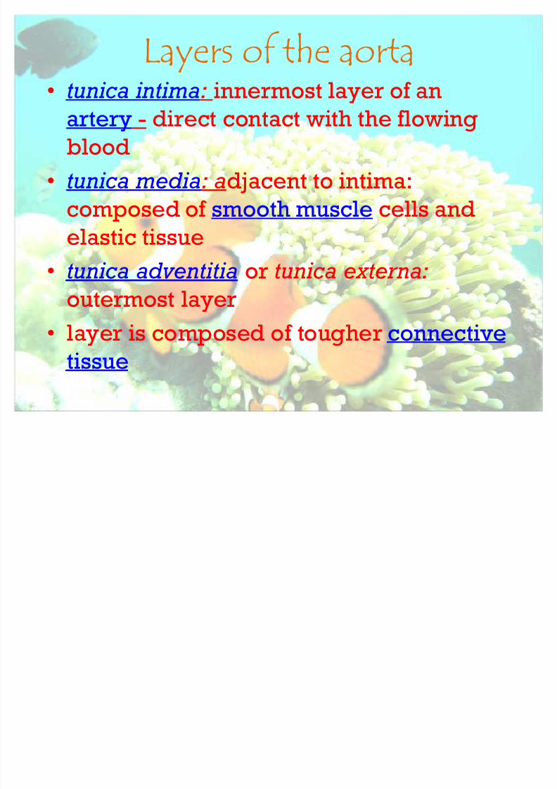
Layers of the aorta tunica intima: innermost layer of an
artery - direct contact with the flowing
blood
tunica media: adjacent to intima:
composed of smooth muscle cells and
elastic tissue
tunica adventitia or tunica externa:
outermost layer
layer is composed of tougher connective
tissue
8/8/2019 Aneurysm Visual Aid
http://slidepdf.com/reader/full/aneurysm-visual-aid 5/29
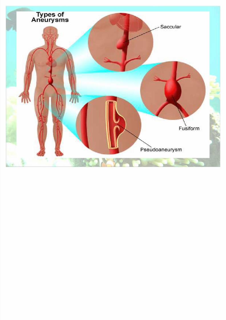
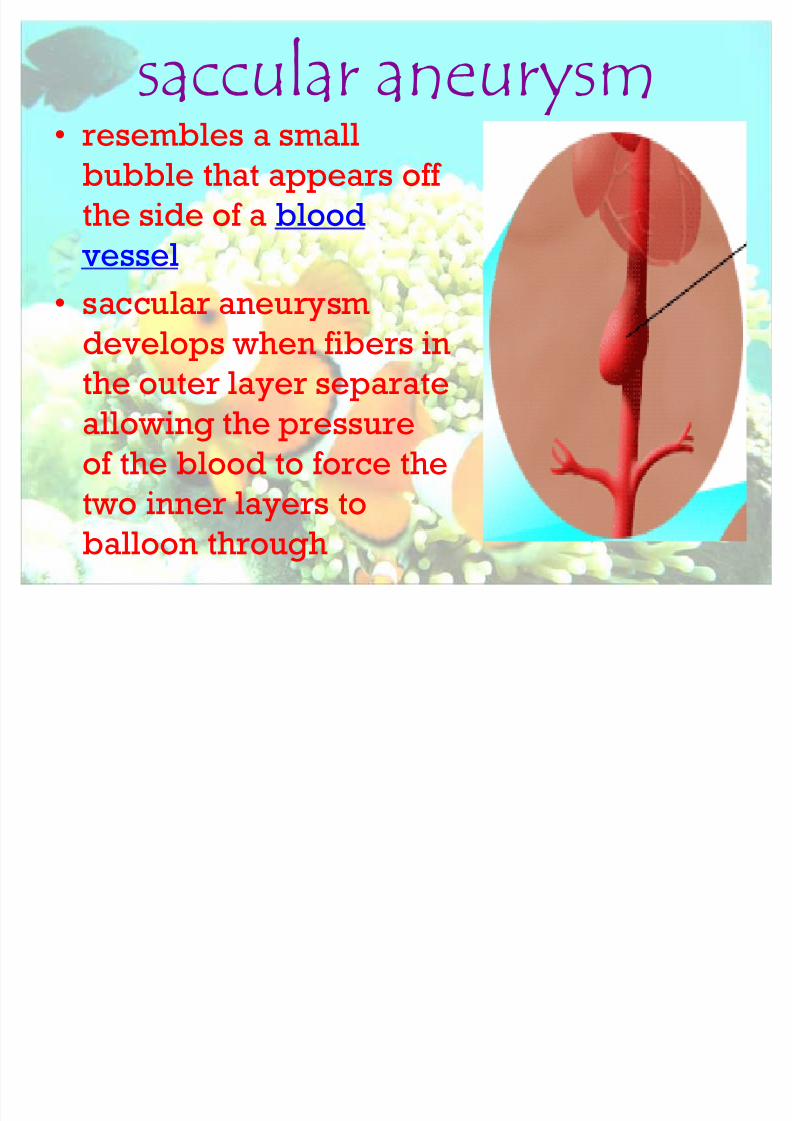
saccular aneurysm resembles a small
bubble that appears off
the side of a blood
vessel
saccular aneurysm
develops when fibers in
the outer layer separate
allowing the pressureof the blood to force the
two inner layers to
balloon through

8/8/2019 Aneurysm Visual Aid
http://slidepdf.com/reader/full/aneurysm-visual-aid 6/29
fusiform aneurysm is a bulging around
the entire
circumference of the vessel without
protrusion of the
inner layers. It is
shaped like afootball or spindle.
8/8/2019 Aneurysm Visual Aid
http://slidepdf.com/reader/full/aneurysm-visual-aid 7/29
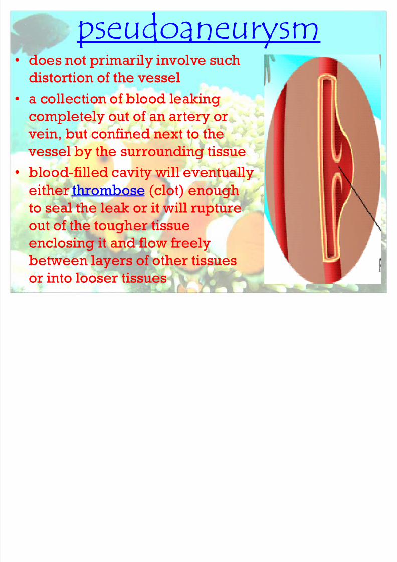
pseudoaneurysm does not primarily involve such
distortion of the vessel
a collection of blood leaking
completely out of an artery or
vein, but confined next to the vessel by the surrounding tissue
blood-filled cavity will eventually
either thrombose (clot) enough
to seal the leak or it will ruptureout of the tougher tissue
enclosing it and flow freely
between layers of other tissues
or into looser tissues

8/8/2019 Aneurysm Visual Aid
http://slidepdf.com/reader/full/aneurysm-visual-aid 8/29
Risk factors Smoking. High blood pressure
Atherosclerosis
Sex Race
Family history
Infection or inflammation (vasculitis) Marfan syndrome

8/8/2019 Aneurysm Visual Aid
http://slidepdf.com/reader/full/aneurysm-visual-aid 9/29
Symp toms A pulsating sensation near the navel
Tenderness or pain in the abdomen or
chest Back pain
8/8/2019 Aneurysm Visual Aid
http://slidepdf.com/reader/full/aneurysm-visual-aid 10/29
Symp toms A small, unchanging aneurysm will
produce no symptoms
Before a larger aneurysm ruptures : ² a sudden and unusually severe headache,
² nausea,
² vision impairment,
² vomiting,
² loss of consciousness
8/8/2019 Aneurysm Visual Aid
http://slidepdf.com/reader/full/aneurysm-visual-aid 11/29
Classification of rup turedaneurysm severity
8/8/2019 Aneurysm Visual Aid
http://slidepdf.com/reader/full/aneurysm-visual-aid 12/29
Hunt and Hess scale of
subarachnoid hemorrhage severity Grade 1: Asymptomatic; or minimal
headache and slight nuchal rigidity.
Approximate survival rate 70%. Grade 2: Moderate to severe
headache; nuchal rigidity; no
neurologic deficit except cranialnerve palsy. 60%.
Grade 3: Drowsy; minimal neurologic
deficit. 50%.

8/8/2019 Aneurysm Visual Aid
http://slidepdf.com/reader/full/aneurysm-visual-aid 13/29
Grade 4: Stuporous; moderate to
severe hemiparesis; possibly early
decerebrate rigidity and vegetativedisturbances. 20%.
Grade 5: Deep coma; decerebrate
rigidity; moribund. 10%. Grade 6: Instant Death
Hunt and Hess scale of subarachnoidhemorrhage severity
8/8/2019 Aneurysm Visual Aid
http://slidepdf.com/reader/full/aneurysm-visual-aid 14/29
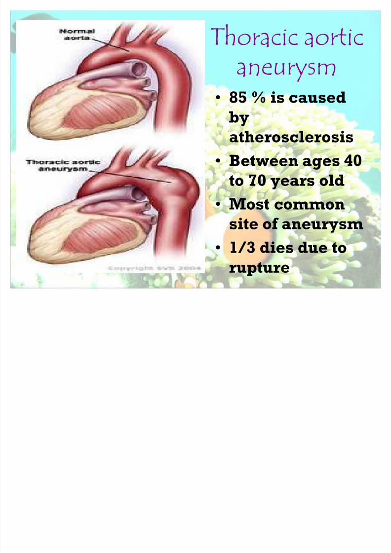
T horacic aortic
aneurysm 85 % is caused
by
atherosclerosis Between ages 40
to 70 years old
Most commonsite of aneurysm
1/3 dies due to
rupture

8/8/2019 Aneurysm Visual Aid
http://slidepdf.com/reader/full/aneurysm-visual-aid 15/29
Clinical Manifestation
Constant boring painduring supine
position
Dyspnea (pressureagainst the trachea)
Cough
Hoarsheness Stidor
Aphonia
Dysphagia

8/8/2019 Aneurysm Visual Aid
http://slidepdf.com/reader/full/aneurysm-visual-aid 16/29
Assessment Superfacial veins of the
chest and neck or arms
are dilated (pressure of
aneurysm) Edematous areas on the
chest wall
C
yanosis Unequal pupil (pressure
in the cervial
sympathetic chain)
Descending
Thoracic
Aneurysm
8/8/2019 Aneurysm Visual Aid
http://slidepdf.com/reader/full/aneurysm-visual-aid 17/29
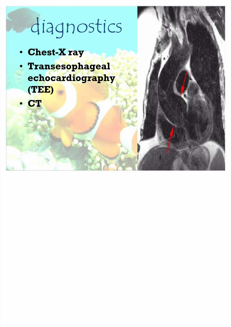
diagnostics Chest-X ray
Transesophageal
echocardiography(TEE)
CT

8/8/2019 Aneurysm Visual Aid
http://slidepdf.com/reader/full/aneurysm-visual-aid 18/29
MEDIC ALMANAGEMEN
T Surgical repair
Control blood
pressure Correct risk factors
8/8/2019 Aneurysm Visual Aid
http://slidepdf.com/reader/full/aneurysm-visual-aid 19/29
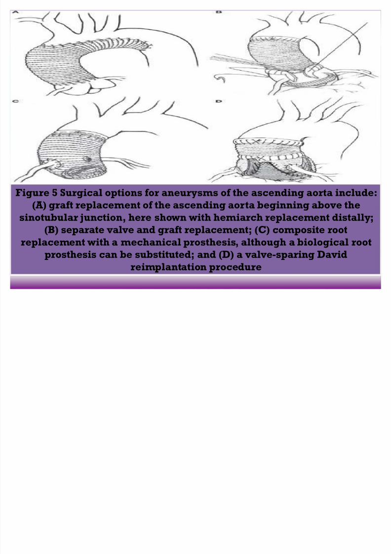
Figure 5 Surgical options for aneurysms of the ascending aorta include:
(A) graft replacement of the ascending aorta beginning above thesinotubular junction, here shown with hemiarch replacement distally;
(B) separate valve and graft replacement; (C) composite root
replacement with a mechanical prosthesis, although a biological root
prosthesis can be substituted; and (D) a valve-sparing David
reimplantation procedure
8/8/2019 Aneurysm Visual Aid
http://slidepdf.com/reader/full/aneurysm-visual-aid 20/29
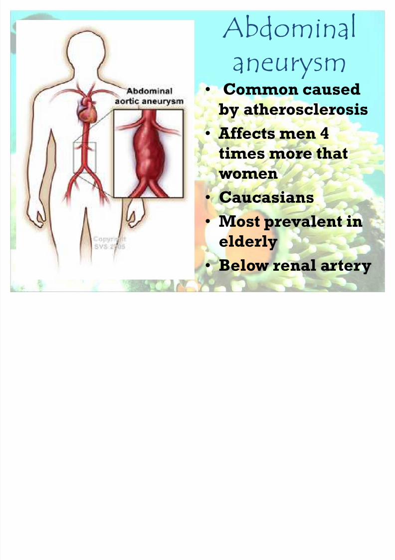
Abdominal
aneurysm Common caused
by atherosclerosis
Affects men 4times more that
women
Caucasians
Most prevalent in
elderly
Below renal artery

8/8/2019 Aneurysm Visual Aid
http://slidepdf.com/reader/full/aneurysm-visual-aid 21/29
Clinical manifestations
40 % have symptoms Heart beating in the
abdomen
Abdominal mass orthrobbing
Cyanosis and mottling
of the toes (cholesterol,platelet or fibrin lodge
in the interosseos or
digital arteries)

8/8/2019 Aneurysm Visual Aid
http://slidepdf.com/reader/full/aneurysm-visual-aid 22/29
Assessment &
diagnostic findings Pulsatile mass in the
middle and upper
abdomen
Can be palpated
Systolic bruit heard
over mass
Duplex
ultrasonography or
CT
8/8/2019 Aneurysm Visual Aid
http://slidepdf.com/reader/full/aneurysm-visual-aid 23/29
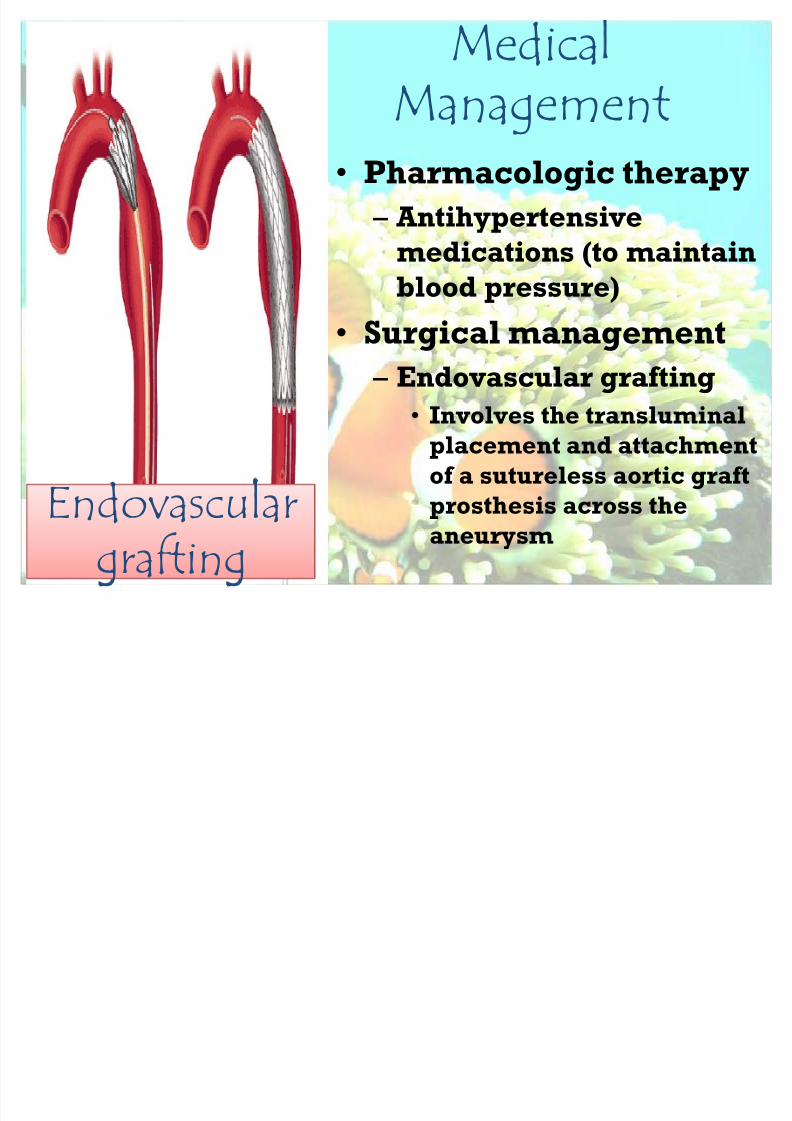
MedicaManagement
Pharmacologic therapy
² Antihypertensive
medications (to maintain
blood pressure)
Surgical management
² Endovascular grafting
Involves the transluminalplacement and attachment
of a sutureless aortic graft
prosthesis across the
aneurysm Endovascular
grafting
8/8/2019 Aneurysm Visual Aid
http://slidepdf.com/reader/full/aneurysm-visual-aid 24/29
N
ursingManagement

8/8/2019 Aneurysm Visual Aid
http://slidepdf.com/reader/full/aneurysm-visual-aid 25/29
Goal: provide emergency care beforesurgery for dissection or rup ture
Vital signs: frequent depending on
severity
I V monitoring
Urine output monitoring 15-30 mins
O2 inhalation
Administer antihypertensive asordered
Transport to OR quickly
Observe the general pre-operative care
8/8/2019 Aneurysm Visual Aid
http://slidepdf.com/reader/full/aneurysm-visual-aid 26/29
Goal: prevent complications post-op
² Vital signsCVP
Peripheral pulses hourly
² Position : initially flat Turn to sides
Note erythema on back from pooled
blood Turn to sides
Note erythema on back from pooled
blood

8/8/2019 Aneurysm Visual Aid
http://slidepdf.com/reader/full/aneurysm-visual-aid 27/29
Goal: promote comfort Position : alignment, comfort,
prevent ulcers
Administer medication: narcotic
8/8/2019 Aneurysm Visual Aid
http://slidepdf.com/reader/full/aneurysm-visual-aid 28/29
Goal: health teaching Minimize recurrence
² avoid trauma,
² infection, ² smoking,
² high cholesterol diet,
² obesity
Regular medical supervision
8/8/2019 Aneurysm Visual Aid
http://slidepdf.com/reader/full/aneurysm-visual-aid 29/29