Agrobacterium Protocols Volume 2 || Eucalyptus
10
125 From: Methods in Molecular Biology, vol. 344: Agrobacterium Protocols, 2/e, volume 2 Edited by: Kan Wang © Humana Press Inc., Totowa, NJ 12 Eucalyptus Zenn-Zong Chen, Cheng-Kuen Ho, In-Suk Ahn, and Vincent L. Chiang Summary Using Eucalyptus camaldulensis as a model system, we describe here a basic Agrobacterium-mediated genetic transformation protocol through organogenesis for the production of transgenic plants. Hypocotyl segments or cotyledon pieces from in vitro seedlings are used as starting materials. The explants are inoculated and cocultivated with a disarmed, binary strain of A. tumefaciens CIB542 harboring a mini Ti plasmid, pBI121. A modified Gamborg’s B5 medium is used as the basal culture medium throughout stages of co-cultivation, callus induction and shoot regeneration. The incorporation of neomycin phosphotransferase II (nptII) and β-glucuronidase (gus) genes into the plant nuclear genome are primarily verified by histochemical analysis and polymerase chain reaction (PCR). Modifications of this protocol to use in mature tissues derived from elite trees and other Eucalyptus species are also described. Key Words: Eucalyptus; organogenesis; genetic transformation; hypocotyl; cotyledon; Agrobacterium tumefaciens; β-glucuronidase (GUS); transgenic trees. 1. Introduction Eucalyptus is one of the most important commercial planting tree species in the production of raw materials for pulp and paper industry. Genetic engi- neering of Eucalyptus trees has been anxiously anticipated to improve wood quality and forest productivity in a limiting land of plantations. Currently, Agrobacterium-mediated transformation still plays the key role in the entire technology of genetic engineering in Eucalyptus species because of its effi- ciency as compared with other transformation methods. In this chapter, a pro- tocol for producing transgenic trees started with juvenile tissues of E. camaldulensis by using Agrobacterium tumefaciens is described step by step. Using this protocol, putative antibiotic resistant callus pieces are produced from 48 out of 100 initially infected explant materials. Of these 48 callus
Transcript of Agrobacterium Protocols Volume 2 || Eucalyptus
125
From: Methods in Molecular Biology, vol. 344: Agrobacterium Protocols, 2/e, volume 2Edited by: Kan Wang © Humana Press Inc., Totowa, NJ
12
Eucalyptus
Zenn-Zong Chen, Cheng-Kuen Ho, In-Suk Ahn, and Vincent L. Chiang
SummaryUsing Eucalyptus camaldulensis as a model system, we describe here a basic
Agrobacterium-mediated genetic transformation protocol through organogenesis for theproduction of transgenic plants. Hypocotyl segments or cotyledon pieces from in vitroseedlings are used as starting materials. The explants are inoculated and cocultivated witha disarmed, binary strain of A. tumefaciens CIB542 harboring a mini Ti plasmid, pBI121.A modified Gamborg’s B5 medium is used as the basal culture medium throughout stagesof co-cultivation, callus induction and shoot regeneration. The incorporation of neomycinphosphotransferase II (nptII) and β-glucuronidase (gus) genes into the plant nucleargenome are primarily verified by histochemical analysis and polymerase chain reaction(PCR). Modifications of this protocol to use in mature tissues derived from elite trees andother Eucalyptus species are also described.
Key Words: Eucalyptus; organogenesis; genetic transformation; hypocotyl; cotyledon;Agrobacterium tumefaciens; β-glucuronidase (GUS); transgenic trees.
1. IntroductionEucalyptus is one of the most important commercial planting tree species
in the production of raw materials for pulp and paper industry. Genetic engi-neering of Eucalyptus trees has been anxiously anticipated to improve woodquality and forest productivity in a limiting land of plantations. Currently,Agrobacterium-mediated transformation still plays the key role in the entiretechnology of genetic engineering in Eucalyptus species because of its effi-ciency as compared with other transformation methods. In this chapter, a pro-tocol for producing transgenic trees started with juvenile tissues of E.camaldulensis by using Agrobacterium tumefaciens is described step by step.Using this protocol, putative antibiotic resistant callus pieces are producedfrom 48 out of 100 initially infected explant materials. Of these 48 callus

lines, 10 proliferate vigorously and regenerate shoots and plants, which allshow positive integration of T-DNA as confirmed by polymerase chain reac-tion (PCR) or Southern hybridization, indicating a 10% transformation effi-ciency of the infected explants (1). This protocol has been modified tosuccessfully use in the transformation of various gene constructs using maturetissues of elite clones in E. camaldulensis, E. grandis as well as E. grandis ×E. urophylla.
2. Materials2.1. Plant Materials
1. Seeds of E. camaldulensis: A seedlot was obtained from Australian Tree SeedCenter, Commonwealth Scientific and Industrial Research Organization (CSIRO),Australia. A 10 g weight gives rise to 5000 viable seeds.
2. Shoot tips, lateral buds, or epicomic shoots from elite trees of E. camaldulensis,E. grandis, E. grandis × E. urophylla grown in test plantations of Taiwan ForestryResearch Institute (TFRI) or in commercial plantations of private pulp and papercompanies, and partially collected from elite clones of rooted cuttings maintainedin greenhouses of TFRI and North Carolina State University.
2.2. Agrobacterium Host Strain and Vector Construct
1. A disarmed binary strain of A. tumefaciens CIB542: This strain was derived fromEHA101 (2) in which the kanamycin resistance gene on the nononcogenic viru-lence (vir) helper plasmid was replaced by a spectinomycin/streptomycin resist-ance gene.
2. A tumor-inducing (Ti) mini plasmid pBI121: This plasmid contains a selectablemarker gene driven by nopaline synthase (NOS) promoter, and a reporter genedriven by cauliflower mosaic virus (CaMV) 35S promoter in a constitutive man-ner (3). The selectable marker, neomycin phosphotransferase-encoding gene II(nptII), confers kanamycin resistance to transformed plant cells. The uidA reportergene encoding β-glucuronidase (GUS) makes transformed plant cells showingblue in the presence of 5-bromo-4-chloro-3-indolyl-β-D-glucuronide (X-Gluc)(see Notes 1 and 2).
2.3. Bacterial Medium and Antibiotics
1. Luria–Bertani (LB) medium: 10 g/L bacto-tryptone, 5 g/L bacto-yeast extract,10 g/L NaCl, and 15 g/L Bacto-agar. Adjust pH to 7.0 with 1 N NaOH. Appropriateantibiotics should be added to the medium after autoclave.
2. Mannitol-Glutamic Acid: 10 g/L mannitol, 2 g/L L-glutamic acid, 0.5 g/LKH2PO4, 0.2 g/L MgSO4·7H2O and 2 μg/L biotin (4).
3. Mannitol-Glutamic Acid:Luria-Bertani (MGL) medium: a 1:1 (v/v) mixture ofmannitol-glutamic acid and LB medium.
4. Stock of 25 mg/mL kanamycin monosulfate dissolved in H2O and 50 mg/L forworking concentration.
126 Chen et al.

5. Stock of 25 mg/mL spectinomycin dihydrochloride hydrate dissolved in H2O and50 mg/L for working concentration.
6. Stock of 250 mg/mL streptomycin sulfate dissolved in H2O and 500 mg/L forworking concentration.
7. Stock of 100 mg/mL carbenicillin dissolved in H2O and 500 mg/L for workingconcentration.
8. Stock of 100 mg/mL cefotaxime dissolved in H2O and 500 mg/L for working con-centration.
9. All antibiotic stocks should be filter-sterilized and stored at −20°C.10. Antibiotics such as carbenicillin, cefotaxime and kanamycin are added in the
media cooled down to about 45°C.
2.4. Tissue Culture and Plant Growth Medium
1. Sterilization solution: 70% (v/v) ethanol, 1% (v/v) sodium hypochlorite (NaOCl),and sterilized distilled water.
2. Phytohormone preparation: 10 mg/mL of 6-benzylaminopurine (BA), 1-naphthy-lacetic acid (NAA), and thidiazuron (TDZ) stocks. Phytohormons are dissolvedusing from 3 to 5 mL of 1N NaOH or 1N KOH and then final volume is adjustedby slowly adding H2O. Keep stirring during preparation. These phytohormonescan be added in the medium before autoclave.
3. 10X stocks of Murashige and Skoog (MS) medium (5), and Gambogs B5medium (6): Basal salt mixtures manufactured commercially (M5524 andG5768, Sigma).
4. 1000X MS vitamin stock: 2 g/L glycine, 500 mg/L nicotinic acid, 500 mg/L pyri-doxine·HCl and 100 mg/L thiamine·HCl dissolved in H2O.
5. 1000X B5 vitamin stock: 1 g/L nicotinic acid, 1 g/L pyridoxine·HCl and 10 g/Lthiamine·HCl dissolved in H2O.
6. 100X myo-inositol stock: 10 g/L myo-inositol dissolved in H2O. All vitamins canbe added in the medium before autoclave.
7. Stock of 100 mM acetosyringone dissolved in dimethylsulfoxide (DMSO).8. MS basal medium (5): MS salts mixture, MS vitamins, myo-inositol.9. B5 basal medium (6): B5 salts mixture, B5 vitamins, myo-inositol
10. In vitro germination medium: MS basal medium containing 3% (w/v) sucrose.11. B5C medium: B5 medium containing 3% (w/v) sucrose, 100 ml/L coconut water,
200 mg/L glutamine, and 100 mg/L casein hydrolysate.12. Callus/shoot induction medium for hypocotyl and cotyledon explants (CSMBA):
B5C medium supplemented with 1 mg/L BA and 3 mg/L NAA.13. Callus/shoot induction medium for explants derived from field-grown elite trees
(CSMTDZ): B5C medium supplemented with 3 mg/L NAA and approx 0.5–1.5 mg/LTDZ (see Note 3).
14. Selection medium I: CSMBA or CSMTDZ containing 40 mg/L kanamycin, 500 mg/Lcefotaxime, and 500 mg/L carbenicillin.
15. Selection medium II: CSMBA or CSMTDZ containing 40 mg/L kanamycin.16. Shoot proliferation medium (SPM): MS basal medium containing 0.1 mg/L BA.
Eucalyptus 127

17. Root induction medium (RIM): Modified MS medium consisting of full strengthmicro elements and vitamins, half strength of macro elements, and 1 mg/L 3-indolebutyric acid (IBA).
18. All media are adjusted to pH 5.6–5.8 using few drops of 1N NaOH or 1N KOH andautoclaved at 121°C (15 psi) for 20 mins. Add 7.5 g/L bacto-agar before autoclave.
19. Artificial soil: A mixture of vermiculite, peat moss and perlite (2:2:1 in volume).
2.5. Transgenic Plant Verification
1. GUS histochemical analysis buffer: 1 mM 5-bromo-4-chloro-3-indolyl-β-D-glucuronide (X-Gluc) in phosphate buffer (100 mM primary sodium phos-phate, pH 7.0) containing 10 mM ethylene diamine tetraacetic acid (EDTA),0.5 mM potassium ferricyanide, 0.5 mM potassium ferrocyanide, and 0.1%Triton X-100.
2. DNA extraction buffer: 20 μM Tris-HCl, pH 7.5, 250 μM NaCl, 25 μM EDTA,pH 8.0, 0.5% sodium dodecyl sulfate (SDS).
3. TE buffer: 10 mM Tris-HCl, pH 8.0, and 1 mM EDTA, pH 8.0.4. Polymerase chain reaction (PCR) related solutions and buffers.5. Reaction buffer (10X): 750 mM Tris-HCl, pH8.8; 200 mM (NH4)2SO4; and 0.1%
(v/v) Tween-20.6. 2.5 mM each of the deoxy nucleotide triphosphates (dNTPs).7. Primers: NPTII gene forward primer (5′-GAACAAGATGGATTGCACGC-3′) and
reverse primer (5′-GAAGAACTCGTCAAGAAGGC-3′).
3. Methods3.1. Explant Preparation
3.1.1. From Seeds of Eucalyptus camaldulensis
1. Weight 0.1 g of Eucalyptus seeds (50 viable seeds) and wrap them with 3 layersof cheesecloth.
2. Immerse the wrapped seeds into 70% ethanol in a 100-mL beaker for 1 min and fol-lowed by immersion in 1% NaOCl with ultrasonic treatment for 15 mins (see Note 4).
3. Move the beaker to a laminar flow hood and rinse the wrapped seeds three timeswith sterile water.
4. Place and spread surface-sterilized seeds evenly on two Petri plates (100 × 25 mmin size) containing 25 mL germination medium.
5. Seal the plates with parafilm and culture them in an incubator at 25 ± 2°C underapprox 140 μE/m2/sec light intensity with 16-h light/8-h dark cycle.
6. Normally, germination is observed within 5 d after plating. Young seedlings ger-minated can be transferred into Magenta GA7 boxes or 125-mL Erlenmeyer flaskscontaining 50 mL of the same germination medium to promote growth.
7. Two days before transformation experiment, isolate hypocotyls or cotyledonsfrom 1-mo-old seedlings germinated in vitro and cut those tissues into smallpieces (approx 5-mm length for hypocotyls and 3 × 3 mm for cotyledons).
8. Pre-culture the segments on CSMBA for 2 d at 25°C in the dark.
128 Chen et al.

Eucalyptus 129
3.1.2. From Shoots or Buds of E. camaldulensis, E. grandis,and E. grandis × E. urophylla
1. Surface-sterilize shoot tips, lateral buds, or epicormic shoots by soaking in 70%ethanol for 30 s followed by a rinse in running tap water for 1 h.
2. Move those tissues to a larminar flow hood and soak them into 2% sodiumhypochlorite (NaOCl) with ultrasonic treatment (T-28, L&R Manufacturing Co,Kearny, NJ) for 10 min and rinse them thoroughly 3 times with sterilized distilledwater.
3. Finally, soak into 100 mg/L filter-sterilized ascorbic acid for 2 h to prevent secre-tion of phenolic compound causing tissue browning.
4. Culture the surface-sterilized materials on the shoot proliferation medium (SPM)to produce multiple shoot clumps and maintain them at 25 ± 2°C under approx140 μE/m2/s light intensity with 16-h light/8-h dark cycle. The buds proliferatewithin 2–3 wk after culturing on the SPM (7–9).
5. Shoot clumps can be maintained by subculturing small shoot tips onto the SPMbiweekly. Excise leaf pieces (approx 3 mm × 3 mm in size) from the in vitro shootclumps and use them as starting materials for transformation.
3.2. Agrobacterium Preparation
1. Three days before transformation experiments, streak a bacterium colony on a LBor MGL plate containing appropriate antibiotics by scrubbing a loop top of bacte-ria glycerol stock stored in −80°C and incubate the plate at 28°C for 2 d. ThisAgrobacterium plate is then become stock plate and stored up to one month at 4°Cfor transformation work.
2. Using a loop or toothpick, pick up a single cell colony from the Agrobacteriumplate and inoculate it into 1 mL LB or MGL liquid broth supplemented withappropriate antibiotics and culture it overnight at 28°C with strong agitation (200 rpm).
3. Pour the overnight-grown bacteria suspension into 25 mL liquid LB or MGL brothin a 125-mL Erlenmeyer flask (see Note 5). The same antibiotics used for 1-mLbacteria culture should be added in 25-mL bacterial culture. Grow the bacteria at28°C for 4–6 h with strong agitation (200 rpm) to reach the cell density to 1.0approx 1.5 OD600.
3.3. Infection and Co-cultivation
1. Pour 25 mL of well grown Agrobacterium suspension into a 100 × 25 mm sizePetri plate. Immerse the explants into the suspension completely and allow theexplants to sit in the inoculum for 10 min with occasional agitation by hand.
2. After inoculation, blot the explants on several layers of sterilized filter paper toremove excess bacteria culture.
3. Transfer the explants onto CSM (CSMBA for tissues from seedlings and CSMTDZfor those from field-grown elite trees) and culture at 25°C in a dark for 2 d for co-cultivation.

3.4. Callus and Shoot Induction
1. After co-cultivation for 2 d, remove the infected segments from the plate and washthem with sterilized distilled water 3 times for 5 min each to remove over-grownbacteria. Five hundred Milligrams per liter cefotaxime and/or 500 mg/L carbeni-cillin can be added in the water for effective removal of bacteria.
2. Blot the segments onto one layer of sterilized filter paper to remove excess waterand transfer them onto selection medium I to select putative transformed cells andprevent proliferation of untransformed cells. For juvenile tissues of E. camaldu-lensis, use selection medium I with 1 mg/L BA. For leaf pieces derived from field-grown elite trees of E. camaldulensis, E. grandis, and E. grandis × E. urophylla,use 0.5–1.5 mg/L TDZ.
3. Incubate the plates at 25 ± 2°C under approx 140 μE/m2/sec light intensity with 16-hlight/8-h dark cycle. Subculture the tissues onto fresh selection medium I biweekly.
4. After four rounds of subcultures, transfer the tissues onto fresh selection mediumII. Keep subculturing the tissues on selection medium II biweekly until putativetransgenic callus appears.
5. Putative transgenic callus can be observed within 30 d and up to 50 d after co-cultivation (see Fig. 1A). Excise the callus from tissues, culture them onto freshselection medium II, and maintain the plate at 25 ± 2°C under approx 140 μE/m2/slight intensity with 16-h light/8-h dark cycle. Ensure you mark the independentcallus line.
6. Subculture the excised callus to the same selection medium II biweekly for thecallus proliferation and shoot regeneration (see Fig. 1B).
3.5. Shoot Proliferation and Elongation
1. Shoots are regenerated within about 4 mo after isolation of callus from infectedtissues and cultured individually on selection medium II (see Notes 6 and 7).
2. Shoot proliferation and elongation can be achieved on the same selection mediumII. However, to promote shoot proliferation, transfer shoot clumps to shoot prolif-eration medium when shoot regeneration is visible (see Fig. 2A,B).
3. Subculture the shoot clumps to fresh shoot proliferation medium biweekly. Thecultures are placed in culture room at 25 ± 2°C under approx 140 μE/m2/s lightintensity with 16-h light/8-h dark cycle. Remove the base part of shoot clumpsduring each subculture.
3.6. Rooting of Shoots and Acclimation of Plantlet
1. Excise the elongated shoots (approx 15 mm in length) from the kanamycin-resistantshoot clumps and transfer them into Magenta GA7 boxes containing 50 mL ofrooting medium.
2. Culture the shoots at 25 ± 2 °C under approx 140 μE/m2/s light intensity with 16-hlight/8-h dark cycle for 2–4 wk until primary and secondary roots appear.
3. Although roots emerge within 10 d after culturing on rooting medium, keep cul-ture until the shoot develops more than two roots.
130 Chen et al.

4. Carefully take the rooted shoots out from the medium and wash medium awayfrom the roots with running tap water.
5. Transplant the rooted plantlet into a small pot (approx 17 cm in inner diameter)containing artificial soil mixture and cover it with a Magenta GA7 box.
6. Move the pot into a greenhouse at 25 ± 2°C under approx 300 μE/m2/s light inten-sity with 10-h light/14-h dark cycle and maintain for 1 wk. Water the pot to wetthe artificial soil completely, no watering is needed during the first wk of acclima-tion. Mortality is less than 10% during the process of acclimation.
Eucalyptus 131
Fig. 1. Putative transgenic callus induction and shoot regeneration. (A) Three puta-tive transgenic calli formed from a tissue cultured on selection medium. (B) Putativetransgenic shoots formation initiated from a callus clump.
Fig. 2. Shoot proliferation and elongation. (A) Multiple shoots proliferated rapidlyafter transfer onto a shoot proliferation medium. (B) Transgenic shoots elongated aftersubculturing twice. Healthy and well-grown shoots are significant.
7. Remove the covered Magenta GA7 box gradually during the period of anotherweek. Water and fertilize the plants regularly.
8. Transgenic plants are cut back at height of 30–100 cm for wood analysis andmaintained by trimming regularly at 8-wk intervals.
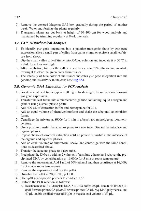
3.7. GUS Histochemical Analysis
1. To identify gus gene integration into a putative transgenic shoot by gus geneexpression, slice a small part of callus from callus clump or excise a small leaf tis-sue from shoot.
2. Dip the small callus or leaf tissue into X-Gluc solution and incubate it at 37°C ina dark for 6 h or overnight.
3. After incubation, transfer the callus or leaf tissue into 95% ethanol and incubateovernight to clear the green color from tissues.
4. The intensity of blue color of the tissues indicates gus gene integration into thegenome and its activity in the cells (see Fig 3A).
3.8. Genomic DNA Extraction for PCR Analysis
1. Isolate a small leaf tissue (approx 50 mg in fresh weight) from the shoot showinggus gene expression.
2. Transfer the leaf tissue into a microcentrifuge tube containing liquid nitrogen andgrind it using a small plastic pestle.
3. Add 400 μL of extraction buffer and homogenize for 30 s.4. Add an equal volume of phenol/chloroform and shake the tube until an emulsion
forms.5. Centrifuge the mixture at 8000g for 1 min in a bench top microfuge at room tem-
perature.6. Use a pipet to transfer the aqueous phase to a new tube. Discard the interface and
organic phases.7. Repeat phenol/chloroform extraction until no protein is visible at the interface of
the organic and aqueous phases.8. Add an equal volume of chloroform, shake, and centrifuge with the same condi-
tions as described above.9. Transfer the aqueous phase to a new tube.
10. Precipitate the DNA by adding 2 volumes of absolute ethanol and recover the pre-cipitated DNA by centrifugation at 16,000g for 5 min at room temperature.
11. Remove the supernatant. Add 1 mL of 70% ethanol and then centrifuge at 16,000gfor 5 min at room temperature.
12. Remove the supernatant and dry the pellet.13. Dissolve the pellet in 20 μL TE, pH 8.0.14. Use nptII gene-specific primers to conduct PCR.15. Perform the PCR reaction as follows:
a. Reaction mixture: 3 μL template DNA, 5 μL 10X buffer, 0.5 μL 10 mM dNTPs, 0.5 μLnptII forward primer, 0.5 μL nptII reverse primer, 0.5 μL Taq-DNA polymerase, and40 μL double distilled water (ddH2O) to make a total volume of 50 μL.
132 Chen et al.

b. PCR conditions: 95°C for 120 s, followed by 30 cycles of 94°C for 30 s, 58°Cfor 20 s, and 72°C for 60 s, and then a final extension at 72°C for 3 min.
16. Electrophorese the PCR product on a 1% agarose gel and visualize the ampliconby staining the gel with ethidium bromide (see Fig. 3B).
4. Notes1. This protocol has been successfully applied to introduce various genes into
Eucalyptus cells including lignin-specific genes such as cinnamate 4-hydroxylase(C4H), 4-coumaric acid:coenzyme A ligase (4CL), coniferaldehyde 5-hydroxylase(CAld5H) and coniferyl alcohol dehydrogenase (CAD), and cold tolerance genessuch as C-repeat (CRT)/dehydration element (DRE) Binding Factor (CBF1) andMyb4.
2. The nptII gene has been successfully replaced with the hygromycin resistance (hpt)gene which confers hygromycin resistance to the transformed cells. To preventescaped shoot production, hygromycin at 15 mg/L is required for the selection.
3. Use 0.5–1.5 mg/L TDZ instead of BA as cytokinin source for callus induction andshoot regeneration. This is one of the most important factors for successful regen-eration of transgenic shoots (10,11) (see also Notes 6 and 7).
4. Healthy seeds are important for germination. Floating seeds in the 70% ethanolare not healthy. Discard them.
5. 100 μM acetosyringone can be added in 25-mL bacteria culture to promote virgene activation. But this is an optional.
6. If no shoots are regenerated with TDZ, zeatin in concentration of 0.5–2 mg/L canbe combined with TDZ.
7. Difficulties in shoot regeneration from putative transformed callus and rooting ofputative transgenic shoots often result from starting explants which are
Eucalyptus 133
Fig. 3. Primary verification of transgenic tissues. (A) Histochemical gus expressionin transgenic shoots. (B) PCR amplification using nptII gene-specific primers. A 780-bpband is observed in all transgenic lines. M maker; lane 1: untransformed leaf; lane 2–8:different transgenic lines.

unrejuvenated or unhealthy. Using leaf pieces excised from in vitro shoot clumpsthat have been frequently subcultured as starting explants for transformationexperiment may solve the problems in shoot regeneration and rooting of shoots.
References1. Ho, C.-K., Chang, S.-H., Tsay, J.-Y., Tsai, C.-J., Chiang, V.L., and Chen, Z.-Z.
(1998) Agrobacterium tumefaciens-mediated transformation of Eucalyptus camal-dulensis and production of transgenic plants. Plant Cell Rep. 17, 675–680.
2. Hood, E.E., Helmer, G.L., Fraley, R.T., and Chilton, M.-D. (1986) The hyperviru-lence of Agrobacterium tumefaciens A281 is encoded in a region of pTiBo542 out-side of T-DNA. J. Bacteriol. 168, 1291–1301.
3. Jefferson, R.A. (1987) Assaying chimeric genes in plants: the GUS gene fusionsystem. Plant Mol. Biol. Rep. 5, 387–405.
4. Roberts, W.P. and Kerr, A. (1974) Crown Gall Induction: serological reaction,isozyme patterns and sensitivity to mitomucin C and to bacteriocin, of pathogenicand non-pathogenic strains of Agrobacterium radiobacter. Physiol. Plant Path. 4,81–91.
5. Murashige, T. and Skoog, F. (1962) A revised medium for rapid bioassays withtobacco tissue cultures. Physiol. Plant. 15, 473–497.
6. Gamborg, O.L., Miller, R.A., and Ojima, K. (1968) Nutrient requirement of sus-pension cultures of soybean root cells. Exp. Cell Res. 50, 151–158.
7. Yang, J.-C., Chung, J.-D., and Chen, Z.-Z. (1995) Vegetative propagation of adultEucalyptus grandis x urophylla and comparison of growth between micropropa-gated plantlets and rooted cuttings. Plant Cell Rep. 15, 170–173.
8. Yang, J.-C., Ho, C.-K., Chang, S.-H., and Chen, Z.-Z. (1996) Micropropagation ofmature trees of Eucalyptus camaldulensis with fast-growing phenotype. Taiwan J.For. Sci. 11, 421–431.
9. Yang, J.-C., Ho, C.-K., Chen, Z.-Z., and Chen, C.-F. (1996) Comparison of differ-ent methods of vegetative propagation of Eucalyptus camaldulensis plus trees.Taiwan J. For. Sci. 11, 433–443.
10. Chen, Z.-Z., Tsay, J.-Y., and Chung, J.-D. (1996) Callus culture of Eucalyptusgrandis x urophylla and preliminary studies on organogenesis and Agrobacterium-mediated transformation. Taiwan J. For. Sci. 11, 43–52.
11. Chen, Z.-Z., Chang, S.-H., Ho, C.-K., Chen, Y.-C., Tsai, J.-B., Chiang, V.L. (2001)Plant production of transgenic Eucalyptus camaldulensis carrying the Populustremuloides cinnamate 4-hydroxylase gene. Taiwan J. For. Sci. 16, 249–258.
134 Chen et al.