A pilot study of regenerative therapy using controlled ... · A pilot study of regenerative therapy...
8
ORIGINAL PAPER A pilot study of regenerative therapy using controlled release of recombinant human fibroblast growth factor for patients with pre-collapse osteonecrosis of the femoral head Yutaka Kuroda 1 & Ryuta Asada 2 & Kazutaka So 1 & Atsushi Yonezawa 3 & Manabu Nankaku 4 & Kumi Mukai 5 & Toshiko Ito-Ihara 5 & Harue Tada 6 & Michio Yamamoto 6 & Toshinori Murayama 5 & Satoshi Morita 6 & Yasuhiko Tabata 8 & Masayuki Yokode 5 & Akira Shimizu 7 & Shuichi Matsuda 1 & Haruhiko Akiyama 9 Received: 11 September 2015 /Accepted: 14 December 2015 /Published online: 29 December 2015 # The Author(s) 2015. This article is published with open access at Springerlink.com Abstract Purpose We evaluated the safety and clinical outcomes of a single local administration of gelatin hydrogel impregnated with recombinant human fibroblast growth factor (rhFGF)-2 for the treatment of the precollapse stage of osteonecrosis of the femoral head (ONFH). Methods Patients with ONFH (precollapse stage ≤2) received a single local administration of 800 μg of rhFGF-2- impregnated gelatin hydrogel and were followed up for one year. The surgery was performed using a minimally inva- sive technique involving a 1-cm skin incision, and walking was allowed from day one postoperatively. The primary outcomes included occurrence of adverse events and complications. The secondary outcomes included changes in the Harris hip scores, visual analog scale for pain scores, University of California, Los Angeles (UCLA) activity scores, and radiological images. Results We included ten patients, of which five experienced 14 adverse events, including one complication from spinal anesthesia. However, patients completely recovered from all adverse events. The mean clinical scores significantly improved by one year postoperatively compared with the pre-operative scores (before vs. after: visual analog score for pain, 21.2 vs. 5.3 mm; UCLA activity score, 5.5 vs. 6.6; Harris hip score, 81.0 vs. 96.9 points). There was only one case of femoral head collapse; however, this occurred in a hip with extensive necrosis. Stage progression and collapse did not occur in the other nine cases. Computed tomography con- firmed bone regeneration in the femoral heads. Conclusions Clinical application of rhFGF-2-impregnated gelatin hydrogel for patients with precollapse ONFH was fea- sible and safe. Keywords Clinical trial . Femoral head . FGF . Growth factor . Osteonecrosis . Regenerative therapy Abbreviations AE Adverse event CT Computed tomography * Yutaka Kuroda [email protected] 1 Department of Orthopaedic Surgery, Graduate School of Medicine, Kyoto University, Shogoin, Kawahara-cho 54, Sakyo-ku, Kyoto 606-8507, Japan 2 Clinical Research Center, Gifu University Hospital, Gifu, Japan 3 Department of Clinical Pharmacology and Therapeutics, Kyoto University Hospital, Kyoto, Japan 4 Rehabilitation Unit, Kyoto University Hospital, Kyoto, Japan 5 Department of Clinical Innovative Medicine, Institute for Advancement of Clinical and Translational Research (iACT), Kyoto University Hospital, Kyoto, Japan 6 Department of Data Science, Institute for Advancement of Clinical and Translational Research (iACT), Kyoto University Hospital, Kyoto, Japan 7 Department of Experimental Therapeutics, Institute for Advancement of Clinical and Translational Research (iACT), Kyoto University Hospital, Kyoto, Japan 8 Department of Biomaterials, Field of Tissue Engineering, Institute for Frontier Medical Sciences, Kyoto University, Kyoto, Japan 9 Department of Orthopaedic Surgery, Gifu University, Gifu, Japan International Orthopaedics (SICOT) (2016) 40:1747–1754 DOI 10.1007/s00264-015-3083-1
Transcript of A pilot study of regenerative therapy using controlled ... · A pilot study of regenerative therapy...
ORIGINAL PAPER
A pilot study of regenerative therapy using controlled releaseof recombinant human fibroblast growth factor for patientswith pre-collapse osteonecrosis of the femoral head
Yutaka Kuroda1 & Ryuta Asada2 & Kazutaka So1 & Atsushi Yonezawa3 &
Manabu Nankaku4& Kumi Mukai5 & Toshiko Ito-Ihara5 & Harue Tada6 &
Michio Yamamoto6 & Toshinori Murayama5 & Satoshi Morita6 & Yasuhiko Tabata8 &
Masayuki Yokode5 & Akira Shimizu7& Shuichi Matsuda1 & Haruhiko Akiyama9
Received: 11 September 2015 /Accepted: 14 December 2015 /Published online: 29 December 2015# The Author(s) 2015. This article is published with open access at Springerlink.com
AbstractPurpose We evaluated the safety and clinical outcomes of asingle local administration of gelatin hydrogel impregnatedwith recombinant human fibroblast growth factor (rhFGF)-2for the treatment of the precollapse stage of osteonecrosis ofthe femoral head (ONFH).Methods Patients with ONFH (precollapse stage ≤2) receiveda single local administration of 800 μg of rhFGF-2-impregnated gelatin hydrogel and were followed up forone year. The surgery was performed using a minimally inva-sive technique involving a 1-cm skin incision, and walking wasallowed from day one postoperatively. The primary outcomesincluded occurrence of adverse events and complications. Thesecondary outcomes included changes in the Harris hip scores,visual analog scale for pain scores, University of California,Los Angeles (UCLA) activity scores, and radiological images.Results We included ten patients, of which five experienced14 adverse events, including one complication from spinalanesthesia. However, patients completely recovered from alladverse events. The mean clinical scores significantly
improved by one year postoperatively compared with thepre-operative scores (before vs. after: visual analog score forpain, 21.2 vs. 5.3 mm;UCLA activity score, 5.5 vs. 6.6; Harriship score, 81.0 vs. 96.9 points). There was only one case offemoral head collapse; however, this occurred in a hip withextensive necrosis. Stage progression and collapse did notoccur in the other nine cases. Computed tomography con-firmed bone regeneration in the femoral heads.Conclusions Clinical application of rhFGF-2-impregnatedgelatin hydrogel for patients with precollapse ONFH was fea-sible and safe.
Keywords Clinical trial . Femoral head . FGF . Growthfactor . Osteonecrosis . Regenerative therapy
AbbreviationsAE Adverse eventCT Computed tomography
* Yutaka [email protected]
1 Department of Orthopaedic Surgery, Graduate School of Medicine,Kyoto University, Shogoin, Kawahara-cho 54,Sakyo-ku, Kyoto 606-8507, Japan
2 Clinical Research Center, Gifu University Hospital, Gifu, Japan
3 Department of Clinical Pharmacology and Therapeutics, KyotoUniversity Hospital, Kyoto, Japan
4 Rehabilitation Unit, Kyoto University Hospital, Kyoto, Japan
5 Department of Clinical Innovative Medicine, Institute forAdvancement of Clinical and Translational Research (iACT), KyotoUniversity Hospital, Kyoto, Japan
6 Department of Data Science, Institute for Advancement of Clinicaland Translational Research (iACT), Kyoto University Hospital,Kyoto, Japan
7 Department of Experimental Therapeutics, Institute forAdvancement of Clinical and Translational Research (iACT), KyotoUniversity Hospital, Kyoto, Japan
8 Department of Biomaterials, Field of Tissue Engineering, Institutefor Frontier Medical Sciences, Kyoto University, Kyoto, Japan
9 Department of Orthopaedic Surgery, Gifu University, Gifu, Japan
International Orthopaedics (SICOT) (2016) 40:1747–1754DOI 10.1007/s00264-015-3083-1
HHS Harris hip scoreJICHW Japanese Investigation Committee of Health
and WelfareMRI Magnetic resonance imagingONFH Osteonecrosis of the femoral headrhFGF Recombinant human fibroblast growth factorTHA Total hip arthroplastyUCLA University of California, Los AngelesVAS Visual analog scale
Introduction
Osteonecrosis of the femoral head (ONFH) is a multifactorialdisease that can cause femoral head collapse, pain, gait disor-ders, and secondary hip osteoarthritis. Several causative fac-tors have been indicated, including hypercoagulation, bonemarrow fat embolisms, elevated internal pressure in the fem-oral head, and vascular endothelial dysfunction; however, thecause has not yet been elucidated completely. ONFH is com-mon among young people in their 30s and 40s, and it bilater-ally occurs in approximately 50 % cases.
Magnetic resonance imaging (MRI) can be useful for earlydiagnosis; however, initial-stage symptoms are usually mild,with pain intensifying after femoral head collapse. In the clinic,although patients are diagnosed, 70–80 % of untreated patientsexperience femoral head collapse and have to undergo total hiparthroplasty (THA) [1]. Thus, research over the past decade hasfocused on identifying a minimally invasive regenerative ther-apy that can help prevent femoral head collapse [2, 3].
Therapies using cells, proteins, and other bone growth-promoting substances have been proposed, and various typesof autologous marrow cell therapy have been used [4–6]. Fur-thermore, non-cellular therapeutic strategies that use growthfactors have also been proposed [2, 3, 7, 8]. In an animal studyusing adult rabbits, we reported that a single local administra-tion of gelatin hydrogel impregnated with recombinant humanfibroblast growth factor (rhFGF)-2 not only promoted the re-generation of the necrotic bone but also suppressed the pro-gression of ONFH [9].
The aim of this preliminary clinical study was to evaluatethe safety and efficacy of a single local administration ofrhFGF-2-impregnated gelatin hydrogel for the treatment ofthe pre-collapse stage of ONFH in humans. Here we providea summary report of the trial and the short-term results.
Materials and methods
Ethics
The treatment protocol was approved by the Ethics Commit-tee of Kyoto University Graduate School and Faculty of
Medicine. Before enrollment, written informed consent wasobtained from each participant included in the study. Thestudy was registered with the public Japanese clinical trialsregistry, the University Hospital Medical Information Net-work Clinical Trials Registry (UMIN000009250).
Study design
We designed this phase II, prospective, open-label clinical trialto evaluate the safety and clinical outcomes of a single localadministration of rhFGF-2-impregnated gelatin hydrogel as atreatment for the precollapse stage of ONFH. The study wasconducted at a single institution over 12 months, with patientsenrolled and treated between March 1, 2013, and March 27,2013, with follow-up ending on June 19, 2014.
Patients
The eligibility criteria were age between 20 and 80 years andpresence of ONFH at precollapse stage 1 or 2 according to theclassification system for ONFH staging developed by the Jap-anese Investigation Committee of Health and Welfare(JICHW) [10]. Factors underlying ONFH were determinedas associated with steroid use, associated with alcohol intake,or idiopathic.
Surgical technique and rehabilitation
Patients were placed in the supine position on a fracture oper-ation table with a C-arm image intensifier. A 1-cm skin incisionwas created over the lateral aspect of the femur near the level ofthe lesser trochanter, and 800 μg of rhFGF-2-impregnated gel-atin hydrogel was embedded percutaneously. In brief, underfluoroscopic control, a 2.4-mm Kirschner guide wire wasinserted in the direction of the ONFH lesion. The first drillingwas carefully directed to the center of the ONFH region using aC-arm image based on preoperative planning. With a soft-tissue protector in place, core decompression was performedvia the guide wire with a 4.8-mm diameter trephine. A 3.5-mmcylinder was inserted into the core decompression site, and thehydrogel was inserted in the ONFH region using a specialpusher (Iso Medical Systems, Tokyo, Japan).
Weight bearing was prohibited on the day of surgery; how-ever, walking was encouraged under the guidance of physicaltherapists on day one post-operatively. Before treatment, thephysical therapist assessed walking capability, including pas-sive hip motion, in all participants.
Preparation of rhFGF-2-impregnated gelatin hydrogel
Recombinant hFGF-2was provided byKaken PharmaceuticalCo. (Tokyo, Japan). For controlled-release, biodegradable gel-atin hydrogel was prepared using glutaraldehyde cross-linking
1748 International Orthopaedics (SICOT) (2016) 40:1747–1754

of acidic gelatin purified from natural bovine bone, as reportedpreviously [11, 12].
Safety assessment
Safety was monitored by the Department of Clinical TrialDesign and Management Translational Center according tothe occurrence and severity of adverse events (AEs). AEswere defined as any sign of worsening of the patients’ condi-tion after treatment, and they were classified as either seriousor non-serious. AEs requiring hospitalization were classifiedas serious and were reported and assessed by an external datamonitoring committee, with the severity classified as mild,moderate, or severe. Causal relationships between the clinicaltrial and resolution of each AE were judged and evaluated.
Clinical assessment
All participants underwent preoperative clinical assessment,and they were prospectively followed-up clinically and radio-graphically. Patients were evaluated at day one, three, six, and12 months post-operatively. The primary endpoint was safetyassessment, and the secondary endpoints were changes in theclinical scores, radiographic stage of ONFH, and bone regen-eration as evaluated by radiography, computed tomography(CT), and MRI.
The following clinical score tools were used at baseline(pre-operative) and at three, six, and 12 months (post-opera-tively). Patients were evaluated using the visual analog scale(VAS) for pain, the University of California, Los Angeles(UCLA) activity score [13], and the Harris hip score (HHS)[14]. VAS for pain is a self-assessment scale for patients,which ranges from 0 mm (no pain) to 100 mm (maximumpain). The UCLA activity score uses a simple scale rangingfrom 1 to 10. A rating of 1 is defined as Bno physical activity,dependent on others^ and 10 is defined as Bregular participa-tion in impact sports.^ HHS ranges from 0 to 100.025 points,with higher scores indicating better outcomes, and has thefollowing subcategories: pain (44 points), limp (11 points),support (11 points), distance walked (11 points), sitting (5points), stairs (4 points), putting on shoes and socks (4 points),absence of deformity (4 points), and range of motion (5.025points). HHS for range of motion was assessed by a physicaltherapist. For patients with bilateral hip involvement, bothhips were examined.
Radiographic assessment
Serial anteroposterior and frog lateral radiographs of the af-fected hip were used to examine radiographic progression ofthe femoral head collapse in hips (before surgery to one yearfollow-up) according to each JICHW classification stage. TheJICHW stage and type classifications were used to describe
ONFH and localization of the necrotic lesion [10]. During pre-operative planning, the drilling route to the necrotic lesion wasplanned using navigation software (Orthomap 3D, Stryker,Kalamazoo, MI, USA; Fig. 1). This software was used toevaluate bone regeneration at the implanted area of the femo-ral head. The fraction of osteonecrotic volume to the wholefemoral head was calculated using integrated data from CTandMRI scans. To clarify the location of bone regeneration inCT images, further radiological analysis (not included in theprotocol of the clinical trial) was performed. The coronal CTimages obtained pre-operatively were compared with thoseobtained at the one year post-operative follow-up afterconverting into a binary data format using Image J software(U.S. National Institutes of Health, Bethesda, MD, USA). Forevaluating binary format data, thresholds for each specimenwere determined at the same condition used to clarify if andwhere the bone regenerated. The difference in the two binaryformat images was defined as the regenerated bone region, asdescribed previously [9].
Statistical analysis
All statistical analyses were performed on an intention-to-treatbasis. Safety and efficacy analyses were conducted on all pa-tients who received treatment. The incidence of AEs and base-line characteristics were described for each patient. Thechanges over time in the VAS for pain scores, UCLA activityscores, and HHSs were summarized and investigated using arepeated-measures linear mixed-effect model containingterms for time with a compound symmetry covariance struc-ture [15]. The effect of time was analyzed with a one-tailed t-test. Advances in the disease stage and the incidence of fem-oral head collapse were evaluated for each patient by radio-graphic evaluation. Statistical analyses were performed usingSAS version 9.3 software (SAS Institute, Inc., Cary, NC,USA). All data are reported as mean ± standard deviation.The linear mixed-effect model was used for statistical analy-ses, unless indicated otherwise. All P values <0.05 were con-sidered significant.
Results
Demographics
We included five men (five hips) and five women (five hips),with a mean age of 39.8 years (range: 29–53 years) at the timeof surgery (Table 1). Although eight patients had bilateralONFH, three had already undergone THA on the contralateralside (cases 1, 2, and 8). At the first medical examination, twopatients with bilateral disease (cases 4 and 10) had alreadybeen scheduled for THA on the contralateral side. Eight pa-tients were receiving treatment with corticosteroid therapy,
International Orthopaedics (SICOT) (2016) 40:1747–1754 1749
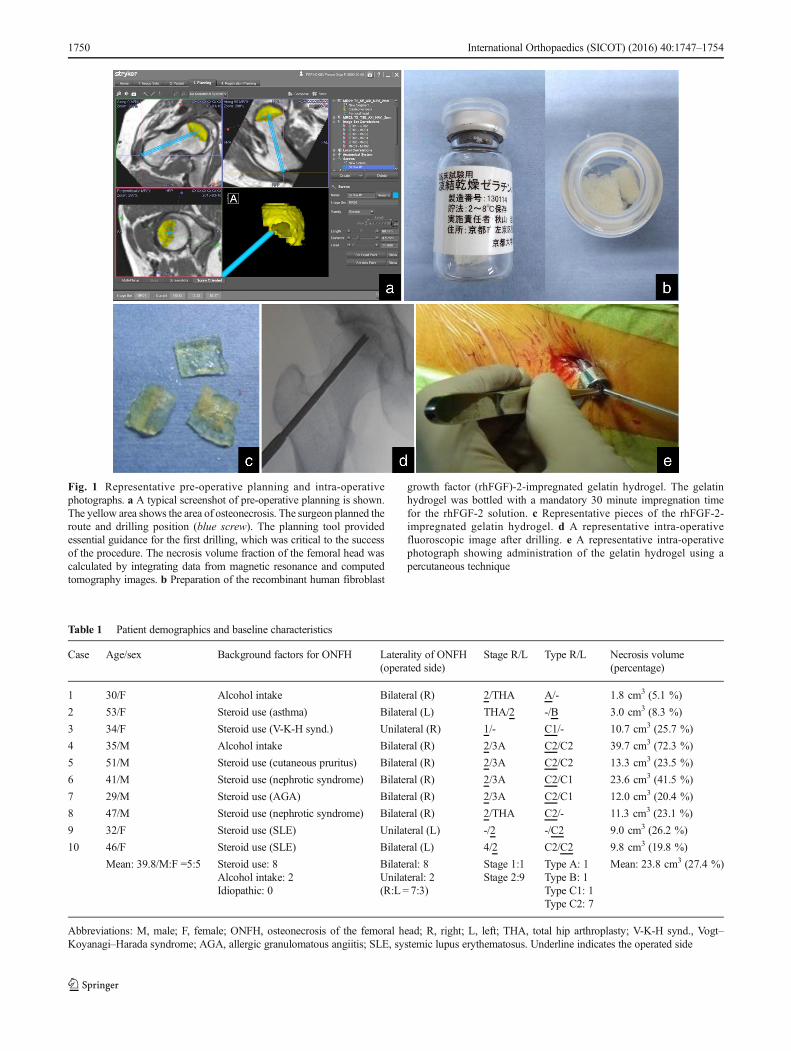
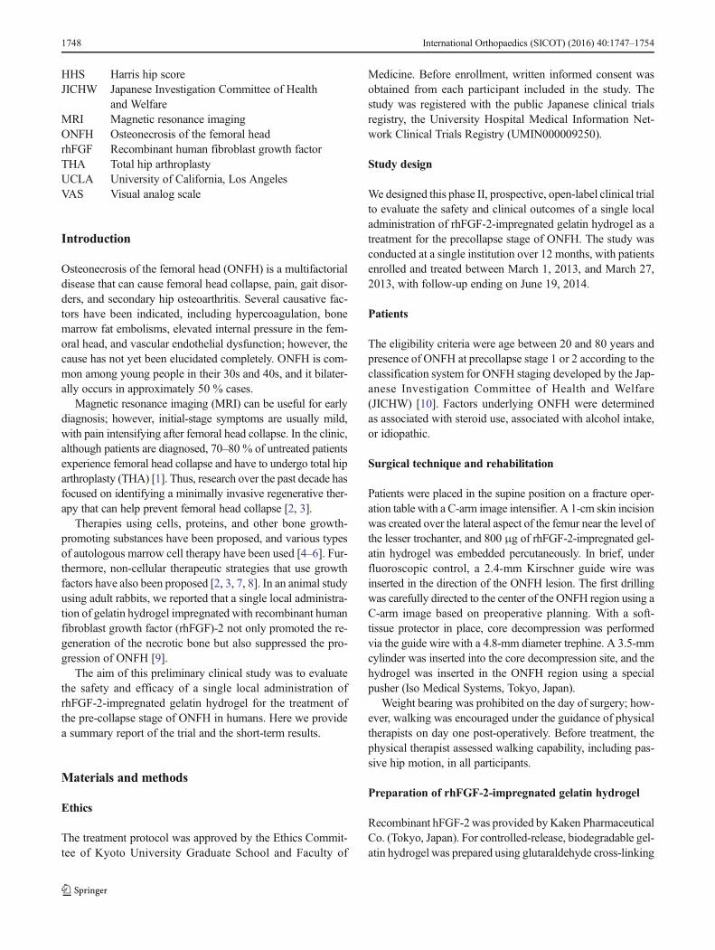
Fig. 1 Representative pre-operative planning and intra-operativephotographs. a A typical screenshot of pre-operative planning is shown.The yellow area shows the area of osteonecrosis. The surgeon planned theroute and drilling position (blue screw). The planning tool providedessential guidance for the first drilling, which was critical to the successof the procedure. The necrosis volume fraction of the femoral head wascalculated by integrating data from magnetic resonance and computedtomography images. b Preparation of the recombinant human fibroblast
growth factor (rhFGF)-2-impregnated gelatin hydrogel. The gelatinhydrogel was bottled with a mandatory 30 minute impregnation timefor the rhFGF-2 solution. c Representative pieces of the rhFGF-2-impregnated gelatin hydrogel. d A representative intra-operativefluoroscopic image after drilling. e A representative intra-operativephotograph showing administration of the gelatin hydrogel using apercutaneous technique
Table 1 Patient demographics and baseline characteristics
Case Age/sex Background factors for ONFH Laterality of ONFH(operated side)
Stage R/L Type R/L Necrosis volume(percentage)
1 30/F Alcohol intake Bilateral (R) 2/THA A/- 1.8 cm3 (5.1 %)
2 53/F Steroid use (asthma) Bilateral (L) THA/2 -/B 3.0 cm3 (8.3 %)
3 34/F Steroid use (V-K-H synd.) Unilateral (R) 1/- C1/- 10.7 cm3 (25.7 %)
4 35/M Alcohol intake Bilateral (R) 2/3A C2/C2 39.7 cm3 (72.3 %)
5 51/M Steroid use (cutaneous pruritus) Bilateral (R) 2/3A C2/C2 13.3 cm3 (23.5 %)
6 41/M Steroid use (nephrotic syndrome) Bilateral (R) 2/3A C2/C1 23.6 cm3 (41.5 %)
7 29/M Steroid use (AGA) Bilateral (R) 2/3A C2/C1 12.0 cm3 (20.4 %)
8 47/M Steroid use (nephrotic syndrome) Bilateral (R) 2/THA C2/- 11.3 cm3 (23.1 %)
9 32/F Steroid use (SLE) Unilateral (L) -/2 -/C2 9.0 cm3 (26.2 %)
10 46/F Steroid use (SLE) Bilateral (L) 4/2 C2/C2 9.8 cm3 (19.8 %)
Mean: 39.8/M:F =5:5 Steroid use: 8Alcohol intake: 2Idiopathic: 0
Bilateral: 8Unilateral: 2(R:L = 7:3)
Stage 1:1Stage 2:9
Type A: 1Type B: 1Type C1: 1Type C2: 7
Mean: 23.8 cm3 (27.4 %)
Abbreviations: M, male; F, female; ONFH, osteonecrosis of the femoral head; R, right; L, left; THA, total hip arthroplasty; V-K-H synd., Vogt–Koyanagi–Harada syndrome; AGA, allergic granulomatous angiitis; SLE, systemic lupus erythematosus. Underline indicates the operated side
1750 International Orthopaedics (SICOT) (2016) 40:1747–1754
and two patients overused alcohol. Stage 1 and 2 disease waspresent in one and nine patients, respectively. One patient eachhad type A, type B, and type C1 disease, whereas seven pa-tients had a type C2 lesion. Pre-operatively, the mean necrosisvolume and necrosis volume fraction (percentage of total tis-sue) was 23.8 cm3 (1.8–39.7 cm3) and 27.4 % (5.1 %–72.3 %), respectively.
Safety
We evaluated all patients over a one year follow-up period(Table 2). Fourteen AEs were experienced by five patientsduring the observation period. There were two serious AEs,classified to be of moderate severity (cases 4 and 10), in whichthe contralateral side was diagnosed as advancing to abovestage 3 and ONFH. However, these were not associated withthe clinical trial, and the patients had already been scheduledfor THA. Regarding causal relationships, one patient (case 10)was judged to have developed headache due to spinal anes-thesia. In all cases, the patients recovered from these AEs overthe follow-up period. There were no complications related tothe surgical technique.
Clinical outcomes
All procedures were performed under spinal anesthesia. Themean operation time was 18 minutes, the skin incision was1 cm, there was little bleeding, walking was encouraged fromday one post-operatively, and the mean hospital stay was6.2 days (range: 5–7 days). Embedding of the rhFGF-2-impregnated gelatin hydrogel in the necrotic area was con-firmed in all cases. The mean clinical scores improved signif-icantly by one year post-operatively (VAS for pain, 5.3 mm;
UCLA activity score, 6.6; HHS, 96.9 points) compared withpre-operative scores (VAS for pain, 21.2 mm; UCLA activityscore, 5.5; HHS, 81.0 points) (Table 3).
Radiographic evaluation
Although one patient (case 4) developed femoral head col-lapse (10 %) at the one year post-operative follow-up, thispatient had the greatest necrosis volume fraction. Moreover,based on post-operative CT images obtained at day one post-operatively, femoral head collapse was considered to haveprogressed from an early collapse that had already been pres-ent at the time of surgery.
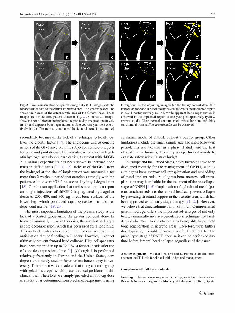
The remaining nine patients neither showed progression ofpre-operative ONFH nor did they exhibit femoral head col-lapse during the follow-up period. Changes in the necrosisvolume fraction and ONFH type were not observed in thenecrotic areas byMRI; however, there was evidence of homo-geneous low-signal areas in postoperative T1-weighted im-ages of the femoral heads (Fig. 2). In radiographs and CTscans, bone regeneration was observed in the necrotic regionsin all cases. Additional radiographic assessment using binaryformat images showed regeneration of the trabecular bonesand subchondral bone (Fig. 3).
Discussion
The minimally invasive treatment described in this studyaimed to prevent femoral head collapse by direct local admin-istration of rhFGF-2, which has both angiogenic and osteo-genic actions. To the best of our knowledge, there have been
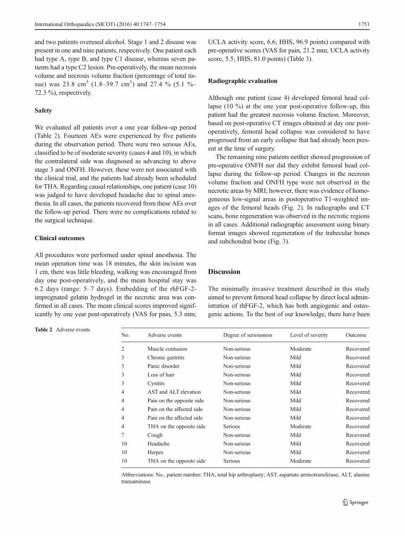
Table 2 Adverse eventsNo. Adverse events Degree of seriousness Level of severity Outcome
2 Muscle contusion Non-serious Moderate Recovered
3 Chronic gastritis Non-serious Mild Recovered
3 Panic disorder Non-serious Mild Recovered
3 Loss of hair Non-serious Mild Recovered
3 Cystitis Non-serious Mild Recovered
4 AST and ALT elevation Non-serious Mild Recovered
4 Pain on the opposite side Non-serious Mild Recovered
4 Pain on the affected side Non-serious Mild Recovered
4 Pain on the affected side Non-serious Mild Recovered
4 THA on the opposite side Serious Moderate Recovered
7 Cough Non-serious Mild Recovered
10 Headache Non-serious Mild Recovered
10 Herpes Non-serious Mild Recovered
10 THA on the opposite side Serious Moderate Recovered
Abbreviations: No., patient number; THA, total hip arthroplasty; AST, aspartate aminotransferase; ALT, alaninetransaminase.
International Orthopaedics (SICOT) (2016) 40:1747–1754 1751

no previous reports regarding the administration of rhFGF-2-impregnated gelatin hydrogel into the femoral head.
During the one year follow-up period, 14 AEs occurred infive patients, and all recovered without problems. There wereno complications related to either the surgical technique ortreatment. Moreover, all clinical scores related to pain, dailyactivity rating, and hip joint functions significantly improvedpost-operatively. Although one patient with stage 3A diseaseat surgery (case 4) developed early femoral head collapse thatwas present on CT images at day one post-operatively, no
other patients showed disease progression or femoral headcollapse. Thus, local administration of rhFGF-2 was safe.
Therapies using cells [4–6], proteins, and other bonegrowth-promoting substances [2, 7, 8] have been proposed,and various cell therapies using autologous marrow cells arealready being attempted. Non-cellular therapeutic strategiesusing growth factors have been proposed; however, therehas been little progress with regard to the verification of ani-mal experiments, primarily because of the absence of an ani-mal model for femoral head-specific necrosis [9, 16] and
Fig. 2 Representative magnetic resonance images of the central coronalplane in the femoral head. a Pre-operative coronal T1-weighted magneticresonance imaging (MRI) showing type C2 osteonecrosis of the femoralhead (ONFH) occupying more than the medial two-thirds of the weight-bearing portion and extending laterally to the acetabular edge. b MRI ofthe pre-operative plan described in Fig. 1. c MRI of the same femoralneck and intertrochanteric region at three months post-operatively,
showing low signal intensity, indicating the drilling route made duringthe surgical procedure. dMRI at six months post-operatively showing thefirst change of signal intensity in the drilling route. The signal intensity ofbone at the drilling site of the femoral neck is normalizing. e MRI at12 months post-operatively showing almost normal signal intensity inthe femoral neck. The area and size of ONFH remains unchanged fromtype 2. Abbreviations: M, months
Table 3 Clinical outcomesScore (range) Pre-operative Post-operative
3 monthsPost-operative6 months
Post-operative12 months
P value
VAS for pain(0–100)
21.2 ± 24.9 6.1 ± 7.8 5.3 ± 5.5 5.3 ± 8.6 0.00210.0 (0–84) 3.5 (0–25) 4.5 (0–14) 1.0 (0–27)
UCLA activity(1–10)
5.5 ± 1.0 6.1 ± 0.7 6.1 ± 0.7 6.6 ± 0.5 <0.0016 (4–7) 6 (5–7) 6 (5–7) 7 (6–7)
HHS (0–100) 81.0 ± 18.1 94.8 ± 4.0 94.5 ± 7.9 96.9 ± 5.0 <0.00191.9 (44.9–
96.0)95.0 (88.0–100.0) 97.0 (74.0–100.0) 100.0 (85.9–
100.0)
Data for each score are expressed as mean ± standard deviation (the upper rows) and median range (the bottomrows). The P value was calculated for the effect of time in a repeated-measures linear mixed-effect model.Abbreviations: VAS, visual analog scale; UCLA, University of California, Los Angeles activity; HHS, Harriship score
1752 International Orthopaedics (SICOT) (2016) 40:1747–1754
secondarily because of the lack of a technique to locally de-liver the growth factor [17]. The angiogenic and osteogenicactions of rhFGF-2 have been the subject of numerous reportsfor bone and joint disease. In particular, when used with gel-atin hydrogel as a slow-release carrier, treatment with rhFGF-2 in animal experiments has been shown to increase bonemass in deficit areas [9, 11, 12]. Release of rhFGF-2 fromthe hydrogel at the site of implantation was measurable formore than 2 weeks, a period that correlates strongly with thepatterns of in vivo rhFGF-2 release and hydrogel degradation[18]. One human application that merits attention is a reporton single injections of rhFGF-2-impregnated hydrogel atdoses of 200, 400, and 800 μg in cut bone surfaces of thelower leg, which produced rapid synostosis in a dose-dependent manner [19, 20].
The most important limitation of the present study is thelack of a control group using the gelatin hydrogel alone. Interms of minimally invasive therapies, the simplest techniqueis core decompression, which has been used for a long time.This method creates a burr hole in the femoral head with theanticipation that self-healing will occur; however, it cannotultimately prevent femoral head collapse. High collapse rateshave been reported in up to 72.7 % of femoral heads after useof core decompression alone [5]. Although it is performedrelatively frequently in Europe and the United States, coredepression is rarely used in Japan unless bone biopsy is nec-essary. Therefore, it was considered that using a control groupwith gelatin hydrogel would present ethical problems in thisclinical trial. Therefore, we simply provided an 800-μg doseof rhFGF-2, as determined from preclinical experiments using
an animal model of ONFH, without a control group. Otherlimitations include the small sample size and short follow-upperiod; this was because, as a phase II study and the firstclinical trial in humans, this study was performed mainly toevaluate safety within a strict budget.
In Europe and the United States, novel therapies have beendeveloped recently for the management of ONFH, such asautologous bone marrow cell transplantation and embeddingof metal implant rods. Autologous bone marrow cell trans-plantation may be reliable for the treatment of the precollapsestage of ONFH [4–6]. Implantation of cylindrical metal (po-rous tantalum) rods into the femoral head can prevent collapseby providing structural support in the necrotic area, which hasbeen approved as an early-stage therapy [21, 22]. However,we believe that direct administration of rhFGF-2-impregnatedgelatin hydrogel offers the important advantages of not onlybeing a minimally invasive percutaneous technique that facil-itates early return to society but also being able to promotebone regeneration in necrotic areas. Therefore, with furtherdevelopment, it could become a useful treatment for theprecollapse stage of ONFH because it can be performed anytime before femoral head collapse, regardless of the cause.
Acknowledgments We thank M. Doi and K. Enomoto for data man-agement and T. Ikeda for clinical trial design and management.
Compliance with ethical standards
Funding This work was supported in part by grants from TranslationalResearch Network Program by Ministry of Education, Culture, Sports,
Fig. 3 Two representative computed tomography (CT) images with thebinary format data of the central implanted area. The yellow dashed lineshows the border of the osteonecrotic area of the femoral head. Theseimages are for the same patient shown in Fig. 2a. Coronal CT imagesshow the bone defect at the implanted region at day one post-operatively(a, b), and apparent bone regeneration is observed one year post-opera-tively (c, d). The normal contour of the femoral head is maintained
throughout. In the adjoining images for the binary format data, thintrabecular bone and subchondral bone can be seen in the implanted regionat day 1 postoperatively (a′, b′), while apparent bone regeneration isobserved in the implanted region at one year post-operatively (yellowarrows, c′, d′). Clear, normal-contour, thick trabecular bone and thicksubchondral bone (yellow arrowheads) can be observed
International Orthopaedics (SICOT) (2016) 40:1747–1754 1753
Science and Technology of Japan and The Uehara Memorial Foundation(to A.H.).
Conflict of interest The authors declare that they have no conflict ofinterest.
Open Access This article is distributed under the terms of the CreativeCommons At t r ibut ion 4 .0 In te rna t ional License (h t tp : / /creativecommons.org/licenses/by/4.0/), which permits unrestricted use,distribution, and reproduction in any medium, provided you give appro-priate credit to the original author(s) and the source, provide a link to theCreative Commons license, and indicate if changes were made.
References
1. Hernigou P, Ooignard A,Nogier A,ManicomO (2004) Fate of verysmall asymptomatic stage-I osteonecrotic lesions of the hip. J BoneJoint Surg Am 86:2589–2593
2. Mont MA, Jones LC, Hngerford DS (2006) Nontraumaticosteonecrosis of the femoral head: Ten years later. J Bone JointSurg Am 88:1117–1132, Review Erratum in: J Bone Joint SurgAm 88A:1602
3. Marker DR, Seylee TM, McGrath MS, Delanois RE, Ulrich SD,Mont MA (2008) Treatment of early stage osteonecrosis of thefemoral head. J Bone Joint Surg Am 90:175–187
4. Hernigou P, Flouzat-Lachaniette CH, Delambre J, Poignard A,Allain J, Chevallier N, Rouard H (2015) Osteonecrosis repair withbone marrow cell therapies: State of the clinical art. Bone 70:102–109
5. Gangji V, De Maertelaer V, Hauzeur JP (2011) Autologous bonemarrow cell implantation in the treatment of non-traumaticosteonecrosis of the femoral head: five year follow-up of a prospec-tive controlled study. Bone 49:1005–1009
6. Houdek MT, Wyles CC, Martin JR, Sierra RJ (2014) Stem celltreatment for avascular necrosis of the femoral head: current per-spectives. Stem Cells Clon 7:65–70
7. Lieberman JR, Conduah A, Urist MR (2004) Treatment ofosteonecrosis of the femoral head with core decompression andhuman bone morphogenetic protein. Clin Orthop Relat Res 429:139–145
8. Sun W, Li Z, Gao F, Shi Z, Zhang Q, Guo W (2014) Recombinanthuman bone morphogenetic protein-2 in debridement and impactedbone graft for the treatment of femoral head osteonecrosis. PLoSOne 9:e100424
9. Kuroda Y, AkiyamaH, Kawanabe K, Tabata Y, Nakamura T (2010)Treatment of experimental osteonecrosis of the hip in adult rabbitswith a single local injection of recombinant human FGF-2 micro-spheres. J Bone Miner Metab 28:608–616
10. Sugano N, Atsumi T, Ohzono K, Kubo T, Hotokebuchi T, TakaokaK (2002) The 2001 revised criteria for diagnosis, classification, and
staging of idiopathic osteonecrosis of the femoral head. J OrthopSci 7:601–605
11. Tabata Y, Ikada Y (1999) Vascularization into a porous sponge bysustained release of basic fibroblast growth factor. Biomaterials 20:2169–2175
12. Tabata Y, Yamada K, Hong L, Miyamoto S, Hashimoto N, Ikada Y(1999) Skull bone regeneration in primates in response to basicfibroblast growth factor. J Neurosurg 91:851–856
13. Amstutz HC, Thomas BJ, Jinnah R, Kim W, Grogan T, Yale C(1984) Treatment of primary osteoarthritis of the hip. A comparisonof total joint and surface replacement arthroplasty. J Bone JointSurg Am 66:228–241
14. Harris WH (1969) Traumatic arthritis of the hip after dislocationand acetabular fractures: treatment by mold arthroplasty. An end-result study using a new method of result evaluation. J Bone JointSurg Am 51-A:737–755
15. Cnaan A, Laird NM, Slasor P (1997) Using the general linear mixedmodel to analyse unbalanced repeated measures and longitudinaldata. Stat Med 16:2349–2380
16. Poignard A, Lebouvier A, Cavet M, Rahmouni A, FlouzatLachaniette CH, Bierling P, Rouard H, Hernigou P, Chevallier N(2014) New preclinical porcine model of femoral headosteonecrosis to test mesenchymal stromal cell efficiency in regen-erative medicine. Int Orthop 38(9):1837–1844
17. Cheng TL, Murphy CM, Cantrill LC, Mikulec K, Carpenter C,Schindeler A, Little DG (2014) Local delivery of recombinant hu-man bone morphogenetic proteins and bisphosphonate via sucroseacetate isobutyrate can prevent femoral head collapse in Legg-Calve-Perthes disease: a pilot study in pigs. Int Orthop 38(7):1527–1533
18. Yamamoto M, Ikeda Y, Tabata Y (2001) Controlled release ofgrowth factors based on biodegradation of gelatin hydrogel. JBiomater Sci Polym Ed 12:77–88
19. Kawaguchi H, Jingushi S, Izumi M, Fukunaga M, Matsushita T,Nakamura T, Mizuno K, Nakamura T, Nakamura K (2007) Localapplication of recombinant human fibroblast growth factor-2 onbone repair: a dose-escalation prospective trial on patients withosteotomy. J Orthop Res 25:480–487
20. Kawaguchi H, Oka H, Jingushi S, Izumi T, Fukunaga M, Sato K,Matsushita T, Nakamura K, TESK Group (2010) A local applica-tion of recombinant human fibroblast growth factor 2 for tibial shaftfractures: a randomized, placebo-controlled trial. J BoneMiner Res25:2735–2743
21. Tsao AK, Roberson JR, Christie MJ, Dore DD, Heck DA,Robertson DD, Poggie RA (2005) Biomechanical and clini-cal evaluations of a porous tantalum implant for the treat-ment of early-stage osteonecrosis. J Bone Joint Surg Am87(Suppl 2):22–27
22. Malizos KN, Papasoulis E, Dailiana ZH, Papatheodorou LK,Varitimidis SE (2012) Early results of a novel technique usingmultiple small tantalum pegs for the treatment of osteonecrosis ofthe femoral head: a case series involving 26 hips. J Bone Joint Surg(Br) 94:173–178
1754 International Orthopaedics (SICOT) (2016) 40:1747–1754