86(3) - SciELO · Arch Cardiol Mex. 2016;86(3):221---232 CLINICAL RESEARCH Clinical management and...
12
Arch Cardiol Mex. 2016;86(3):221---232 www.elsevier.com.mx CLINICAL RESEARCH Clinical management and hospital outcomes of acute coronary syndrome patients in Mexico: The Third National Registry of Acute Coronary Syndromes (RENASICA III) Carlos Martinez-Sanchez a,c , Gabriela Borrayo a,d , Jorge Carrillo a,e , Ursulo Juarez a,f , Juan Quintanilla a,g , Carlos Jerjes-Sanchez a,b,h,i,* , on the behalf of RENASICA III Investigators ♦ a Executive Committee of RENASICA III, Mexico b National Coordinator, Mexico c Emergency Department and Coronary Critical Care, Instituto Nacional de Cardiología ‘‘Ignacio Chavez’’, Mexico d Hospital de Cardiologia, Centro Medico Nacional Siglo XXI, Mexico e Cardiology Department in Hospital Central Dr. Ignacio Morones Prieto, San Luis Potosi, Mexico f Instituto Nacional de Cardiologia Ignacio Chavez, Mexico g Hemodynamic Laboratory, Hospital San Jose and Instituto de Cardiologia y Medicina Vascular, TEC Salud, Mexico h Instituto de Cardiologia y Medicina Vascular, TEC Salud, Mexico i Translational Research Center, Escuela de Medicina, Tecnologico de Monterrey, Mexico Received 25 December 2015; accepted 22 April 2016 KEYWORDS Acute coronary syndromes; ST-elevation myocardial infarction; Non-ST elevation myocardial infarction; Unstable angina; Mexico Abstract Objective: To describe current management and clinical outcomes in patients hospitalized with an acute coronary syndrome (ACS) in Mexico. Methods: RENASICA III was a prospective multicenter registry of consecutive patients hospital- ized with an ACS. Patients had objective evidence of ischemic heart disease; those with type II infarction or secondary ischemic were excluded. Study design conformed to current quality recommendations. Results: A total of 123 investigators at 29 tertiary and 44 community hospitals enrolled 8296 patients with an ACS (4038 with non-ST-elevation myocardial infarction/unstable angina [NSTEMI/UA], 4258 with ST-elevation myocardial infarction [STEMI]). The majority were younger (62 ± 12 years) and 76.0% were male. On admission 80.5% had ischemic chest pain lasting >20 min and clinical stability. Left ventricular dysfunction was more frequent in NSTEMI/UA than in those Clinicaltrials.gov: NCT02141750. ∗ Corresponding author at: Centro de Investigacion Traslacional. Escuela de Medicina, Tecnologico de Monterrey, San Patricio, San Pedro Garza Garcia, NL, Mexico. Tel.: +52 81 8888 0000. E-mail addresses: [email protected], [email protected] (C. Jerjes-Sanchez). ♦ A list of the RENASICA III investigators is provided in Appendix A. http://dx.doi.org/10.1016/j.acmx.2016.04.007 1405-9940/© 2016 Instituto Nacional de Cardiolog´ ıa Ignacio Ch´ avez. Published by Masson Doyma M´ exico S.A. This is an open access article under the CC BY-NC-ND license (http://creativecommons.org/licenses/by-nc-nd/4.0/).
Transcript of 86(3) - SciELO · Arch Cardiol Mex. 2016;86(3):221---232 CLINICAL RESEARCH Clinical management and...

Arch Cardiol Mex. 2016;86(3):221---232
www.elsevier.com.mx
CLINICAL RESEARCH
Clinical management and hospital outcomes of acute
coronary syndrome patients in Mexico: The Third
National Registry of Acute Coronary Syndromes
(RENASICA III)�
Carlos Martinez-Sanchez a,c, Gabriela Borrayo a,d, Jorge Carrillo a,e, Ursulo Juarez a,f,Juan Quintanilla a,g, Carlos Jerjes-Sanchez a,b,h,i,∗, on the behalf of RENASICA IIIInvestigators♦
a Executive Committee of RENASICA III, Mexicob National Coordinator, Mexicoc Emergency Department and Coronary Critical Care, Instituto Nacional de Cardiología ‘‘Ignacio Chavez’’, Mexicod Hospital de Cardiologia, Centro Medico Nacional Siglo XXI, Mexicoe Cardiology Department in Hospital Central Dr. Ignacio Morones Prieto, San Luis Potosi, Mexicof Instituto Nacional de Cardiologia Ignacio Chavez, Mexicog Hemodynamic Laboratory, Hospital San Jose and Instituto de Cardiologia y Medicina Vascular, TEC Salud, Mexicoh Instituto de Cardiologia y Medicina Vascular, TEC Salud, Mexicoi Translational Research Center, Escuela de Medicina, Tecnologico de Monterrey, Mexico
Received 25 December 2015; accepted 22 April 2016
KEYWORDSAcute coronarysyndromes;ST-elevationmyocardialinfarction;Non-ST elevationmyocardialinfarction;Unstable angina;Mexico
Abstract
Objective: To describe current management and clinical outcomes in patients hospitalized withan acute coronary syndrome (ACS) in Mexico.Methods: RENASICA III was a prospective multicenter registry of consecutive patients hospital-ized with an ACS. Patients had objective evidence of ischemic heart disease; those with typeII infarction or secondary ischemic were excluded. Study design conformed to current qualityrecommendations.Results: A total of 123 investigators at 29 tertiary and 44 community hospitals enrolled8296 patients with an ACS (4038 with non-ST-elevation myocardial infarction/unstable angina[NSTEMI/UA], 4258 with ST-elevation myocardial infarction [STEMI]). The majority were younger(62 ± 12 years) and 76.0% were male. On admission 80.5% had ischemic chest pain lasting >20 minand clinical stability. Left ventricular dysfunction was more frequent in NSTEMI/UA than in those
� Clinicaltrials.gov: NCT02141750.∗ Corresponding author at: Centro de Investigacion Traslacional. Escuela de Medicina, Tecnologico de Monterrey, San Patricio, San Pedro
Garza Garcia, NL, Mexico. Tel.: +52 81 8888 0000.E-mail addresses: [email protected], [email protected] (C. Jerjes-Sanchez).
♦ A list of the RENASICA III investigators is provided in Appendix A.
http://dx.doi.org/10.1016/j.acmx.2016.04.0071405-9940/© 2016 Instituto Nacional de Cardiologıa Ignacio Chavez. Published by Masson Doyma Mexico S.A. This is an open access articleunder the CC BY-NC-ND license (http://creativecommons.org/licenses/by-nc-nd/4.0/).
222 C. Martinez-Sanchez et al.
with STEMI (30.0% vs. 10.7%, p < 0.0001). In STEMI 37.6% received thrombolysis and 15.0% pri-mary PCI. PCI was performed in 39.6% of NSTEMI/UA (early strategy in 10.8%, urgent strategy in3.0%). Overall hospital death rate was 6.4% (8.7% in STEMI vs. 3.9% in NSTEMI/UA, p < 0.001). Thestrongest independent predictors of hospital mortality were cardiogenic shock (odds ratio 22.4,95% confidence interval 18.3---27.3) and ventricular fibrillation (odds ratio 12.5, 95% confidenceinterval 9.3---16.7).Conclusion: The results from RENASICA III establish the urgent need to develop large-scaleregional programs to improve adherence to guideline recommendations in ACS, including ratesof pharmacological thrombolysis and increasing the ratio of PCI to thrombolysis.© 2016 Instituto Nacional de Cardiologıa Ignacio Chavez. Published by Masson Doyma MexicoS.A. This is an open access article under the CC BY-NC-ND license (http://creativecommons.org/licenses/by-nc-nd/4.0/).
PALABRAS CLAVESíndromes coronariosagudos;Infarto del miocardiocon elevación del ST;Infarto del miocardiosin elevación del ST;Angina inestable;México
Abordaje clínico y evolución hospitalaria en pacientes mexicanos con sindrome
coronario agudo: El Tercer Registro Nacional de Síndromes Coronarios Agudos
(RENASICA III)
Resumen
Objetivo: describir abordaje terapéutico actual y evolución en pacientes hospitalizados con unsíndrome coronario agudo (SCA) en México.Métodos: RENASICA III registro multicéntrico prospectivo de pacientes consecutivos con un SCA.Todos tuvieron demostración objetiva de enfermedad coronaria; se excluyeron infarto tipo II oisquemia secundaria. El diseno incluyó recomendaciones actuales de calidad.Resultados: 123 investigadores en 29 hospitales de tercer nivel y en 44 de segundo ingresaron8296 pacientes, 4038 con infarto del miocardio sin elevación del ST/angina inestable (IMSEST/AI)y 4258 con infarto del miocardio y elevación del ST (IMEST). La mayoría fueron jóvenes (62 ± 12anos) y el 76% del sexo masculino. Al ingreso 80.5% tuvo dolor torácico con perfil isquémico >20minutos y estabilidad clínica. Se observó mayor disfunción ventricular en grupo con IMSEST/AIque en aquellos con IMEST (30.0% vs 10.7%, p <0.0001). En IMEST el 37.6% recibió trombolisisy el 15% angioplastía primaria. Este procedimiento se realizó en el 39.6% de los pacientes conIMSEST/AI (estrategia temprana 10.8%, estrategia urgente 3.0%). La mortalidad hospitalaria fuedel 6.4% (8.7% IMEST vs. 3.9% IMSEST/AI, p <0.001). Los predictores independientes con mayorpoder para mortalidad fueron choque cardiogénico (RM 22.4, 95% IC 18.3---27.3) y fibrilaciónventricular (RM 12.5, 95% IC 9.3---16.7).Conclusión: los resultados del RENASICA III establecen la urgente necesidad de desarrollar enSCA programas regionales a gran escala para mejorar el apego a la guías y recomendaciones,incluyendo mayor porcentaje de trombolisis e incrementar la proporción de angioplastia pri-maria.© 2016 Instituto Nacional de Cardiologıa Ignacio Chavez. Publicado por Masson Doyma MexicoS.A. Este es un artıculo Open Access bajo la CC BY-NC-ND licencia (http://creativecommons.org/licencias/by-nc-nd/4.0/).
Introduction
Knowledge of the epidemiological characteristics, therapeu-tic trends, and risk stratification of patients presenting withacute coronary syndromes (ACS) in Mexico is derived fromthe national registries: RENASICA I (Registro Nacional deSindromes Coronarios Agudos),1 RENASICA II,2 and RENASCA(National Registry of patients with ACS in the IMSS).3 Theresults from the ACCESS (ACute Coronary Events --- a multi-national Survey of current management Strategies) registry,4
conducted in 11,731 ACS patients in Africa, Latin Amer-ica, and the Middle East, confirmed the results from thenational Mexican registries,1---3 highlighting persistent under-use of reperfusion therapies.
In the current era of therapeutic transition in ACS, itis important to identify changes in practice in terms of
reperfusion approaches and antithrombotic strategies.RENASICA III was a prospective observational Mexican reg-istry, the aims of which are to describe current managementand clinical outcomes in patients with the broad spectrum ofACS types treated in everyday clinical practice. The resultsfrom RENASICA III will expand on the information providedby RENASICA I and RENASICA II.
Methods
Registry design
RENASICA III was established by the Mexican Cardiology Soci-ety, with unrestricted support provided by Sanofi. The fullprotocol, including definitions, has been published.5 In brief,

Clinical management and hospital outcomes of acute coronary syndrome patients in Mexico 223
RENASICA III was a prospective, multicenter registry involv-ing adult men and women hospitalized with ACS in Mexico.In-hospital data were collected from patients in tertiaryand community hospitals across both the public and pri-vate healthcare systems. The principal investigators wereselected from community and tertiary hospitals locatedthroughout Mexico.
The study population comprised a consecutive, prospec-tive cohort of patients with a high clinical suspicion of ACSwho were admitted to hospital. The initial diagnosis wasestablished by the physician-in-charge on the basis of thepatient’s clinical and electrocardiographic characteristics.Four groups of patients were identified according to theelectrocardiographic findings: normal or non-specific elec-trocardiogram (ECG), ST depression, transient ST elevation,6
and persistent ST elevation. Patients with a final diag-nosis of ACS and objective evidence of ischemic heartdisease (identified using invasive or non-invasive tests) wereincluded in the registry and those with type II infarctionwere excluded. ACS nomenclature (ST-elevation myocardialinfarction [STEMI] and non---ST-elevation myocardial infarc-tion [NSTEMI] or unstable angina [UA]) was used at admissionand, for epidemiologic reasons, UA or myocardial infarctionat discharge.
The registry structure, data collection, and data analysiswere based on current quality recommendations, includ-ing 11 criteria proposed by Gitt et al.7 The registry wasconducted in accordance with national and internationalregulations. The protocol was approved by the institu-tional ethics committees in all participating centers and allpatients provided informed consent.
The data were sent to the registry-coordinating cen-ter via a web site (http://www.renasica.mx/). Electroniccase report forms were reviewed by the coordinating centerto determine data quality. All participating sites had reg-ular access to a data-entry clerk. Data-quality monitoringwas conducted at 20% of the centers, chosen at random.Technical support was provided through periodic softwareupdates. Investigators at all sites underwent training inthe use of the electronic database and in the study defi-nitions, and had real-time access to technical advice fromthe coordinating center through a 01-800 number. Follow-upby telephone and during site visits was performed throughthe coordinating center. In specific cases follow-up was doneby the physician-in-charge. An executive committee set outthe publication policies to allow an expedited process forpublication or presentation at national and internationalmeetings.
Sample size determination
A sample size with statistical significance was obtained usingdata from RENASCA3 and RENASICA II.2 Fifty percent of thepatients in RENASCA3 did not receive adequate treatmentand only 8% underwent percutaneous coronary intervention(PCI); consequently, 30% of patients developed left ven-tricular dysfunction. In RENASICA II, 15% of the patientshad PCI and 17% developed left ventricular failure as anearly complication. This difference between the two studiesallowed us to calculate the smaller sample size necessary toevaluate this outcome (i.e. left ventricular dysfunction). A
sample of 182 patients in each group (STEMI and NSTEMI/UA)would give a statistical power of 80%, with an alpha valueof 0.05, delta value of 13%, 1:1 ratio of exposed: unex-posed, and a confidence interval (CI) of 95%. Allowing foran estimated 20% loss to follow-up, the final number ofpatients chosen per group was 220. A lower delta value formortality of 2% required 3975 patients to be treated (ornot) with PCI; thus 8000 subjects had to be enrolled in theregistry.
Statistical analysis
Data are expressed as percentages, mean ± standard devia-tion (SD), and odds ratios (ORs) with 95% CIs. Differencesbetween continuous variables with a normal distributionwere examined using Student’s t test. The Wilcoxon rank-sum test was used for non-normal continuous variables. Thechi-square test, Fisher’s exact test, or Yates’ correction wasused to analyze categorical variables, as appropriate. A two-tailed test with a p-value ≤0.05 was considered statisticallysignificant. Logistic regression analysis was used to identifyindependent predictors of hospital mortality on the basisof those with a p-value of ≤0.05 on univariate regressionanalysis, with adjustment for potential confounders to avoidconfusion, the relationship between historical variables foratherosclerosis and cardiovascular events was examinedthrough logistic regression. With the Cox proportional riskmultivariable model, the relationship between each of thesevariables was analyzed. Kaplan---Meier survival curves wereused to determine the risk of mortality, and differencesbetween the curves were determined using the log-rankstatistic. The Cox proportional risk model was used foradjusted survival analysis.
Results
Study population
Between November 2012 and November 2013, 8296 consec-utive ACS patients with proven ischemic heart disease wereenrolled by 123 investigators at 29 tertiary and 44 commu-nity hospitals (63% government health system centers, 29%private practices, and 8% other health systems; hospitalswere located across 91% of the Mexican territory). Of thesepatients, 4038 were diagnosed with NSTEMI/UA and 4258with STEMI. All tertiary hospitals had the capability to per-form percutaneous coronary intervention (PCI) and coronaryartery bypass graft (CABG) surgery.
The mean ± SD age of the population was 62.0 ± 12years, 73.8% were male, and the mean body mass indexwas 27.8 ± 4.0 kg/m2 (Table 1). The prevalence of majorrisk factors for ischemic heart disease was high in bothACS groups. Patients with STEMI were younger than thosewith NSTEMI/UA and a higher percentage smoked tobacco.Patients with NSTEMI/UA were more likely to have angina,dyslipidemia, heart failure, hypertension, previous myocar-dial infarction, or to have undergone a previous PCI or CABG(all p < 0.0001).

224 C. Martinez-Sanchez et al.
Table 1 Characteristics of study population, overall and according to type of acute coronary syndrome.
Characteristic All patientsn = 8296
NSTEMI/UAn = 4038
STEMIn = 4258
P-valuea
Age, mean ± SD, years 62 ± 12 63.1 ± 11.7 61.4 ± 11.9 <0.0001Men, % 73.8 68.5 78.9 <0.0001Weight, mean ± SD, kg 76.0 ± 12 75.5 ± 11.9 76.4 ± 12.1 0.001Body mass index, mean ± SD, kg/m2 27.8 ± 4 27.9 ± 3.9 27.8 ± 4 0.457
Medical history, %
Hypertension 62.1 68.2 56.3 <0.0001Smoking 53.3 49.6 56.8 <0.0001Diabetes 45.8 46.3 45.3 0.355Dyslipidemia 36.9 39.5 34.4 <0.0001Myocardial infarction 25.5 33.3 18.2 <0.0001Angina 22.1 28.7 15.9 <0.0001Heart failure 2.3 3.2 1.4 <0.0001Cerebro vascular event/TIA 2.3 2.4 2.2 0.660Percutaneous coronary intervention 12.1 16.5 7.9 <0.0001Coronary artery bypass graft 3.3 5.0 1.6 <0.0001
Clinical characteristics, %
Chest pain on admission
Ischemic profile 86.0 83.8 88.4 <0.0001Non-ischemic profile 13.8 16.2 11.6 <0.0001
Duration, minutes, %
<20 19.5 26.0 13.4 <0.0001>20 80.5 74.0 86.6 <0.0001
Killip class II---IV 20.7 30.0 10.7 <0.0001SBP, mean ± SD, mmHg 124.1 ± 26 126.8 ± 25.4 121.7 ± 26.3 <0.0001Heart rate, mean ± SD, beats/min 77.7 ± 29.9 76.8 ± 34.4 78.6 ± 24.8 0.013Jugular distention, % 14.7 13.5 15.9 0.003Third heart sound, % 6.9 6.2 7.4 0.033Normal peripheral pulses, % 90.1 91.3 88.9 <0.001
Electrocardiogram findings, %
Anterior ST-elevation 24.8 9.9 38.9 <0.0001Inferior ST-elevation 20.3 7.2 32.7 <0.0001Lateral ST-elevation 10.8 3.8 17.6 <0.0001Anterior ST depression 11.1 15.0 7.5 <0.0001Inferior ST depression 5.2 6.5 3.9 <0.0001Lateral ST depression 10.5 14.2 7.0 <0.0001Atrioventricular block 10.7 7.0 14.3 <0.0001Right bundle branch block 6.8 7.5 6.2 0.030Left bundle branch block 5.6 6.5 4.7 <0.0001Ventricular tachycardia 0.6 0.7 0.4 0.041Ventricular fibrillation 0.3 0.2 0.4 0.154Atrial fibrillation 1.6 1.9 1.2 0.008Supraventricular tachycardia 0.4 0.3 0.4 0.600
a P-value comparing NSTEMI/UA with STEMI by X2 and Student’s t-test.SD, standard deviation; TI, transient ischemic attack.
Presenting symptoms, clinical characteristics, andbiomarkers
On admission 80.5% of patients had ischemic chest pain last-ing >20 min and clinical stability (Table 1). Killip class II---IV5
(Appendix B) was more frequent in patients with NSTEMI/UAthan in those with STEMI (p < 0.0001). Mitral and septal rup-ture murmur and cardiac rub were infrequent in NSTEMI/UA(1.5%, 0.1%, 0.1%, respectively) and STEMI (1.1%, 0.2%,0.1%).
Patients with STEMI more likely have had anterior, infe-rior, and lateral ST-elevation, whereas anterior, inferior, andlateral ST depressions were more frequent in patients withNSTEMI/UA (Table 1). The prevalence of atrial fibrillationwas low in both groups, although it was numerically morefrequent in patients with NSTEMI/UA.
The plasma concentrations of various biomarkers, includ-ing hemoglobin, hematocrit, leucocytes, glucose, creatinephosphokinase, CK-MB, troponin I and NT-proBNP, werehigher in patients with STEMI compared to those with
Clinical management and hospital outcomes of acute coronary syndrome patients in Mexico 225
Table 2 Biomarker measurements.
Biomarker All patientsn = 8296
NSTEMI/UAn = 4038
STEMIn = 4258
P-valuea
Hemoglobin, mg/dL 14.1 ± 3.0 13.9 ± 3.3 14.3 ± 2.7 <0.0001Hematocrit 41.4 ± 7.6 40.9 ± 7.4 41.8 ± 7.7 <0.0001Platelets, t/m 238.4 ± 80.5 240.3 ± 80.4 236.7 ± 80.5 0.075Leucocytes, miles/mm3 10.4 ± 5.0 9.5 ± 4.7 11.3 ± 5.1 <0.0001Glucose, mg/dL 170.5 ± 103.7 158.5 ± 98.8 181.2 ± 106.7 <0.0001Urea, mg/dL 41.6 ± 26.9 42.7 ± 29.2 40.7 ± 24.5 0.010Creatinine, mg/dL 1.3 ± 1.2 1.3 ± 1.2 1.2 ± 1.1 0.073CPK, U/Lb 217.0 (90.0---720.0) 131.0 (66.0---327.0) 374.0 (130.0---1154.4) <0.0001CK-MB, U/Lb 29.0 (14.8---79.0) 20.0 (11.1---43.0) 44 (20.0---120.0) <0.0001Troponin I, ng/mLb 2.5 (0.28---14.3) 1.1 (0.1---7.9) 6.1 (0.8---19.3) 0.004BNP, pg/mLc 191.0 (53.2---469.0) 184.5 (43.8---457.0) 197.0 (61.4---501.0) 0.439Pro-BNP, pg/mLc 251.0 (182.0---453.8) 242.0 (179.5---381.5) 261.0 (187.5---642.5) 0.001Fibrinogen, mg/dL 492.4 ± 193.1 484.0 ± 194.8 498.9 ± 191.5 0.155
a P-value comparing NSTEMI/UA with STEMI by Student’s t-test.Data given as mean ± standard deviation.
b Data given as median (percentiles 25---75) by Mann---Whitney U test.c pg/mL, picograms per milliliter.CK-MB, creatinine kinase MB fraction; CPK, total creatinine phosphokinase; BNP, brain natriuretic peptide; Pro-BNP, pro-brain natriureticpeptide --- N terminal. NSTEMI, non-ST-segment elevation myocardial infarction; STEMI, ST-segment elevation myocardial infarction; UA,unstable angina; UL, units per liter.
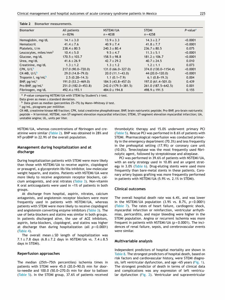
NSTEMI/UA, whereas concentrations of fibrinogen and cre-atinine were similar (Table 2). BNP was obtained in 28% andNT-proBNP in 22.9% of the overall population.
Management during hospitalization and atdischarge
During hospitalization patients with STEMI were more likelythan those with NSTEMI/UA to receive aspirin, clopidogrelor prasugrel, a glycoprotein IIb/IIIa inhibitor, low-molecularweight heparin, and statins. Patients with NSTEMI/UA weremore likely to receive angiotensin receptor blockers, cal-cium antagonists, and oral nitrates (Table 3). Non-vitaminK oral anticoagulants were used in <1% of patients in bothgroups.
At discharge from hospital, aspirin, nitrates, calciumantagonists, and angiotensin receptor blockers were morefrequently used in patients with NSTEMI/UA, whereaspatients with STEMI were more likely to receive clopidogreland angiotensin converting enzyme inhibitors (Table 3). Theuse of beta-blockers and statins was similar in both groups.In patients discharged alive, the use of ACE inhibitors,aspirin, beta-blockers, clopidogrel, and statins was higherat discharge than during hospitalization (all p < 0.0001)(Table 4).
The overall mean ± SD length of hospitalization was7.1 ± 7.8 days (6.8 ± 7.2 days in NSTEMI/UA vs. 7.4 ± 8.5days in STEMI).
Reperfusion approaches
The median (25th---75th percentiles) ischemia times inpatients with STEMI were 45.0 (20.0---90.0) min for door-to-needle and 100.0 (50.0---270.0) min for door to balloon(Table 5). In the STEMI group, 37.6% of patients received
thrombolytic therapy and 15.0% underwent primary PCI(Table 5). Rescue PCI was performed in 8.6% of patients withSTEMI. Pharmacological reperfusion was conducted primar-ily in the emergency department (70.3%) and less frequentlyin the prehospital setting (17.9%) or coronary care unit(10.0%). Tenecteplase was the most frequently used fibri-nolytic agent, followed by streptokinase and alteplase.
PCI was performed in 39.6% of patients with NSTEMI/UA,with an early strategy used in 10.8% and an urgent strat-egy in 3.0% (Table 6). Drug-eluting stents were used morefrequently than bare-metal stents in these patients. Coro-nary artery bypass grafting was more frequently performedin patients with NSTEMI/UA (5.9% vs. 2.1% in STEMI).
Clinical outcomes
The overall hospital death rate was 6.4%, and was lowerin the NSTEMI/UA population (3.9% vs. 8.7%, p < 0.0001)(Table 7). The rates of heart failure, cardiogenic shock,myocardial infarction or reinfarction, ventricular arrhyth-mias, pericarditis, and major bleeding were higher in theSTEMI population. Angina or recurrent ischemia was morefrequent in patients with NSTEMI/UA (p < 0.0001). The inci-dences of renal failure, sepsis, and cerebrovascular eventswere similar.
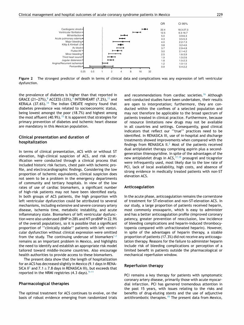
Multivariable analysis
Independent predictors of hospital mortality are shown inTable 8. The strongest predictors of hospital death, based onrisk factors and cardiovascular history, were STEMI diagno-sis, left ventricular dysfunction, and age >65 years (Fig. 1).The strongest predictor of death in terms of clinical dataand complications was any expression of left ventricu-lar dysfunction (Fig. 2). Ventricular and supraventricular
226 C. Martinez-Sanchez et al.
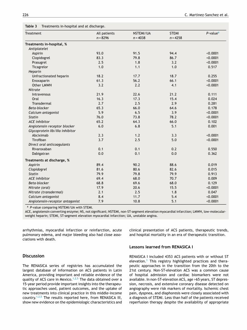
Table 3 Treatments in-hospital and at discharge.
Treatment All patientsn = 8296
NSTEMI/UAn = 4038
STEMIn = 4258
P-valuea
Treatments in-hospital, %
Antiplatelet
Aspirin 93.0 91.5 94.4 <0.0001Clopidogrel 83.3 79.8 86.7 <0.0001Prasugrel 2.5 1.8 3.2 <0.0001Ticagrelor 1.0 1.1 1.0 0.517
Heparin
Unfractionated heparin 18.2 17.7 18.7 0.255Enoxaparin 61.3 56.2 66.1 <0.0001Other LMWH 3.2 2.2 4.1 <0.0001
Nitrate
Intravenous 21.9 22.6 21.2 0.111Oral 16.3 17.3 15.4 0.024Transdermal 2.7 2.5 2.9 0.281
Beta-blocker 65.3 66.0 64.6 0.178Calcium antagonist 5.9 6.5 3.9 <0.0001Statin 76.0 73.8 78.2 <0.0001ACE inhibitor 65.2 64.3 66.0 0.102Angiotensin receptor blocker 6.0 6.8 5.1 0.001Glycoprotein IIb/IIIa inhibitor
Abciximab 2.3 1.2 3.3 <0.0001Tirofiban 3.7 2.5 5.0 <0.0001
Direct oral anticoagulants
Rivaroxaban 0.1 0.1 0.2 0.550Dabigatran 0.0 0.1 0.0 0.362
Treatments at discharge, %
Aspirin 89.4 90.2 88.6 0.019Clopidogrel 81.6 80.6 82.6 0.015Statin 79.9 79.8 79.9 0.913ACE inhibitor 69.4 68.0 70.7 0.009Beta-blocker 68.8 69.6 68.0 0.129Nitrate (oral) 17.9 20.6 15.5 <0.0001Nitrate (transdermal) 2.1 2.5 1.8 0.047Calcium antagonist 8.4 11.7 5.4 <0.0001Angiotensin-receptor antagonist 7.9 10.8 5.1 <0.0001
a P-value comparing NSTEMI/UA with STEMI.ACE, angiotensin converting enzyme; NS, not significant; NSTEMI, non-ST-segment elevation myocardial infarction; LMWH, low-molecular-weight heparin; STEMI, ST-segment elevation myocardial infarction; UA, unstable angina.
arrhythmias, myocardial infarction or reinfarction, acutepulmonary edema, and major bleeding also had close asso-ciations with death.
Discussion
The RENASICA series of registries has accumulated thelargest database of information on ACS patients in LatinAmerica, providing important and reliable evidence of thequality of ACS care in Mexico.1,2,5 The data obtained over a15-year period provide important insights into the therapeu-tic approaches used, patient outcomes, and the uptake ofnew treatments into clinical practice in this middle-incomecountry.1,2,5 The results reported here, from RENASICA III,show new evidence on the epidemiologic characteristics and
clinical presentation of ACS patients, therapeutic trends,and hospital mortality in an era of therapeutic transition.
Lessons learned from RENASICA I
RENASICA I included 4353 ACS patients with or without STelevation.1 This registry highlighted practices and thera-peutic approaches in the transition from the 20th to the21st century. Non-ST-elevation ACS was a common causeof hospital admission and cardiac biomarkers were notavailable. In non-ST-elevation ACS, age >65 years, ST depres-sion, necrosis, and extensive coronary disease detected onangiography were risk markers of mortality. Ischemic chestpain, dyspnea, and diaphoresis were closely associated witha diagnosis of STEMI. Less than half of the patients receivedreperfusion therapy despite the availability of appropriate

Clinical management and hospital outcomes of acute coronary syndrome patients in Mexico 227
Table 4 Comparing treatments in-hospital and atdischarge.
Treatment, % In-hospitaln = 8296
At dischargea
n = 7769P-valueb
Aspirin 93.0 95.5 <0.0001Clopidogrel 83.3 86.9 <0.0001Beta-blocker 65.3 73.3 <0.0001ACE inhibitor 65.2 73.9 <0.0001Statin 76.0 85.1 <0.0001
a Not considering death.b P-value comparing in-hospital with at discharge.
ACE, angiotensin converting enzyme.
Table 5 Pharmacological and mechanical reperfusion inpatients with STEMI.
Variable Patients (n = 4258)
Ischemia time, minutes
Door to electrocardiogram 13.0 (5.0---50.0)ECG to specific therapy 18.0 (5.0---65.0)Door-to-needlea 45.0 (20.0---90.0)Door-to-balloona 100.0 (50.0---270.0)Total ischemic time 200.0 (90---350)
Thrombolytic therapy, % 37.6Tenecteplase 20.7Streptokinase 6.6Alteplase 4.9Not specified 3.9Other 1.4
Reperfusion criteria, %
Pain resolution 45.5ST resolution >70% 36.4Reperfusion arrhythmia 15.1
Percutaneous coronary intervention 23.6Primary 15.0Rescue 8.6
Data given as median (percentiles 25---75).a P = 0.006 (comparing Door to needle with Door to balloon by
Mann---Whitney U test).STEMI, ST-segment elevation myocardial infarction.
facilities in 90% of hospitals. Standard antiplatelet andantithrombin treatments were used in 70% of patients; low-molecular-weight heparin and glycoprotein IIb/IIIa receptorblockers were used fewer patients. The RENASICA I registryprovided valuable information for Mexican health authori-ties to better apply health resources for the management ofACS in the 21st century.1
Lessons learned from RENASICA II
The second RENASICA registry included 8098 patients withfinal diagnosis of ACS. This registry had a higher ratio ofSTEMI to NSTEMI/UA (1.3:1) compared with RENASICA I (35%vs. 65%), attributed to the availability of mechanical reper-fusion facilities in participating hospitals, as well as a highprevalence of diabetes in patients with STEMI. The averagelength of hospital stay was 8.1 days, independent of the
Table 6 Early invasive strategy in patients withNSTEMI/UA.
Strategy Patients (n = 4038)
Percutaneous coronary intervention, % 39.6Conservative 17.1Early 10.8Urgent 3.0Other 8.7
Type of stent, %
Bare-metal stent 13.7Drug-eluting stent 25.3
Number of stents, %
1 22.42 7.13 1.7>3 0.1
NSTEMI, non-ST-segment elevation myocardial infarction; UA,unstable angina.
final diagnosis. The incidence of diabetes (42%) was slightlylower than that in RENASICA I (50%). This metabolic disor-der was a strong predictor of hospital mortality in patientswith NSTEMI/UA. Tests to assess patients’ glucometabolicstate were not done, so the true prevalence of diabetes,impaired glucose tolerance, and insulin resistance are likelyto be higher than indicated, and the rate of obesity wasnot determined.2 The clinical presentation of ACS was sim-ilar to that observed in RENASICA I and use of troponin waslow. Optimal pharmacological treatment (e.g. glycoproteinIIb/IIIa inhibitors and statins) was lower than expected inboth groups of ACS patients. The rate of pharmacologicalthrombolysis decreased from 50% in RENASICA I to 37% inRENASICA II, with streptokinase being the most commonlyused drug. The rate of PCI was 15%. Consequently, a consid-erable proportion of patients with STEMI received no form ofreperfusion therapy. Left ventricular dysfunction (AppendixB) was the most important hospital complication and thestrongest predictor of death, which occurred with a fre-quency of 10% in STEMI and 4% in NSTEMI/UA.2
RENASICA III
The profile of patients in RENASICA III in terms of age andsex is similar to that in RENASICA II,2 suggesting that thehigh incidence of ACS persists in this high-productivity socialgroup and in male patients. Of note, the rate of tobaccosmoking decreased from 60% in RENASICA I1 and 64% in RENA-SICA II2 to 53.3% in RENASICA III, signifying that the healthpolicy toward reducing rates of tobacco consumption hashad some effect. Conversely, the prevalence of traditionalrisk factors such as hypertension, diabetes, and hypercho-lesterolemia increased, and the rate of diabetes remainedunchanged, at nearly 50%. The proportion of men and theprevalence of hypertension in RENASICA III (73.8% and 62.1%,respectively) are similar to those reported in the GRACEregistry (62---72% and 50---65%, respectively),8 the NationalRegistry of Myocardial infarction (NRMI),9---11 and the EuroHeart Survey (64---72% and 52---64%, respectively).12 However,

228 C. Martinez-Sanchez et al.
Table 7 In-hospital major cardiovascular adverse events, adverse events, and death.
Event All (n = 8296) NSTEMI/UA (n = 4038) STEMI (n = 4258) P-valuea
Death, % 6.4 3.9 8.7 <0.0001
Cardiovascular complications, %
Angina or recurrent ischemia 19.9 21.8 18.0 <0.0001Heart failure 10.9 9.8 11.9 0.002Acute pulmonary edema 8.2 8.2 8.3 0.905Cardiogenic shock 8.2 6.1 10.2 <0.0001MI/reinfarction 6.1 3.9 8.2 <0.0001Ventricular tachycardia 4.6 2.8 6.4 <0.0001Mechanical complications 3.0 2.7 3.2 0.174Ventricular fibrillation 2.4 1.3 3.6 <0.0001Pericarditis 1.0 0.6 1.4 0.001
Bleeding complications, %
Minor bleeding 2.2 2.0 2.4 0.203Major bleeding 0.8 0.6 1.0 0.026
Non-cardiovascular complications, %
Renal failure 6.6 6.2 7.0 0.158Sepsis 2.0 2.0 2.1 0.938Cerebrovascular event 1.0 0.8 1.1 0.258
a P-value comparing NSTEMI/UA with STEMI.MI, myocardial infarction; NSTEMI, non-ST-segment elevation myocardial infarction; STEMI, ST-segment elevation myocardial infarction;UA, unstable angina.
Table 8 Multivariate analysis to early mortality.
Variable (n = 8296) B Sig.* Exp(B) CI 95%
Cardiogenic shock 2.5 <0.0001 12.8 10.0---16.4Ventricular fibrillation 1.5 <0.0001 4.6 3.1---6.8MI/reinfarction 1.2 <0.0001 3.4 2.6---4.6Ventricular tachycardia 1.0 <0.0001 2.7 2.0---3.7Major bleeding 0.7 0.081 2.0 0.9---4.5Diagnosis 0.5 <0.0001 1.7 1.3---2.1AV block 0.5 0.001 1.7 1.2---2.3Hypertension 0.3 0.003 1.4 1.1---1.7Killip and Kimball ≥2 0.3 0.011 1.4 1.1---1.7
* Statistical significance by Wald test for logistic regression.CI, confidence interval; MI, myocardial infarction.
Stemi
Previous heart failure
Age>65
Hypertension
Female
Smoking
Oyslipidemia
Previous angina
Previous myocardial infarction
Previous PCI
Previous CABG
0.25 0.5 1 2 4
OR Cl 95%
2.3 1.9-2.8
2.0 1.2-3.1
1.9 1.6-2.3
1.2 1.0-1. 5
1.1 1.1-1.2
1.1 1.0-1.2
1.0 0.9-1.0
1.0 0.8-1.3
0.9 0.7-1.1
0.8 0.6-1.0
0.6 0.3-1.1
Figure 1 The strongest predictors of hospital death, based on risk factors and cardiovascular history, were STEMI diagnosis, leftventricular dysfunction, and age >65 years.
Clinical management and hospital outcomes of acute coronary syndrome patients in Mexico 229
0.25
Cardiogenic shock
Ventricular fibrillation
Ml/reinfaction
Acute pulmonary edema
Major bleedingKillip & Kimball ≥2
Av block
Flutter/AF
Minor bleeding
Third heart sound
Jugular distension
Angina/Recurrent ischemia
Pericarditis
0.5 1 2 4 8 16 32
22.4
OR CI 95%
18.3-27.3
12.5 9.3-16.7
5.0 3.9-6.3
4.3 3.5-5.3
4.0 2.2-7.3
3.8 3.2-4.6
3.7 2.9-4.8
3.0 2.1-4.2
2.5 1.6-3.9
1.9 1.5-2.6
1.8 1.5-2.3
1.2 1.0-1.5
0.9 0.4-2.2
Figure 2 The strongest predictor of death in terms of clinical data and complications was any expression of left ventriculardysfunction.
the prevalence of diabetes is higher than that reported inGRACE (21---27%),8 ACCESS (33%),4 INTERHEART (7.2%),13 andKERALA (37.6%).14 The Indian CREATE registry found thatdiabetes prevalence was related to socioeconomic status,being lowest amongst the poor (18.7%) and highest amongthe most affluent (40.9%).15 It is apparent that strategies forprimary prevention of diabetes and ischemic heart diseaseare mandatory in this Mexican population.
Clinical presentation and duration ofhospitalization
In terms of clinical presentation, ACS with or without STelevation, high-clinical suspicion of ACS, and risk strat-ification were conducted through a clinical process thatincluded historic risk factors, chest pain with ischemic pro-file, and electrocardiographic findings. Considering the lowproportion of ischemic equivalents, clinical suspicion doesnot seem to be a problem in the emergency departmentsof community and tertiary hospitals. In view of the lowrates of use of cardiac biomarkers, a significant numberof high-risk patients may not have been identified early.In both groups of ACS patients, the high proportion withleft ventricular dysfunction could be attributed to severalmechanisms, including extensive and severe coronary arterydisease, ischemia time, metabolic instability, and acuteinflammatory state. Biomarkers of left ventricular dysfunc-tion were also underused (BNP in 28% and NT-proBNP in 22.9%of the overall population), so it is possible that a significantproportion of ‘‘clinically stable’’ patients with left ventri-cular dysfunction without clinical expression were omittedfrom the study. The continuing underuse of biomarkers1,2
remains as an important problem in Mexico, and highlightsthe need to identify and establish an appropriate risk modeltailored toward middle-income countries. Also encouragehealth authorities to provide access to these biomarkers.
The present data show that the length of hospitalizationfor an ACS has decreased slightly in Mexico (9.1 days in RENA-SICA II2 and 7.1 ± 7.8 days in RENASICA III), but exceeds thatreported in the NRMI registries (4.3 days).9---11
Pharmacological therapies
The optimal treatment for ACS continues to evolve, on thebasis of robust evidence emerging from randomized trials
and recommendations from cardiac societies.16 Althoughwell-conducted studies have been undertaken, their resultsare open to interpretation; furthermore, they are con-ducted within the confines of a selected population andmay not therefore be applicable to the broad spectrum ofpatients treated in clinical practice. Furthermore, becauseof resource limitations new drugs may not be availablein all countries and settings. Consequently, good clinicalindicators that reflect our ‘‘true’’ practices need to beidentified. In RENASICA III, use of in-hospital and dischargetreatments showed improvements when compared with thefindings from RENASICA II.2 Most of the patients receiveddual antiplatelet therapy comprising aspirin plus a second-generation thienopyridine. In spite of the advantages of thenew antiplatelet drugs in ACS,17,18 prasugrel and ticagrelorwere infrequently used, most likely due to the low rate ofPCI, lack of local availability, high costs, and absence ofstrong evidence in medically treated patients with non-STelevation ACS.
Anticoagulation
In the acute phase, anticoagulation remains the cornerstoneof treatment for ST-elevation and non-ST-elevation ACS. Inour study, a large proportion of patients received heparin,most commonly enoxaparin, which is easy to administerand has a better anticoagulation profile (improved coronarypatency, greater prevention of reocclusion, low incidenceof bleeding complications and heparin-induced thrombocy-topenia compared with unfractionated heparin). However,in spite of the advantages of heparin therapy, a sizableproportion of patients (17.3%) did not receive any anticoagu-lation therapy. Reasons for the failure to administer heparininclude risk of bleeding complications or perception of alimited benefit in patients outside the pharmacological ormechanical reperfusion window.
Reperfusion therapy
PCI remains a key therapy for patients with symptomaticcoronary artery disease, primarily those with acute myocar-dial infarction. PCI has garnered tremendous attention inthe past 15 years, with issues relating to the risks andbenefits of drug-eluting stents and the use of adjunctiveantithrombotic therapies.19 The present data from Mexico,

230 C. Martinez-Sanchez et al.
spanning nearly two decades,1---3 show that pharmacologi-cal reperfusion remains the most frequently used form ofreperfusion in STEMI patients. Primary PCI in STEMI andearly cardiac catheterization (within 48---72 h) in NSTEMI/UAwere infrequent. When compared with the earlier RENA-SICA registries,1,2 the most notable finding from RENASICAIII was the transition in STEMI patients from first-generationto third-generation thrombolytic therapy and from unfrac-tionated heparin to low-molecular weight heparin.
In terms of ischemia time, delays from first medi-cal contact to electrocardiogram or to starting a specifictherapy were within acceptable ranges, whereas delaysto reperfusion exceeded the current recommendations.16
While quality-improvement initiatives that target bothphysicians and patients should help to reduce these delays,a national program is far from being a reality; therefore itis important to reinforce the need for regional programs inorder to improve the care of ACS patients. One such pro-gram is currently in place at the Cardiology National Institute‘‘Ignacio Chavez’’ in Mexico City. In this program, staff in theemergency departments of community hospitals have beentrained to administer pharmacological thrombolysis. Elec-trocardiograms that are difficult to interpret are sent forevaluation, and recommendations on the best treatment areprovided. This program intent evaluating the efficacy andsafety of a pharmaco-invasive strategy.
Hospital outcomes
The hospital mortality rate in RENASICA III was 6.4% (8.7%in STEMI and 3.9% in NSTEMI/UA), which shows a reassuringdecrease from RENASICA II (10% in STEMI, 4% in NSTEMI/UA).2
However, it is higher than the 30-day mortality reported inACCESS (3.6% all ACS; 5.0% in STEMI, 2.4% in NSTE-ACS)4 andhospital mortality in GRACE (4.6% in STEMI, 2.2% in NSTE-ACS).20 The GRACE registry also demonstrated a statisticallysignificant decline in hospital death over the course of thestudy (from 8.4% to 4.6%, p < 0.001, for STEMI; and from2.9% to 2.2%, p = 0.02, for NSTE-ACS), which correspondedwith increasing use of medications and primary PCI anddecreasing use of pharmacological reperfusion.20 The rateof bleeding complications in RENASICA III was low and can beattributed to the low rates of use of PCI, glycoprotein IIb/IIIainhibitors, and new antiplatelets and oral anticoagulants.
The most important hospital complication in our studywas the expression of left ventricular dysfunction (AppendixB). In the multivariable analysis, as observed previously,1,2
cardiogenic shock was the strongest predictor of death, aswell as myocardial infarction or reinfarction. All of thesemajor adverse cardiovascular events are closely related toprolonged ischemic times and low rate of PCI. Althoughbleeding complications and ventricular arrhythmias werescarce, both were related with hospital mortality.
Limitations
RENASICA III was not a population-based epidemiologicalstudy; therefore, some bias may have been introduced withrespect to the selection of participating centers. Other lim-itations include the restricted number of visits to monitordata accuracy. Patients who died early, before admission
to hospital or while in the emergency department, wereexcluded. The inclusion of community hospitals may explainthe low rate of PCI.
Conclusion
The results from RENASICA III establish the urgent needto develop effective regional programs to improve ratesof reperfusion therapy, to increase the ratio of PCI tothrombolysis, and to reduce ischemia times. Through theachievement of these targets, we can ensure a better qualityof life for many Mexican patients presenting with an ACS.
Ethical disclosures
Protection of human subjects and animals in research.
The authors declare that the procedures followed were inaccordance with the regulations of the responsible ClinicalResearch Ethics Committee and in accordance with those ofthe World Medical Association and the Helsinki Declaration.
Confidentiality of data. The authors declare that they havefollowed the protocols of their work centre on the publi-cation of patient data and that all the patients includedin the study have received sufficient information and havegiven their informed consent in writing to participate in thatstudy.
Right to privacy and informed consent. The authors haveobtained the informed consent of the patients and/orsubjects mentioned in the article. The author for correspon-dence is in possession of this document.
Funding
An unrestricted grant was provided by Sanofi.
Conflict of interest
The authors declare no conflict of interest.
Acknowledgments
Sophie Rushton-Smith, PhD (Medlink Healthcare Communi-cations Ltd), provided editorial support.
Appendix A. RENASICA III Investigators
Rafael Barraza Félix, Luis Alberto Cervantes Chávez, VaniaQuisbert Vattuone, Centro Médico Nacional ‘‘La Raza’’,IMSS, México, D. F. (1413); Anabel García Sosa, Juan Ger-ardo Campos Cassio, Aarón Jasso Guido, Esteban Luis ReyesCerezo, Hospital de Cardiologia 34, IMSS, Monterrey, NuevoLeón (1188); Rodolfo Parra Michel, Héctor Raúl BarajasCampa, Juan Ángel Campos Aguilar, Jaime Chávez Michel,Luis Alfredo Pelayo Jiménez, Silvano Vela Alarcón, CentroMédico Nacional de Occidente, IMSS, Guadalajara, Jalisco(985). Alexandra Arias Mendoza, Alfredo Altamirano Castillo,Amada Alvarez San Gabriel, Francisco Azar Manzur, JoséLuis Briseno De la Cruz, Gustavo Rojas Velasco, Instituto

Clinical management and hospital outcomes of acute coronary syndrome patients in Mexico 231
Nacional de Cardiología ‘‘Ignacio Chávez’’, México, D. F.(951); Belinda González Díaz, Oracio González Ortiz, IvánAlfonso González Rosas, Ismael Hernández Santamaría, Hos-pital Juárez de México, México, D. F. (658); Ambrosio CruzDíaz, Hospital Central de Alta Especialidad Sur, PEMEX,México, D.F. (342); Rocío Camacho Casillas, Antonio Gar-cía Espino, UMAE Hospital de Especialidades No. 71, IMSS,Torreón, Coahuila (315); José Luis Leiva Pons, HospitalCentral ‘‘Dr. Ignacio Morones Prieto’’, San Luis Potosí,San Luis Potosí (230); Amanda M. Castelán Ojeda, UMAEHospital de Especialidades No. 2, IMSS, Ciudad Obregón,Sonora (221); Juan Carlos Osnaya Martínez, Sergio SalasPadilla Hospital Regional ‘‘Lic. Adolfo López Mateos’’,ISSSTE, México, D. F. (184); Alfredo Comparan Núnez, Hos-pital Regional No. 1, IMSS, Tijuana, Baja California (169);Lidia E. Betancourt Hernández, UMAE Hospital de Espe-cialidades No. 14, IMSS, Veracruz, Veracruz (135); SergioGonzález Romero, Miguel Ángel Luna Calvo, Juan CarlosNúnez Fragoso, Hospital General de Durango, Durango,Durango (127); Raúl Alberto Rivas Lira, Hospital Centraldel Norte, PEMEX, México, D.F. (122); Manuel AlfonsoBanos González, Hospital Regional de Alta Especialidad‘‘Dr. Juan Grauss’’, Villahermosa, Tabasco (112); Rodolfode Jesús Castano Guerra, Roberto López Rosas, HospitalGeneral de México, México, D.F. (82); Anabella C. DelgadoSánchez, Sergio Luna Ramírez, Oscar Samuel Medina Tor-res, UMAE Hospital de Especialidades No 1 Bajío, León,Guanajuato (67); Norberto Matadamas Hernández, Hospi-tal General de Acapulco, ISSSTE, Acapulco, Guerrero (45);Guillermo Llamas Esperón, Rolando Chávez Martínez, Eliz-abeth Espinoza Garza, Monserrat González Limón, AnaPatricia Grajales Alonso, Miguel Ángel Ramos Guzmán,Martha Vacio Olguín, Alberto Zamora Mucino-Arroyo, Cardi-ológica Aguascalientes, Aguascalientes, Aguascalientes (42);Marco Antonio Alcocer Gamba, Pablo Jaime Arias Fajardo,Karla Verónica Cárdenas Mejía, Eliodoro Castro Montes,Enrique García Hernández, Salvador León González, AbelLinares Rodríguez, Wendy Munoz Rosales, José ErnestoPombo Bartelt, Instituto de Corazón de Querétaro, Queré-taro, Querétaro (37); Laura Román Herrera, Guadalupe RenéRuiz Avila, Hospital General de Zona No.6, IMSS, CiudadJuárez, Chihuahua (36); Leocadio Munoz Beltrán, Hospi-tal Ángeles, Ciudad Juárez, Chihuahua (34); Jesús ZúnigaSedano, Luis Virgen Carrillo, Hospital Ángeles del Carmen,Guadalajara, Jalisco (34); Jorge H. Jiménez Orozco, Unidadde Medicina Física ‘‘Magdalena de las Salinas’’, IMSS, Méx-ico, D. F. (34); Salvador Facundo Bazaldua, Hospital GeneralVictoria, Ciudad Victoria, Tamaulipas (32); Manuel Odín Delos Ríos Ibarra, Alberto Zenón Banos Velasco, Hospital Gen-eral de Culiacán, Culiacán, Sinaloa (32); Ángel LeovigildoAlberto Delgado, María del Rocío Blázquez Cruz, Luis EnriqueBerumen Domínguez, Alberto Cortez Benítez, Luis ManuelPáez Lizárraga, Hospital Central Militar, México, D.F. (27);Víctor Armando Varguez Argüelles, Hospital Regional dePuebla, ISSSTE, Puebla, Puebla (27); Manuel de los ReyesBarrera Bustillos, Clínica Mérida, Mérida, Yucatán (26); Nor-berto Matadamas Hernández, Hospital General de Acapulco,Acapulco, Guerrero (25); José Fabián Hernández Díaz, Hos-pital Regional de Alta Especialidad de Oaxaca; Oaxaca,Oaxaca (25); Ma. Isabel Sánchez Ramírez, Hospital GeneralLa Paz, ISSSTE, La Paz, Baja California Sur (25); Francisco
Javier Redding Escalante, Star Médica ‘‘Lomas Verdes’’,Lomas Verdes, Estado de México (25); José Luis GonzálezPena, Hospital Regional Reynosa, PEMEX, Reynosa, Tamauli-pas (24); Ramón José Cué Carpio, Instituto Mexicano deTrasplantes, Cuernavaca, Morelos (24); Raúl Isaac Márquez,Hospital General de Zona No.3, IMSS, Cancún, QuintanaRoo (22); Federico Rodríguez Weber, Hospital Ángeles delPedregal, México, D.F. (21); Oswaldo Lagunas Uriarte, Hos-pital Civil de Culiacán, Culiacán, Sinaloa (20); Raúl TenienteValente, Hospital Regional de Alta Especialidad del Bajío,León, Guanajuato (20); Juan Carlos Núnez Fragoso, Hos-pital Regional No. 1, IMSS, Durango, Durango (20); HazelGonzález Castro, Juan Carlos Pérez Alva, Hospital Generalde Puebla, Puebla, Puebla (19); Carlos A. Arean Martínez,Hospital General ‘‘Dr. Miguel Silva’’, Morelia, Michoacán(18); Miguel E. Beltrán Gámez, ISSSTECALI, ISSSTE, Mexi-cali, Baja California (18); Pánfilo Mauricio Saucedo, HospitalGeneral de Zacatecas, Zacatecas, Zacatecas (17); VladimirRuiz Ronquillo, Hospital General de Zona No.18, IMSS; Playadel Carmen, Quintana Roo (15); Agustín Vallecillo Gómez,Hospital Regional Salamanca, PEMEX, Salamanca, Guana-juato (15); Reynaldo Magana Magana, Hospital General deUruapan, Uruapan, Michoacán (14); Jaime Barragan Luna,Hospital Regional Poza Rica, PEMEX, Poza Rica, Veracruz(14); Jorge G. Chávez Páez, Amerimed, Puerto Vallarta,Jalisco (13); Ma. Isabel Sánchez Ramírez, Hospital Generalcon Especialidades ‘‘Juan María de Salvatierra’’, La Paz,Baja California Sur (12); José Luis González Pena, Hospi-tal ‘‘Christus Muguerza’’, Reynosa, Tamaulipas (11); JorgeG. Chávez Páez, Hospital Cornerstone, Puerto Vallarta,Jalisco (11); Alfredo Comparan Núnez, Hospital Florence,Tijuana, Baja California (11); Jesús Manuel Acosta Gra-jeda, Hospital General de Zona No.1, IMSS, Chihuahua,Chihuahua (11); Luis Delgado Leal, Centenario Hospital‘‘Miguel Hidalgo’’, Aguascalientes, Aguascalientes (11);Orlando Henne Otero, Hospital Ángeles de Villahermosa, Vil-lahermosa, Tabasco (10); Víctor Páramo Bautista, HospitalGeneral ‘‘Adolfo Prieto’’, Taxco, Guerrero (10); César Ger-ardo Martínez Hernández, Hospital General de Zona No.50,IMSS, San Luis Potosí, San Luis Potosí (10); José ManuelEnciso Munoz, Pánfilo Mauricio Saucedo, Hospital ‘‘SanAgustín’’, Zacatecas, Zacatecas (10); Alberto Zenón BanosVelasco, Hospital Ángeles de Culiacán, Culiacán, Sinaloa(9); Francisco Guerrero Martínez, Hospital General de León,León, Guanajuato (9); Mario Morales Medina, Policlínicade Especialidades, Tuxtla, Chiapas (9); Luis Sánchez Tru-jillo, Hospital Metropolitano, Monterrey, Nuevo León (7);Roberto Domínguez López, Centro de Especialidades Médicas‘‘Dr. Rafael Lucio’’, Xalapa, Veracruz (6); Julio Iván FarjatRuiz, Sergio Alonso Villarreal Umana, Centro Médico ‘‘LasAméricas’’, Mérida, Yucatán (6); José Luis Novelo Del Valle,Hospital General de Zona No.1, IMSS, Campeche, Campeche(6); Rosa Elena Limas Rodríguez, Hospital General, ISSSTE,Tijuana, Baja California (6); Ivette Y. Alferez Jiménez,Hospital Ángeles de Lindavista, México, D.F. (5); MelissaJiménez Madrigal, Centro de diagnóstico y tratamientoCardiológico, Mexicali, Baja California (5); Víctor PáramoBautista, Clínica de Especialidades Taxco, Taxco, Guerrero(5); Jorge G. Chávez Páez, Hospital CMQ, Puerto Vallarta,Jalisco (5); Alfredo Comparan Núnez, Hospital del Prado,Tijuana, Baja California (5); José Luis Novelo Del Valle,

232 C. Martinez-Sanchez et al.
Hospital General de Especialidades INDESALUD, Campeche,Campeche (5).
Appendix B.
Myocardial infarction clinical classification Uncomplicated:normal blood pressure without left ventricular dysfunc-tion signs, EF >40%, brain natriuretic peptide type-B (BNP)<100 pg/dL, or both5. Complicated with left ventriculardysfunction without clinical manifestations: normal bloodpressure or lower limits, no third left ventricular heartsound, EF <40%, or both, BNP >100 pg/dL, or both5.Complicated with ventricular dysfunction with clinical mani-festations: acute pulmonary edema or pre-cardiogenic shocksuspicion (borderline blood pressure, hypotension, or both),clinical expression of --- renin-angiotensin-aldosterone vaso-pressin system activity, oliguria, delayed capillary refill,lower lobes crackling rales, EF <30%, BNP >600 pg/dL5.
Killip and Kimball classification
I: without left ventricular dysfunction signs; II: left ventri-cular third heart sound, basal crackling rales, or both; III:acute pulmonary edema IV: cardiogenic shock.5
Pulmonary edema: increased work of breathing with orwithout severe desaturation (saturation <90%), left ventri-cular gallop or tachycardia, crackling rales or wheezes >50%in lung fields. Chest X-ray with interstitial, alveolar, or mixedpulmonary edema.5
Cardiogenic shock: (1) systolic blood pressure <90 mmHgwithout support of vasoactive substances or 100 mmHg withvasopressors use; (2) clinical, radiographic expression of pul-monary venocapillary hypertension, or both; (3) signs ofperipheral vascular hypoperfusion; (4) metabolic acidosis;(5) cardiac index <2.2 L/min/m2; (6) pulmonary capillarypressure >18 mmHg; and (7) arteriovenous oxygen difference>5.5 ml/dL.5
References
1. Lupi Herrera E, The RENASICA Cooperative Group. NationalRegistry of Acute Ischemic Coronary Syndromes (RENASICA).Mexican Cardiology Society. Arch Cardiol Mex. 2002;72:S45---64.
2. Garcia-Castillo A, Jerjes-Sanchez C, Martinez Bermudez P, et al.Mexican Registry of Acute Coronary Syndromes. Arch CardiolMex. 2005;75:S6---32.
3. Borrayo-Sanchez G, Madrid-Miller A, Arriaga-Nava R, et al. Riskstratified in the National Registry of Acute Coronary Syndromesat the IMSS. Rev Med Inst Mex Seguro Soc. 2010;48:259---64.
4. ACCESS Investigators. Management of acute coronary syn-dromes in developing countries: acute coronary events --- amultinational survey of current management strategies. AmHeart J. 2011;162, 852---859 e822.
5. Jerjes-Sanchez C, Martinez-Sanchez C, Borrayo G, et al. Thirdnational registry of acute coronary síndromes (RENASICA III).Arch Cardiol Mex. 2015;85:207---14.
6. García-Sosa A. Which is the best therapeutic approach in tran-sitory ST elevation patients? In: Verdejo J, editor. ClínicasMexicanas de Cardiología. 2012. p. 33---8.
7. Gitt AK, Bueno H, Danchin N, et al. The role of cardiac registriesin evidence-based medicine. Eur Heart J. 2010;31:525---30.
8. Steg PG, Goldberg RJ, Gore JM, et al. Baseline character-istics, management practices, and in-hospital outcomes ofpatients hospitalized with acute coronary syndromes in theGlobal Registry of Acute Coronary Events (GRACE). Am J Cardiol.2002;90:358---63.
9. Rogers WJ, Canto JG, Lambrew CT, et al., Every N. Tempo-ral trends in the treatment of over 1.5 million patients withmyocardial infarction in the US from 1990 through 1999: theNational Registry of Myocardial Infarction 1, 2 and 3. J Am CollCardiol. 2000;36:2056---63.
10. Curtis JP, Portnay EL, Wang Y, et al. National Registry ofMyocardial I. The pre-hospital electrocardiogram and timeto reperfusion in patients with acute myocardial infarction,2000---2002: findings from the National Registry of MyocardialInfarction-4. J Am Coll Cardiol. 2006;47:1544---52.
11. Nallamothu BK, Bates ER, Herrin J, et al. Times to treat-ment in transfer patients undergoing primary percutaneouscoronary intervention in the United States: National Reg-istry of Myocardial Infarction (NRMI)-3/4 analysis. Circulation.2005;111:761---7.
12. Hasdai D, Behar S, Wallentin L, et al. A prospective survey ofthe characteristics, treatments and outcomes of patients withacute coronary syndromes in Europe and the Mediterraneanbasin; the Euro Heart Survey of Acute Coronary Syndromes (EuroHeart Survey ACS). Eur Heart J. 2002;23:1190---201.
13. Yusuf S, Hawken S, Ounpuu S, et al. Effect of potentially mod-ifiable risk factors associated with myocardial infarction in 52countries (the INTERHEART study): case---control study. Lancet.2004;364:937---52.
14. Mohanan PP, Mathew R, Harikrishnan S, et al. Presentation, man-agement, and outcomes of 25,748 acute coronary syndromeadmissions in Kerala, India: results from the Kerala ACS Registry.Eur Heart J. 2013;34:121---9.
15. Xavier D, Pais P, Devereaux PJ, et al. Treatment and outcomesof acute coronary syndromes in India (CREATE): a prospectiveanalysis of registry data. Lancet. 2008;371:1435---42.
16. Steg PG, James SK, Atar D, et al. ESC Guidelines for the man-agement of acute myocardial infarction in patients presentingwith ST-segment elevation. Eur Heart J. 2012;33:2569---619.
17. Wiviott SD, Braunwald E, McCabe CH, et al. Prasugrel versusclopidogrel in patients with acute coronary syndromes. N EnglJ Med. 2007;357:2001---15.
18. Roe MT, Armstrong PW, Fox KA, et al. Prasugrel versus clopido-grel for acute coronary syndromes without revascularization. NEngl J Med. 2012;367:1297---309.
19. Roe MT, Messenger JC, Weintraub WS, et al. Treatments, trends,and outcomes of acute myocardial infarction and percutaneouscoronary intervention. J Am Coll Cardiol. 2010;56:254---63.
20. Fox KA, Steg PG, Eagle KA, et al. Decline in rates of death andheart failure in acute coronary syndromes, 1999---2006. JAMA.2007;297:1892---900.