
3 Cran Vault Dura 12(1)
60
1 Cranial Vault & Dura Cranial Vault & Dura Paul F. Aravich, Ph.D. Paul F. Aravich, Ph.D. Medical Gross Anatomy & Embryology Medical Gross Anatomy & Embryology 2012 2012 Department of Pathology & Anatomy Glennan Center for Geriatrics & Gerontology & Department of Physical Medicine & Rehabilitation Eastern Virginia Medical School Norfolk, VA [email protected] Eastern Virginia Medical School
-
Upload
myungdong-shinsa -
Category
Documents
-
view
10 -
download
0
description
cran
Transcript of 3 Cran Vault Dura 12(1)

1
Cranial Vault & DuraCranial Vault & DuraCranial Vault & DuraCranial Vault & Dura
Paul F. Aravich, Ph.D.Paul F. Aravich, Ph.D.Medical Gross Anatomy & Embryology 2012Medical Gross Anatomy & Embryology 2012
Department of Pathology & AnatomyGlennan Center for Geriatrics & Gerontology &
Department of Physical Medicine & RehabilitationEastern Virginia Medical School
Norfolk, [email protected]
aste
rn V
irg
inia
Med
ical
Sch
oo
l

2
Cranial Vault & DuraCranial Vault & Dura
Objectives: Concept map:Objectives: Concept map:Cranial Vault & DuraCranial Vault & Dura
Objectives: Concept map:Objectives: Concept map: The organization of the scalp and its danger zone.The organization of the scalp and its danger zone. Blood supply to the scalpBlood supply to the scalp Openings for the cranial nervesOpenings for the cranial nerves Dura mater & its reflections in the cranial vaultDura mater & its reflections in the cranial vault Dural sinuses & the venous drainage of the brainDural sinuses & the venous drainage of the brain Internal carotid, vertebral & middle meningeal aaInternal carotid, vertebral & middle meningeal aa

3
Clinical correlatesClinical correlatesClinical correlatesClinical correlates
Scalp infections & lacerationsScalp infections & lacerations Periorbital Ecchymosis: Black eye vs. Raccoon eyesPeriorbital Ecchymosis: Black eye vs. Raccoon eyes CSF rhinorrhea & CSF otorrhea CSF rhinorrhea & CSF otorrhea Basilar venous plexus & metastatic/infection spreadBasilar venous plexus & metastatic/infection spread Internal carotid artery aneurysm & CN II optic nerveInternal carotid artery aneurysm & CN II optic nerve Fracture of the pterion and type of hemorrhageFracture of the pterion and type of hemorrhage Cavernous sinus thrombusCavernous sinus thrombus
Clinical correlates

4
Outline: Outline: Cranial Vault & DuraCranial Vault & DuraOutline: Outline: Cranial Vault & DuraCranial Vault & Dura
ScalpScalp Cranial nerve openingsCranial nerve openings Dura materDura mater Dural sinusesDural sinuses Internal carotid, vertebral & middle meningeal aaInternal carotid, vertebral & middle meningeal aa

5
Scalp & its layersScalp & its layers spell S.C.A.L.P.spell S.C.A.L.P.Scalp & its layersScalp & its layers spell S.C.A.L.P.spell S.C.A.L.P.
Fig
. 7.1
8 A
, p. 5
39, G
rant
's A
tlas
1999
Skin
Connective tissue (sub. cut. tissue): vessels/nn
Aponeurosis, epicranial see next slide
Loose connective tissue to allow movement of 1st 3 layers
Pericranium (periostium) tissue lining bone
Scalp: Soft tissue covering cranial vault. Usually has hair.
Sca
lp p
rope
r
Factory/machine accidents entangling hair: Often pull-off scalp proper, causing a scalping injury
Clinical correlate

6
Epicranial aponeurosisEpicranial aponeurosis: : for for occipitofrontalis moccipitofrontalis mEpicranial aponeurosisEpicranial aponeurosis: : for for occipitofrontalis moccipitofrontalis m
Aponeurosis = broad flat t.Aponeurosis = broad flat t. Connects 2 bellies of the:Connects 2 bellies of the:
Occipitofrontalis mOccipitofrontalis m A A m of facial expressionm of facial expression, e.g., e.g. frontal frontal belly belly wrinkles foreheadwrinkles forehead & &
-elevates eyelids -elevates eyelids “I can’t believe that”“I can’t believe that”
OccipitalOccipital belly smoothes belly smoothes forehead skin forehead skin
CN VII facial nCN VII facial n
-innervates both bellies-innervates both bellies Fig
. 7.1
2 A
, p. 5
35, G
rant
's A
tlas
1999
Occipital belly Frontal belly
Occipitofrontalis m
Epicranial aponeurosis
FYI: other mm attach to the epicranial aponeurosis, e.g., superior auricular m, which elevates the auricle (external ear)
FYI. AKA galea aponeurosis
7
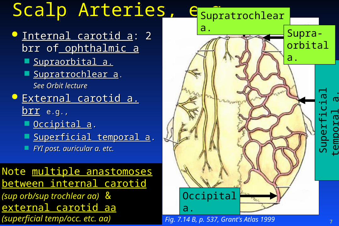
Scalp Arteries, e.g.Scalp Arteries, e.g.Scalp Arteries, e.g.Scalp Arteries, e.g. Internal carotid aInternal carotid a: 2 brr of: 2 brr of
ophthalmic aophthalmic a Supraorbital a.Supraorbital a. Supratrochlear aSupratrochlear a..
See Orbit lectureSee Orbit lecture
External carotid a. brrExternal carotid a. brr e.g.,e.g.,
Occipital aOccipital a.. Superficial temporal aSuperficial temporal a.. FYI post. auricular a. etc.FYI post. auricular a. etc.
Fig. 7.14 B, p. 537, Grant's Atlas 1999
Sup
erfic
ial t
empo
ral a
.
Occipital a.
Supratrochlear a.
Supra-orbital a.
Note multiple anastomoses between internal carotid (sup orb/sup
trochlear aa) & external carotid aa (superficial temp/occ. etc. aa)

8
Laceration of scalpLaceration of scalp aa: aa:Laceration of scalpLaceration of scalp aa: aa: Bleed like crazyBleed like crazy
Bleed from Bleed from bothboth ends of laceration ends of laceration Due to lots of anastomosesDue to lots of anastomoses FYI. Also, held open by subcutaneous collagen fibersFYI. Also, held open by subcutaneous collagen fibers FYI. Good news: washes away infections better than other sites FYI. Good news: washes away infections better than other sites
Scalp lacerationsScalp lacerations Most common head laceration requiring surgeryMost common head laceration requiring surgery
Clinical correlate
http://www.aafp.org/afp/2008/1015/p945.html
FY
I. H
air
ap
posi
tion
te
chni
que
w/ t
issu
e
glu
e fo
r su
per
ficia
l, n
on-b
lee
din
g w
oun
d
FYI. For hemostasis (stoppage of bleeding): provide local anesthetic w/ epinephrine & manual pressure; clip/clamp larger aa

9
Diploic veins of skullDiploic veins of skull:: Diploic veins of skullDiploic veins of skull::
Fig
. 7.1
6, p
. 538
, Gra
nt's
Atla
s 19
99
They are in the diploe “Di-Plo-E” (AKA spongy layer or cancellous bone) of the calvaria (skullcap). Bone marrow is also in the diploe By contrast, the external & internal surfaces of the calvaria are made of compact (cortical) bone.

10
Emissary veinsEmissary veins: connect the skull : connect the skull surface w/ cranial vaultsurface w/ cranial vaultEmissary veinsEmissary veins: connect the skull : connect the skull surface w/ cranial vaultsurface w/ cranial vault
Pla
te 1
02 u
pper
, Net
ter
Emissary v.
They drain into Dural sinus: part of the venous drainage system of brain; discussed later
Diploic v.
Superficial v

11
CalvariaCalvaria has has foramina for emissary vvforamina for emissary vv..CalvariaCalvaria has has foramina for emissary vvforamina for emissary vv..
Fig
. 7.4
B, p
. 841
, Moo
re 1
999
Emissary v. foramina (present in various skull bones)
FYI. Indentations for arachnoid granulations are bigger: Granulations shunt CSF into dural sinuses
anterior
posterior

12
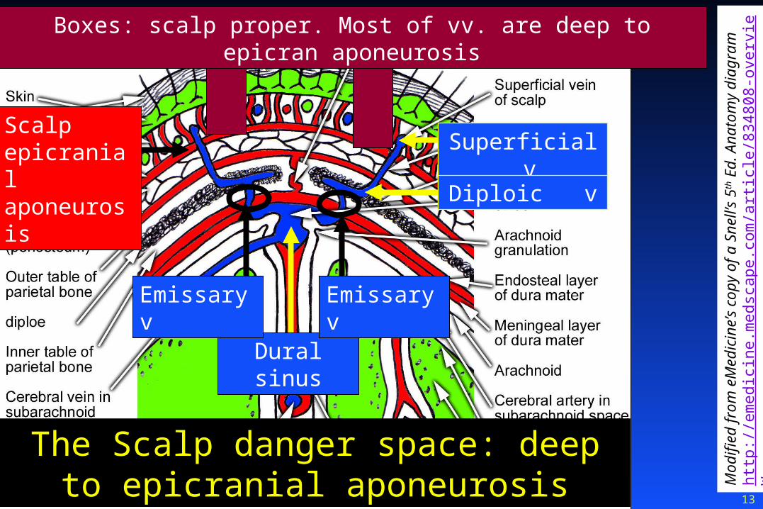
Epicranial aponeurosis:Epicranial aponeurosis:scalp danger zonescalp danger zoneEpicranial aponeurosis:Epicranial aponeurosis:scalp danger zonescalp danger zone Infections Infections deep to epicranial aponeurosisdeep to epicranial aponeurosis
Can gain access to cranial vaultCan gain access to cranial vault Via Emissary veinsVia Emissary veins Scalp infection then becomes intracranial infectionScalp infection then becomes intracranial infection
Infections Infections superficial to epicranial aponeurosissuperficial to epicranial aponeurosis Much less likely to cause intracranial infectionMuch less likely to cause intracranial infection
Clinical correlates
Q. Name another “danger space?” Ans. Retropharyngeal danger space. A fascial plane behind the visceral compartment of the neck
13
Mod
ified
from
eM
edic
ine’
s co
py o
f a S
nell’
s 5th
Ed.
Ana
tom
y di
agra
m
http
://em
edic
ine.
med
scap
e.co
m/a
rtic
le/8
3480
8-ov
ervi
ew
Boxes: scalp proper. Most of vv. are deep to epicran aponeurosis
Dural sinus
Emissary vEmissary v
The Scalp danger space: deep to epicranial aponeurosis
Scalp epicranial aponeurosis
Superficial v
Diploic v

14
Periorbital Ecchymosis Periorbital Ecchymosis ec-key-mosis ec-key-mosis
“P“Peri:” around eyes. “Ecchymosis:” subcutaneous hematomaeri:” around eyes. “Ecchymosis:” subcutaneous hematomaPeriorbital Ecchymosis Periorbital Ecchymosis ec-key-mosis ec-key-mosis
“P“Peri:” around eyes. “Ecchymosis:” subcutaneous hematomaeri:” around eyes. “Ecchymosis:” subcutaneous hematoma
Occipitofrontalis muscle: attachments:Occipitofrontalis muscle: attachments: Posterior attachment: mostly occipital bone Posterior attachment: mostly occipital bone & part of temporal bone & part of temporal bone
-prevents scalp bleeding/infection from going into neck-prevents scalp bleeding/infection from going into neck Lateral attachment: mostly zygomatic archesLateral attachment: mostly zygomatic arches
-prevents scalp bleeding/infection from going laterally-prevents scalp bleeding/infection from going laterally Anterior attachmentAnterior attachment: : skin of forehead/eyelidsskin of forehead/eyelids, , not bonenot bone
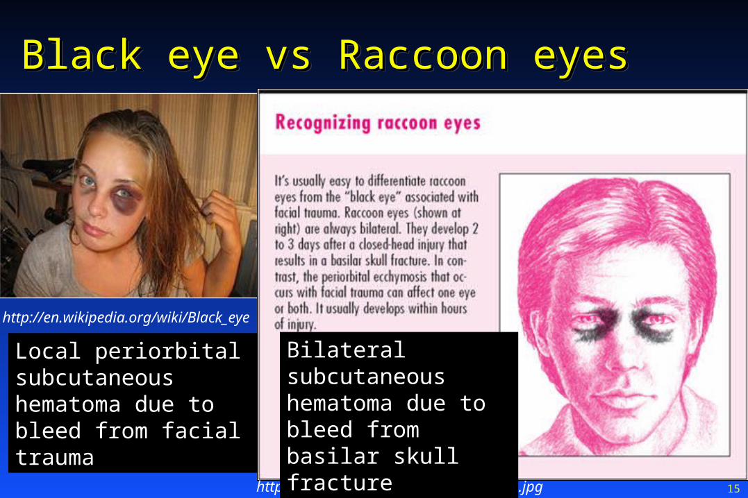
Scalp blood Scalp blood can can drain drain into into loose connective tissue of eyelids/root noseloose connective tissue of eyelids/root nose ““Black eye: Black eye: locallocal periorbital bleed; occurs periorbital bleed; occurs w/in hrs post traumaw/in hrs post trauma ““Raccoon Raccoon eyes: eyes:
Occurs Occurs 2-3 days post trauma2-3 days post trauma Always BilateralAlways Bilateral One cause: One cause: bleed from anterior basilar skull Fx can drain into lidsbleed from anterior basilar skull Fx can drain into lids
(i.e., fracture to cribriform plate in floor of anterior cranial fossa)(i.e., fracture to cribriform plate in floor of anterior cranial fossa)
Clinical correlate
15
Black eye vs Raccoon eyesBlack eye vs Raccoon eyesBlack eye vs Raccoon eyesBlack eye vs Raccoon eyes
http://en.wikipedia.org/wiki/Black_eye
http://hgimg.com/bookimages/5/2461.1.jpg
Local periorbital subcutaneous hematoma due to bleed from facial trauma
Bilateral subcutaneous hematoma due to bleed from basilar skull fracture

16
Scalp lacerationsScalp lacerationsScalp lacerationsScalp lacerations
Lacerations Lacerations superficial to epicranial aponeurosissuperficial to epicranial aponeurosis Do not gape (aponeurosis holds edges close)Do not gape (aponeurosis holds edges close)
Lacerations Lacerations deep to epicranial aponeurosisdeep to epicranial aponeurosis Gape widely; Gape widely; much more bleedingmuch more bleeding
FYI: especially in coronal plane due to opposite pulls of 2 bellies of occipitofront mFYI: especially in coronal plane due to opposite pulls of 2 bellies of occipitofront m
Suture to adjacent epicranial aponeurosisSuture to adjacent epicranial aponeurosis
Clinical correlates

17
Outline: Outline: Cranial Vault & DuraCranial Vault & DuraOutline: Outline: Cranial Vault & DuraCranial Vault & Dura
ScalpScalp Cranial nerve openingsCranial nerve openings Dura materDura mater Dural sinusesDural sinuses Internal carotid, vertebral & middle meningeal aaInternal carotid, vertebral & middle meningeal aa
18
Fig
. 7. 4
B, p
. 527
, G
rant
’s A
tlas
1999
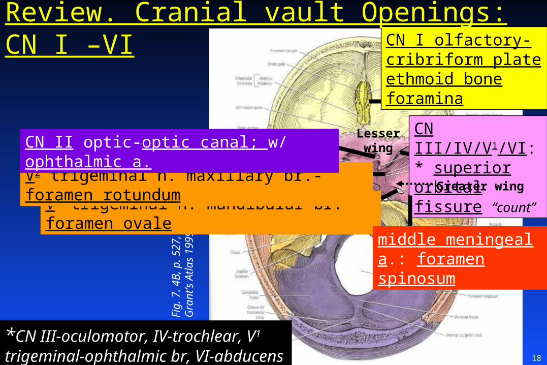
CN III/IV/V1/VI:* superior orbital fissure “count”
V3 trigeminal n. mandibular br.-foramen ovale
middle meningeal a.: foramen spinosum
CN I olfactory-cribriform plate ethmoid bone foramina
*CN III-oculomotor, IV-trochlear, V1 trigeminal-ophthalmic br, VI-abducens
V2 trigeminal n. maxillary br.-foramen rotundum
Review. Cranial vault Openings: CN I –VI
CN II optic-optic canal; w/ ophthalmic a.Lesser wing
Greater wing

19
Fig
. 7.
4B
, p.
527
, G
rant
’s A
tlas
1999
CN VII/VIII-facial/vestibulocochlear: internal auditory meatus on upper posterior face of petrous part of temporal bone
CN IX/X/XI:* jugular foramen on post. lower face of petrous; “count”
CN XII hypoglossal: hypoglossal canal in occipital bone*CN’s: IX-glossopharyngeal, X-vagus,
XI-spinal accessory
Review. Openings: CN VII - XII
20
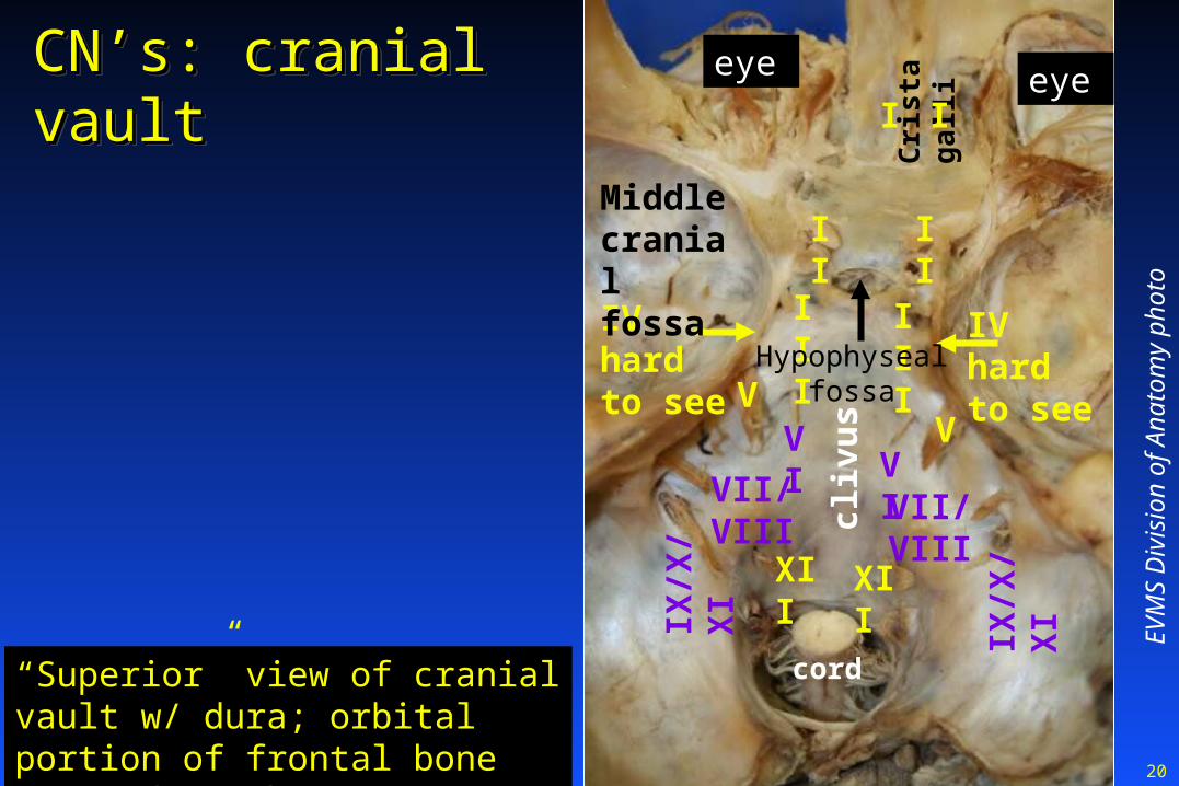
CN’s: cranial vaultCN’s: cranial vaultCN’s: cranial vaultCN’s: cranial vault
EV
MS
Div
isio
n of
Ana
tom
y ph
oto
Cri
sta
ga
lli
“Superior” view of cranial vault w/ dura; orbital portion of frontal bone removed to show eye
eye eyeI I
II II
III IIIIV hard to see
IV hard to see
VV
VIVI
VII/VIII VII/VIII
IX/X
/XI
IX/X
/XI
cord
XII XII
Hypophyseal fossa
cliv
us
Middle cranial fossa

21
CN II optic n: CN II optic n: nearnearcerebral part ofcerebral part of internal internal carotid acarotid a
CN II optic n: CN II optic n: nearnearcerebral part ofcerebral part of internal internal carotid acarotid a
Aneurysm hereAneurysm here Can affect visionCan affect vision
EV
MS
Div
isio
n of
Ana
tom
y ph
oto
Cri
sta
ga
lli
“Superior” view of cranial vault w/ dura; orbital portion of frontal bone removed to show eye
eye eye
III III
V
Internal carotid a cerebral segment
Internal carotid a
CN II CN II
cord
XII
Hypophyseal fossa
IX/X
/XI
VII/VIII
VIClinical correlate

22
10 CN’s 10 CN’s connect connect w/ w/ brainstembrainstem
10 CN’s 10 CN’s connect connect w/ w/ brainstembrainstem
Div
. A
nato
my
EV
MS
Hemisected head
Clivus: mostly made from basilar part of occipital bone*
Squamous (“plate-like”) part of Occipital bone Dotted line:
foramen magnum
brai
nste
m
*AKA “basiocciput”
All but CN’s CN I olfactory, & XI spinal acc

23
Outline: Outline: Cranial Vault & DuraCranial Vault & DuraOutline: Outline: Cranial Vault & DuraCranial Vault & Dura
ScalpScalp Cranial nerve openingsCranial nerve openings Dura materDura mater Dural sinusesDural sinuses Internal carotid, vertebral & middle meningeal aaInternal carotid, vertebral & middle meningeal aa

24
Review. Review. MeningesMeninges: “3 mothers” or : “3 mothers” or coverings for the central nervous systemcoverings for the central nervous systemReview. Review. MeningesMeninges: “3 mothers” or : “3 mothers” or coverings for the central nervous systemcoverings for the central nervous system Tuff mother: Tuff mother: Dura materDura mater
Superficial/outer layerSuperficial/outer layer Adheres directly to inside surface of cranial vaultAdheres directly to inside surface of cranial vault
Spider mother: Spider mother: Arachnoid materArachnoid mater IntermediateIntermediate
Tender mother: Tender mother: Pia materPia mater DeepestDeepest Adheres directly to brain/cordAdheres directly to brain/cord
LeptomeningesLeptomeninges: arachnoid mater + pia mater: arachnoid mater + pia mater
25
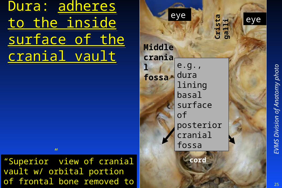
Dura: Dura: adheres to adheres to the inside surface the inside surface of the cranial vaultof the cranial vault
Dura: Dura: adheres to adheres to the inside surface the inside surface of the cranial vaultof the cranial vault
EV
MS
Div
isio
n of
Ana
tom
y ph
oto
Cri
sta
ga
lli
“Superior” view of cranial vault w/ orbital portion of frontal bone removed to show eye
eye eye
cord
Hypophyseal fossa
cliv
us
Middle cranial fossa e.g., dura
lining basal surface of posterior cranial fossa
26
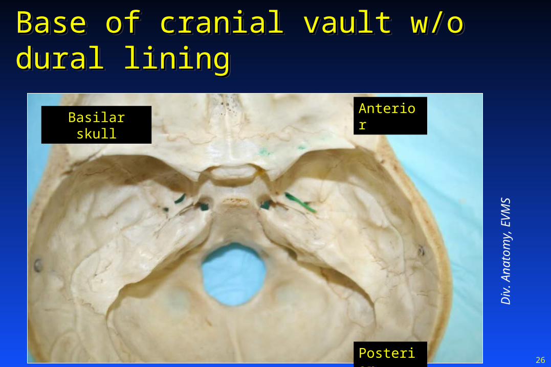
Base of cranial vault w/o dural liningBase of cranial vault w/o dural liningBase of cranial vault w/o dural liningBase of cranial vault w/o dural lining
Div
. A
nato
my,
EV
MS
Basilar skullAnterior
Posterior
27
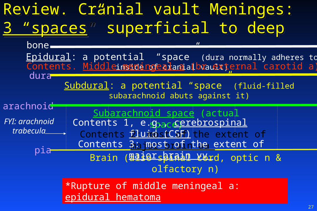
Review. Cranial vault Meninges: Review. Cranial vault Meninges: 3 “spaces3 “spaces” superficial to deep” superficial to deepReview. Cranial vault Meninges: Review. Cranial vault Meninges: 3 “spaces3 “spaces” superficial to deep” superficial to deep
Epidural: a potential “space” (dura normally adheres to inside of cranial vault)
Subdural: a potential “space” (fluid-filled subarachnoid abuts against it)
Subarachnoid space (actual space)
Brain (also spinal cord, optic n & olfactory n)
bone
Contents. Middle meningeal a (br external carotid a)*dura
arachnoid
pia
Contents 1, e.g., cerebrospinal fluid (CSF)FYI: arachnoid trabecula
*Rupture of middle meningeal a: epidural hematoma
Contents 2: most of the extent of major brain aa Contents 3: most of the extent of major brain vv
28
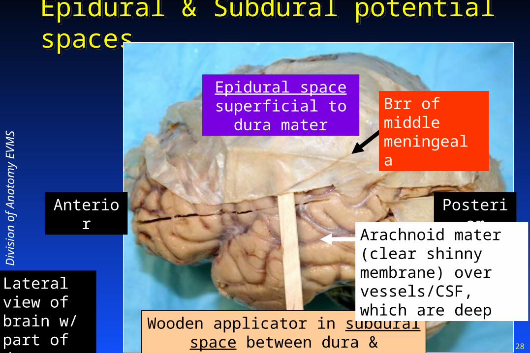
Epidural & Subdural potential spacesEpidural & Subdural potential spacesEpidural & Subdural potential spacesEpidural & Subdural potential spaces
Anterior Posterior
Epidural space superficial to dura
mater
Wooden applicator in subdural space between dura & arachnoid
Arachnoid mater (clear shinny membrane) over vessels/CSF, which are deep
Div
isio
n of
Ana
tom
y E
VM
S
Brr of middle meningeal a
Lateral view of brain w/ part of dura removed

29
Dural repairDural repairDural repairDural repair
Suture/seal after cranial surgerySuture/seal after cranial surgery Fascia lata from pt’s own thighFascia lata from pt’s own thigh
Fascia lata is deep fascia on lateral thighFascia lata is deep fascia on lateral thigh Synthetic duraSynthetic dura Bovine pericardial sacBovine pericardial sac 2011 FDA advisory2011 FDA advisory11 cautioning against certain cautioning against certain
Cadaveric dura transplantsCadaveric dura transplants b/c of risk of fatal prion infection b/c of risk of fatal prion infection Creutzfeldt-Jakob DiseaseCreutzfeldt-Jakob Disease
1http://www.fda.gov/MedicalDevices/Safety/AlertsandNotices/PublicHealthNotifications/ucm241829.htm
Sutures + “Dura Seal”
http
://m
edga
dget
.com
/200
5/04
/dur
asea
l.htm
l

30
FYI P
ost-c
entra
l gyr
us
FY
I Pre
-cen
tral
gyr
us
Blue pin through arachnoid mater. Note how the arachnoid jumps over sulci (valleys), unlike deeper pia mater, which adheres to brain and penetrates sulci
FYI gyri
Div
isio
n of
Ana
tom
y. E
VM
S

31
Meninges: 3Meninges: 3 Differences between Cord/Brain Differences between Cord/BrainMeninges: 3Meninges: 3 Differences between Cord/Brain Differences between Cord/Brain
Spinal Cord
vertebrae
duraarachnoid
pia
FYI: arachnoid trabecula
1. Unlike Brain: Actual epidural space exists with cord
2. Unlike Brain: Contains fat & internal vertebral venous plexus
Subarachnoid space (actual space)
3. Unlike Brain: Subdural space is NOT clinically relevant in cord
32
Dural tears & basilar skull FxDural tears & basilar skull FxDural tears & basilar skull FxDural tears & basilar skull Fx Trauma- (or surgically) induced torn dura/arachnoidTrauma- (or surgically) induced torn dura/arachnoid CSF rhinorrheaCSF rhinorrhea
CSF (clear fluid) leaks out of CSF (clear fluid) leaks out of nosenose Following Head injury via:Following Head injury via:
Basal skull FxBasal skull Fx of of cribriform plate of ethmoid cribriform plate of ethmoid (i.e., Fx of base of (i.e., Fx of base of anterior cranial fossa) anterior cranial fossa)
CSF otorrheaCSF otorrhea
CSF leaks out of CSF leaks out of earear Following head injury viaFollowing head injury via Basal skull FxBasal skull Fx of of petrous part temporal bone petrous part temporal bone Ruptured ear drum Ruptured ear drum also neededalso needed for CSF for CSF to to leak out of earleak out of ear
Clinical correlates
33
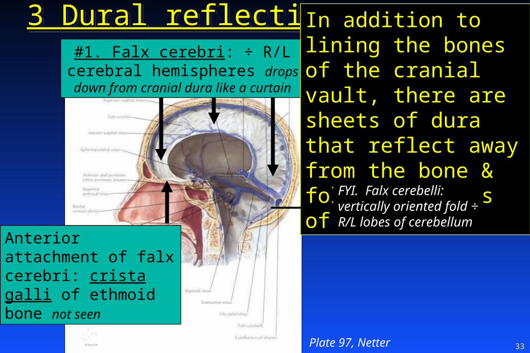
3 Dural reflections3 Dural reflections3 Dural reflections3 Dural reflections
Plate 97, Netter
Anterior attachment of falx cerebri: crista galli of ethmoid bone not seen
#1. Falx cerebri: ÷ R/L cerebral hemispheres drops down from
cranial dura like a curtain
In addition to lining the bones of the cranial vault, there are sheets of dura that reflect away from the bone & follow contours of brain
FYI. Falx cerebelli: vertically oriented fold ÷ R/L lobes of cerebellum

34
Mod
ified
fro
m P
late
98,
Net
ter
1997
3 Folds of dura, cont.
From, e.g., top of petrous portion of temporal bone and A/P clinoid processes
To falx cerebri, cut here
#3. Diaphragma sellae: separates pituitary from brain
#2. Tentorium cerebelli: makes a tent above cerebellum to separate it from cortex
Superior view of cranial vault

35
Diaphragma sellae Diaphragma sellae “diaphragm of the saddle”“diaphragm of the saddle”Diaphragma sellae Diaphragma sellae “diaphragm of the saddle”“diaphragm of the saddle”
EV
MS
Div
isio
n of
Ana
tom
y ph
oto
Cri
sta
ga
lli
“Superior” view of cranial vault w/ orbital portion of frontal bone removed to show eye
eye eye
cord
pituitary
cliv
us
Middle cranial fossa
Dotted line: Diaphragma sellae separating pituitary from brain

36
Div
. A
nato
my
EV
MS
Falx cerebri
cerebellumTentorium cerebelli
Hemisected head
Other views

37
Div
isio
n of
Ana
tom
y E
VM
SFalx cerebri
Tentorium cerebelli running “down hill”
Falx cerebri and tentorium cerebelliFalx cerebri and tentorium cerebelliFalx cerebri and tentorium cerebelliFalx cerebri and tentorium cerebelliBrain cut away at brainstem
38
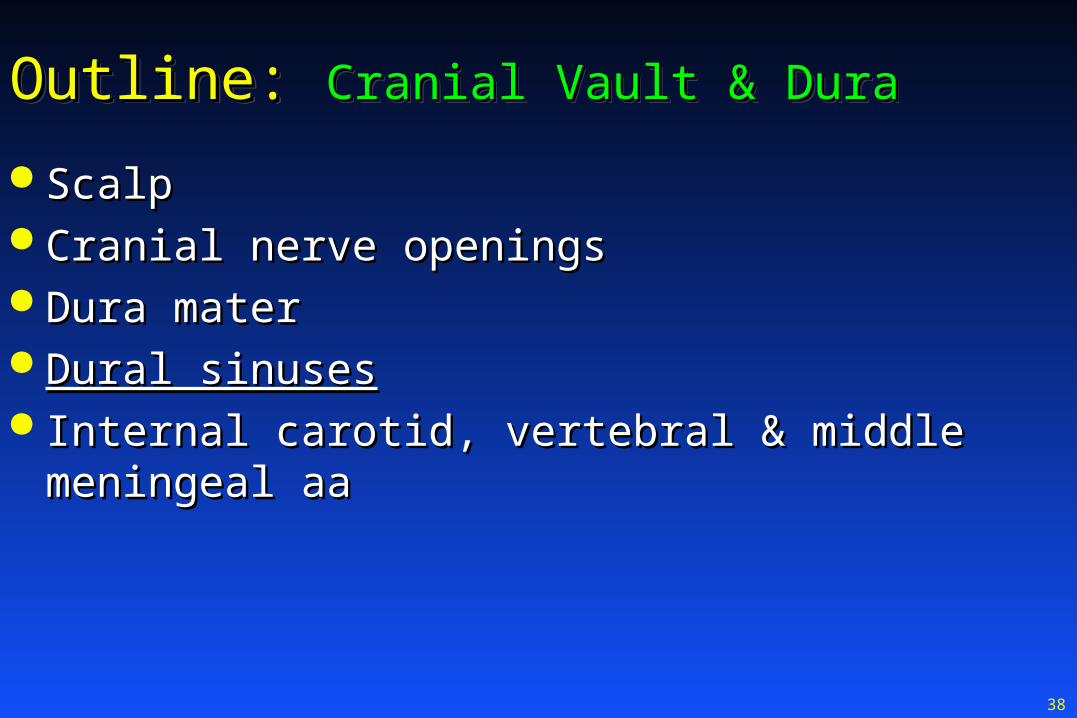
Outline: Outline: Cranial Vault & DuraCranial Vault & DuraOutline: Outline: Cranial Vault & DuraCranial Vault & Dura
ScalpScalp Cranial nerve openingsCranial nerve openings Dura materDura mater Dural sinusesDural sinuses Internal carotid, vertebral & middle meningeal aaInternal carotid, vertebral & middle meningeal aa

39
Dural sinusesDural sinusesDural sinusesDural sinuses There are There are 2 layers of dura2 layers of dura
Periosteal layerPeriosteal layer (superficial; against bone) (superficial; against bone) Meningeal layerMeningeal layer (deeper) (deeper)
Layers split to form dural sinusesLayers split to form dural sinusesA dural sinus is, therefore, A dural sinus is, therefore, a tube of duraa tube of dura
2 Functions of Dural sinuses2 Functions of Dural sinuses:: Drain Drain venous bloodvenous blood from brain from brain
-To -To internal jugular vvinternal jugular vv FYI: R is bigger than LFYI: R is bigger than L
Drain Drain cerebral spinal fluid from braincerebral spinal fluid from brain
Dural sinus
40
6 Selected dural sinuses6 Selected dural sinuses6 Selected dural sinuses6 Selected dural sinuses Superior sagittal sinusSuperior sagittal sinus Inferior sagittal sinusInferior sagittal sinus Straight sinusStraight sinus Transverse sinusTransverse sinus Sigmoid sinusSigmoid sinus Cavernous sinusCavernous sinus

41
Dural sinusesDural sinusesDural sinusesDural sinuses
Pla
te 9
7, N
ette
r
Falx cerebriInferior sagittal sinus; in lower part of falx cerebri
Superior sagittal sinus; in upper part of falx cerebri
Straight sinus; in medial part of tentorium cerebelli
Confluence of sinuses
42
Mod
ified
from
Pla
te 9
8, N
ette
r
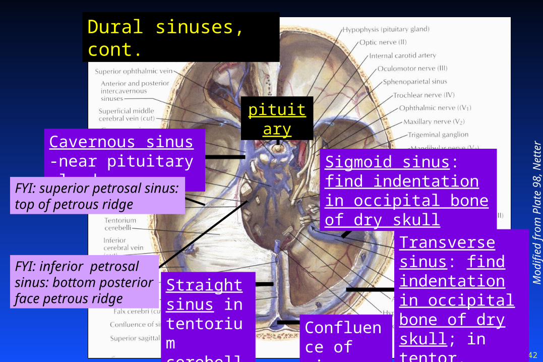
Transverse sinus: find indentation in occipital bone of dry skull; in tentor. cerebelli
Cavernous sinus -near pituitary gland Sigmoid sinus: find
indentation in occipital bone of dry skull
Straight sinus in tentorium cerebelli
Confluence of sinuses
Dural sinuses, cont.
FYI: superior petrosal sinus: top of petrous ridge
FYI: inferior petrosal sinus: bottom posterior face petrous ridge
pituitary

43
Indentations: transverse & sigmoid sinusesIndentations: transverse & sigmoid sinusesIndentations: transverse & sigmoid sinusesIndentations: transverse & sigmoid sinuses
Blue arrow: transverse sinus
Black arrow: sigmoid sinus
Div
. Of a
nato
my.
EV
MS
Orange arrow: jugular foramen for IJV

44
Div
isio
n of
Ana
tom
y E
VM
S
Green wire traveling in opened superior sagittal sinus on top of falx cerebri
Green wire continuing into opened transverse sinus within tentorium cerebelli
General area of the confluence of sinuses
Occipital sinus deep
Posterior view of brain

45
Superior sagittal sinus
Confluence of sinusesTransverse Sinus
Sigmoid Sinus
Div
. of
Ana
tom
y, E
VM
S
Dural sinuses & internal jugular v posterior view
Internal jugular v
angiogram

46
Mod
ified
fro
m P
late
98,
Net
ter
Cavernous sinus -near pituitary gland
Cavernous sinus: 3, 4, 51,52,6, int carotid a., & post. ganglionic. SNS on ICA “count” or say “O TOM CAS”
In the wall of the
sinus: CN III, IV, V1 & V2 “O TOM”34
`
51 52
Within the sinus itself; the int. carotid artery and CN VI. “CA” plus
sympathetics on the ICA “S.” “O TOM CAS”
6
C
The unusual instance of an artery inside a venous system

47
Cavernous sinus: Cavernous sinus: Internal carotid a & CN’s 3,4,5Internal carotid a & CN’s 3,4,511,5,522,6 ,6 “count”“count”Cavernous sinus: Cavernous sinus: Internal carotid a & CN’s 3,4,5Internal carotid a & CN’s 3,4,511,5,522,6 ,6 “count”“count”
Div
isio
n of
Ana
tom
y E
VM
SStick: sup orbital fissure w/ III, IV, V1, VI
Orange: V2 heading for foramen rotundum
Pipe cleaner: internal carotid a w/ post. gang. SNS fibers from sup cerv gang
Sella turcica
A: Paralyze all eye movement: III, IV & VI
Box: Carotid sinus
cliv
us
Superior view of the middle cranial fossa
Q: A cavernous sinus thrombus would have what affect on eye movement?
Cavernous sinus also has ophthalmic vv draining into it from orbit
Clinical correlate

48
Cavernous Cavernous sinus sinus white boxwhite box
Cavernous Cavernous sinus sinus white boxwhite box
Black circle: hypophyseal fossa
IIII
IIIIII
VI VI VV1/2
Middle fossa
Posterior fossa
Div
. Of a
nato
my.
EV
MS
cordcl
ivus
Superior view of the base of the cranial vault
Green wires entering sinus w/ CN III
not seen-IV
ICA* ICA*
*ICA exiting sinus w/ SNS fibers
49
Mod
ified
fro
m P
late
98,
Net
ter
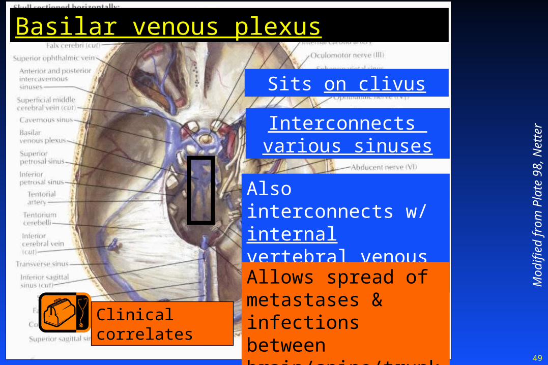
Basilar venous plexus
Sits on clivus
Interconnects various sinuses
Also interconnects w/ internal vertebral venous plexus of spine
Allows spread of metastases & infections between brain/spine/trunk
Clinical correlates
50
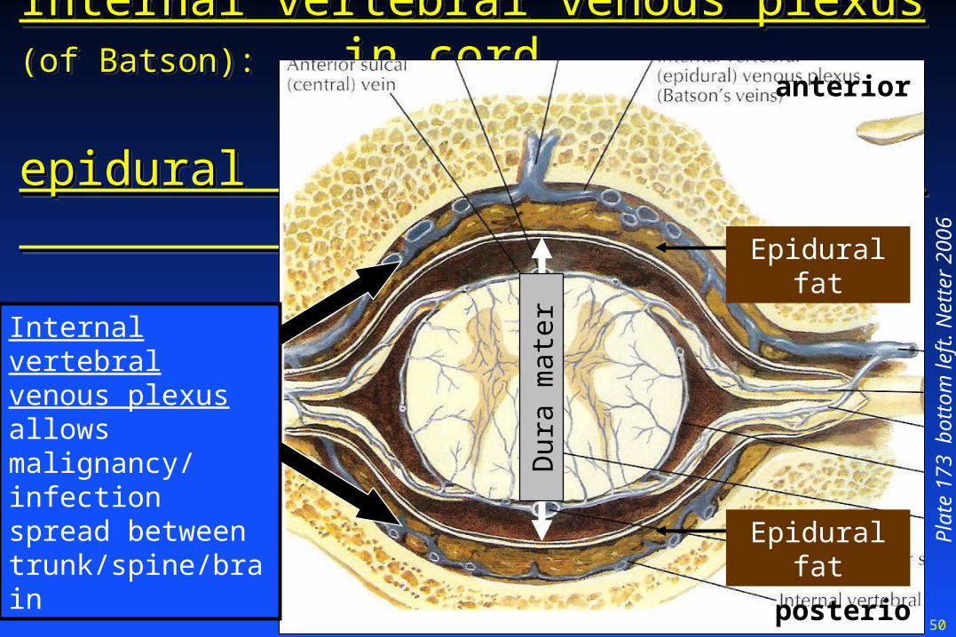
Internal vertebral venous plexusInternal vertebral venous plexus (of Batson):(of Batson): in cord in cord epidural epidural spacespace
Internal vertebral venous plexusInternal vertebral venous plexus (of Batson):(of Batson): in cord in cord epidural epidural spacespace
Pla
te 1
73
bott
om le
ft.
Net
ter
2006
anterior
posterior
Internal vertebral venous plexus allows malignancy/ infection spread between trunk/spine/brain
Dur
a m
ater
Epidural fat
Epidural fat

51
Outline: Outline: Cranial Vault & DuraCranial Vault & DuraOutline: Outline: Cranial Vault & DuraCranial Vault & Dura
ScalpScalp Cranial nerve openingsCranial nerve openings Dura materDura mater Dural sinusesDural sinuses Internal carotid, vertebral & middle meningeal aaInternal carotid, vertebral & middle meningeal aa

52
Brain: Blood SupplyBrain: Blood SupplyBrain: Blood SupplyBrain: Blood Supply
Fig. 10.2, p. 369, Blumenfeld. Neuroanatomy Through Clinical Cases. Sinauer, 2002
FYI Common carotid aa
FY
I L. s
ubc
lavi
an a
FYI External carotid a
FYI Arch of aorta
Ver
tebr
al-b
asila
r sy
stem
Internal carotid aInternal carotid a Br common carotid aBr common carotid a Most strokesMost strokes
Vertebral aaVertebral aa 11stst ascending br subclavian a ascending br subclavian a Travel through: Travel through:
--transverse foramina of 1transverse foramina of 1stst 6 cerv 6 cerv vertvert & through& through
--foramen magnumforamen magnum
-make -make basilar abasilar aIn
tern
al c
arot
id a

53
Internal carotid arteryInternal carotid artery (red): (red): segmentssegmentsInternal carotid arteryInternal carotid artery (red): (red): segmentssegments
Grant’s Atlas 2005 Fig. 7.23B p. 621
Cervical seg. That part in
the neck. NO brr in neck
Petrous seg. That part inside carotid canal, which is in petrous part of temp bone
Cavernous seg. That part inside cavernous sinus
Cerebral seg

54
Slide 4
Internal carotid artery segments. AP MRA (magnetic resonance angiogram). Does not “capture” stationary tissue
Cervical seg.
Petrous seg.
Cavernous seg.
Cerebral seg.
Klioze SD. The Brain. Dept. of Anatomy & Neurobiology & Dept. Radiology, EVMS

55
Vertebral aa Vertebral aa dotted linesdotted linesVertebral aa Vertebral aa dotted linesdotted lines
Posterior view of clivus
Div
. Of a
nato
my.
EV
MS
cliv
usVIVI
VV
XI
III III
IXX
VII VIII
XII
Vertebral aa (reflected inferiorly) as they enter foramen magnum
56
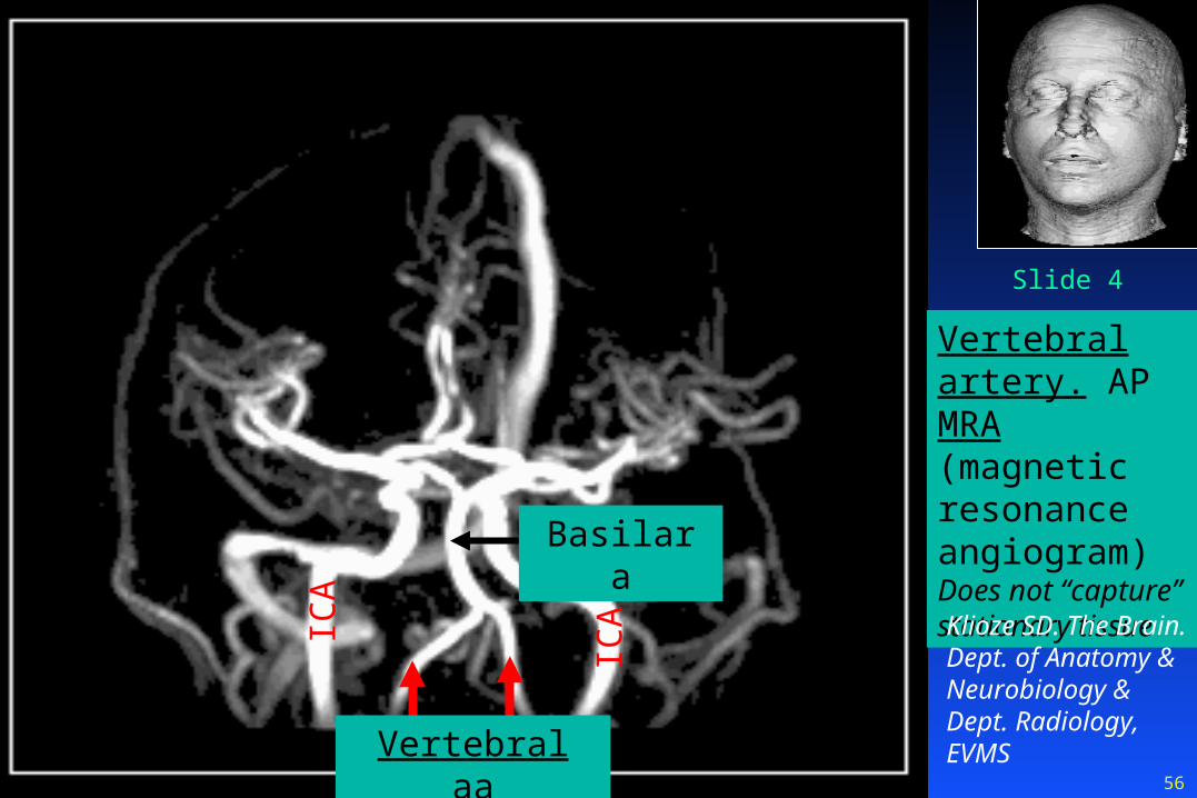
Slide 4
Vertebral artery. AP MRA (magnetic resonance angiogram) Does not “capture” stationary tissue
Vertebral aa
ICA
ICA Klioze SD. The
Brain. Dept. of Anatomy & Neurobiology & Dept. Radiology, EVMS
Basilar a

57
Middle Middle meningeal ameningeal aMiddle Middle meningeal ameningeal a
Q: A Q: A Fx of the Fx of the pterionpterion will will rupture what a. rupture what a. & cause what & cause what kind of bleed?kind of bleed?
A: A: ant. br ant. br middle middle meningeal ameningeal a; ; epidural bleedepidural bleed
Grant’s Atlas 2005 Fig. 7.12A p. 609
Posterior br of middle
meningeal a
Anterior br of middle meningeal artery is deep to Pterion “H” encircled in black
FYI. A/P brr sometimes called Frontal/Parietal brr
External carotid a
Maxillary a
Middle meningeal a
Clinical correlate
Medial view

58
Craniotomy & CranioplastyCraniotomy & CranioplastyCraniotomy & CranioplastyCraniotomy & Cranioplasty CraniotomyCraniotomy: temporary removal of a bone flap: temporary removal of a bone flap
Epidural Video Epidural Video http://www.youtube.com/watch?v=dLMCwGmWvrw&feature=related
Store bone flap in abdominal subcutaneous space orStore bone flap in abdominal subcutaneous space or Store under the epicranial aponeurosis Store under the epicranial aponeurosis see next slide see next slide oror Freeze & store at a bone bankFreeze & store at a bone bank
CranioplastyCranioplasty: repair of damaged skull: repair of damaged skull Replace bone flap or useReplace bone flap or use Plastic implantPlastic implant
http://www.unifesp.br/dneuro/neurociencias/229_revisao.pdf
http://www.medscape.com/viewarticle/472974_4

59
Middle meningeal aMiddle meningeal aMiddle meningeal aMiddle meningeal a
Div
. A
nato
my,
EV
MS
Basilar skull
Anterior
Posterior
Enters cranial vault via foramen spinosum in greater wing of sphenoid
Petrous part
temp bone
Foramen ovale
Foramen magnum
Indentation made by post br of middle meningeal a

60
Cranial Vault & DuraCranial Vault & Dura
Objectives: Concept map:Objectives: Concept map:Cranial Vault & DuraCranial Vault & Dura
Objectives: Concept map:Objectives: Concept map: The organization of the scalp and its danger zone.The organization of the scalp and its danger zone. Blood supply to the scalpBlood supply to the scalp Openings for the cranial nervesOpenings for the cranial nerves Dura mater & its reflections in the cranial vaultDura mater & its reflections in the cranial vault Dural sinuses & the venous drainage of the brainDural sinuses & the venous drainage of the brain Internal carotid, vertebral & middle meningeal aaInternal carotid, vertebral & middle meningeal aa