Ari Marttila Engineering of Charge, Biotin-binding and Oligomerization of Avidin
Upload
david-chung-tiang-liangCategory
view
62download
0
J. Comp. Path. 2007,Vol.136, 57^64
A Non-biotin Polymerized Horseradish-peroxidaseMethod for the Immunohistochemical
Diagnosis of Canine Distemper
C. T. Liang*,y, L. L. Chuehy, V. F. Pangy, Y. X. Zhuo*, S. C. Liang*,z, C. K. Yu*,H. Chiang*, C. C. Leey and C. H. Liuy
*National LaboratoryAnimal Center, National Applied Research Laboratories, Nan-Kang,Taipei 115,yDepartment and Graduate Institute ofVeterinaryMedicine, College of Bioresources and Agriculture,
NationalTaiwan University,Taipei 106,zInstitute of Biology and Anatomy, National DefenseMedical Center,Taipei 114, and yShin KongWu-Ho-SuMemorial Hospital,
Taipei 111,Taiwan
Summary
This report describes a modi¢ed non-biotin polymerized horseradish peroxidase (HRP) immunohistochemicalmethod for the diagnosis of canine distemper virus (CDV) infection from formalin-¢xed, para⁄nwax-embeddedtissues. This method con¢rmed infection in seven of eight (87.5%) suspected cases. Labelled CDVantigen wasobserved in the following sites: cerebrum, cerebellum, meninges, glial cells, neurons, vascular endothelium, peri-ventricular areas and pericytes, and choroid plexus; grey and white matter and central canal of the spinal cord;renal pelvis and tubular epithelium, and urinary bladder epithelium; macrophages and lymphocytes in splenicwhite pulp and lymph nodes; skin epidermis; bronchiolar epithelium and alveolar macrophages; hepatic Kup¡ercells, andgastric and intestinalmucosal epithelium; strati¢ed squamous epitheliumof the tongue andoesophagus.With the non-biotin HRP detection system, pretreatment by autoclaving followed by microwave heating gavebetter labelling results than did microwave pretreatment alone. No obvious di¡erence was noted between thelabelling results produced by the non-biotin HRP detection system and the Super SensitiveTM Link-Label IHCdetection system.
r 2006 Elsevier Ltd. All rights reserved.
Keywords: canine distemper; dog; non-biotin HRP; viral infection
Introduction
Canine distemper (CD), which is caused by a morbilli-virus (genus Morbillivirus, family Paramyxoviridae), pro-duces systemic or central nervous system (CNS)infections of dogs and related species and is associatedwith high mortality inTaiwan (Wu et al., 2000;Yu et al.,2001).The characteristic CNS changes include polioen-cephalomalacia, white matter demyelination, astro-gliosis, eosinophilic intranuclear and cytoplasmicinclusionbodies, gemistocytes and occasional multinu-
cleated syncytial giant cells (Summers et al.,1984; Sum-mers and Appel, 1985). However, syncytial giant cellswere demonstrated in only 9^28% of canine distempervirus (CDV)- infected brains (Palmer et al., 1990; Wuet al., 2000), and eosinophilic intranuclear or cytoplas-mic inclusion bodies in only 17% (Haines et al., 1999),33% (Stanton et al., 2002), 38.1% (Wu et al., 2000)or 72% (Palmer et al., 1990). Thus, histological exami-nation of lesions does not appear to be a reliable indica-tor of CDV infection. Immunohistochemical detectionof CDV antigen in tissue sections was reported byPalmer et al. (1990) to be superior to the demonstrationof inclusion bodies or syncytial cells for the diagnosisof canine distemper encephalitis. In infected dogs,
www.elsevier.com/locate/jcpa
ARTICLE IN PRESS
0021-9975/$ - see front matter r 2006 Elsevier Ltd. All rights reserved.
doi:10.1016/j.jcpa.2006.11.002
Correspondence to: C.- H. Liu, Department of Veterinary Medicine,
NationalTaiwan University, No.1, Section 4, Roosevelt Road,Taipei,Taiwan
106 (e-mail: [email protected]).

immunohistochemical examinationdemonstrates CDVantigen in glial cells, neurons, ependymal cells, in£am-matory macrophages, choroid plexus cells and menin-geal cells (Mitchell et al., 1991); in haired skin andfootpad epithelium (Haines et al., 1999; Koutinas et al.,2004); and in lung, spleen, kidney and lymph nodes(Iwatsuki et al., 1995). The traditional avidin^biotincomplex (ABC) immunohistochemical labelling meth-od has several disadvantages. First, avidin has a highisoelectric point. At pH 7.4, it is positively charged andtends to bind to certain proteins (Haines and Chelack,1991).The hydrophobic and electrostatic characteristicsof the avidin conjugates appear to play a role in the non-speci¢c binding (Masuoka et al., 2002). Second, kidney,pancreas, liver, lymphoid tissue and nervous systemtissue may contain lectin-like, negatively charged,endogenous, biotin-rich material which may adherenon-speci¢cally to the ABC, resulting in false positivereactions (True, 1990; Ramos-Vara, 2005). Althoughthe background can be greatly reduced by replacingavidinwith streptavidin, background from endogenousbiotin is still a problem with streptavidin methods(Ramos-Vara, 2005).This report describes the development of a novel
method of non-biotin chain polymer conjugate label-ling combined with heat-induced epitope retrieval(Shi et al., 2001; Ramos-Vara, 2005) for CDV diagnosis.
Materials and Methods
Animals
Eight dogs (nos1^8), from either an animal shelter or aprivate clinic, with clinical signs of CDwere used in thestudy. All animals had at least one sign of each ofthe following: CNS infection (seizure, ataxia, circlingand myoclonus); digestive tract infection (diarrhoea,vomiting, depression and anorexia); and respiratorytract infection (nasal and ocular discharge, cough, dys-pnoea and sneezing). The animals were either huma-nely destroyed or died naturally.
Pathological Examination
Samples taken post mortem consisted of brain (includingmedulla oblongata, cerebrum and cerebellum), spinalcord, lung, trachea, lymph nodes, heart, liver, spleen,large and small intestine, stomach, kidneys, urinarybladder, adrenal gland, and skin. These samples were¢xed in 10% neutral bu¡ered formalin for at least36^48 h and no more than 7 days. They were thenprocessed by routine methods and embedded in paraf-¢n wax. Sections (6 mm) were either stained with hae-matoxylin and eosin (HE) for histopathologicalexamination or examined immunohistochemically, to
determine the type of lesion and the presence of inclu-sion bodies, syncytial cells and CDVantigen.
Immunohistochemistry (IHC)
The Super SensitiveTMNon-BiotinHRPDetection Sys-tem (BioGenex Laboratories, San Ramon, CA, USA)was used.The primary antibody was mouse anti-CDV(MCA 1893, Clone DV2-12; Serotec, Kidlington,Oxford, UK).Tissue sections were dewaxed in xylene and rehy-
drated in a graded alcohol series. Antigen unmaskingwas performed by immersion of sections inTrilogyTM
(Cell Marque, Hot Springs, Arkansas, USA) 5% in Qwater (Milli-Q, UF Plus; Millipore Co. Billerica,MA, USA) and boiling for 15min at 121 1C in anautoclave(SA-252F; Sturdy Industrial Co., Taipei,Taiwan). Sections were then immediately transferredto fresh hot TrilogyTM solution in a second stainingdish, heated by a 1450-W microwave oven (RE-C102;Sampo Co., Taipei, Taiwan). This was left to stand for10min at 80 1C. The sections were then immersed incoolTris-bu¡ered saline (TBS; DakoCytomation, Car-pinteria, CA, USA) for10min, and then rinsed inTBS.For evaluation of the antigen retrieval e¡ect, the ¢rstautoclave treatment was omitted and the sections wereplaceddirectly in fresh hotTrilogyTM solution as above.Endogenous peroxidase activity was quenched with
hydrogen peroxide 3% in methanol (Merck, Darm-stadt, Germany) for 10min at room temperature.Thiswas followed by rinsing in TBS and incubation inPower BlockTM solution (BioGenex) for10min at roomtemperature.The primary antibody diluted 1 in 500 inantibody diluent (Ventana Medical System, Tucson,AZ, USA) was then applied for 24 h at 4 1C.Substitution of TBS or negative mouse serum for the
primary antibody on sections of CDV-infected cere-brum served as a non-speci¢c negative control. Cere-bral sections from dogs with no evidence of distemperinfection served as a speci¢c negative control.The sec-tions were then rinsed in TBS, incubated with SuperEnhancerTM reagent (BioGenex) for 60min at roomtemperature, rinsed in TBS, and incubated in Poly-mer-HRP reagent (BioGenex) for 60min at room tem-perature. To compare these results with conventionalIHC, the Super EnhancerTM reagent was replaced bySuper Sensitive Multilinks reagent (BioGenex) andthe Polymer-HRP reagent was replaced by streptavi-din peroxidase reagent (Super SensitiveTMLink-LabelIHC Detection System; BioGenex). The subsequentprocedures were identical for each of the two methodscompared. These subsequent procedures consistedof rinsing in TBS once more, incubation with AEC(3-amino, 9 ethyl-carbazole) or DAB (3, 30- diamino-benzidine tetrahydrochloride) (BioGenex) chromogen
ARTICLE IN PRESS
C.T. Liang et al.58
solution for 3^5min at room temperature, counter-staining with Mayer’s haematoxylin, and mountingwith Super Mounts (BioGenex). The sections werethen examined microscopically.
CDV Antigen Test
The RapiGens Canine Distemper Antigen rapid testkit (RapiGen Inc., Keumjung, Gunpo, Kyonggi, Kor-ea) was used, according to the manufacturer’s instruc-tions. Brie£y, serum, plasma or ocular discharge fromsuspected cases was dispensed into the sample wells.The results were read after waiting for 5^10min. Onered or purple band in the control line (C) with no ap-parent band in the test line (T) indicated a negative re-sult for CDV infection. One red or purple band in thecontrol line (C) and one band in the test line (T) indi-cated a positive result.
Reverse Transcription Polymerase Chain Reaction (RT-PCR)
RNA was extracted from serum, heparinized wholeblood, cerebro-spinal £uid (CSF; without centrifuga-tion), nasal swab in 1ml of phosphate-bu¡ered saline(PBS), ocular swab in1ml of PBS, anda10-fold dilutionof vomitus or faeces fromdogswith clinically suspectedCD. The RNAwas extracted by theTRIzols reagent(Invitrogen, Carlsbad, CA, USA) phenol-chloroformextraction method. Brie£y, a 500 ml volume of homoge-nized tissue sample was placed in a 2-ml sterile micro-centrifuge tube and 1ml of TRIzols reagent wasadded.The tubewas left to stand for 5minat roomtem-perature. After vigorous shaking, followed by the addi-tion of 200 ml of chloroform (Merck) and vortexing toensure an even mix, the tube was left to stand for2^3min at room temperature before being centrifugedat13000 rpm (12000 g) for10min at 2^8 1C.The super-nate was then transferred to another sterile microcen-trifuge tube. An equal volume of isopropyl alcohol(Merck) wasmixedwith the supernate, whichwas thenkept at ÿ20 1C for 30min before being centrifuged at13000 rpm (12000 g) again for 15min at 2^8 1C. Theresultant supernate was discarded and the pellet mixedwith1ml of 75% cold ethanol (Merck). Centrifugationwas performed once again, as above for10min.The pel-let was air dried in a lamina £ow apparatus for 20min.
Finally, 20 ml of RNase free diethyl pyrocarbonate(DEPC) water were added to the RNA pellet. Primersspeci¢c to CDVAF378705 strain phosphoprotein (P)(Table 1) were used. cDNA synthesis was carried outin 20 ml of RT-PCR reaction cocktail mixture contain-ing1 ml of 10 mM primary F2 primer set mixture,10 mlof RNA template, 5X ¢rst strand bu¡er (including250mM Tris-HCl [pH 8.3], 375mM KCl, 15mMMgCl2 ), 2 ml of 0.1M dithiothreitol (DTT), 1 ml of10mM deoxynucleotide triphosphate mixture(dNTP),1 ml of reverse transcriptase 200 IU/ml (Super-ScriptTM II Reverse Transcriptase) (Invitrogen).cDNAwas synthesizedwith athermocycler (GeneAmpPCR System 2400; Applied Biosystems, Foster City,CA, USA) at 42 1C for 1h, and the sample was dena-tured by the same thermocycler at 70 1C for15min.The resulting cDNA product from each sample was
used as a template in the following PCR procedure.The PCR reaction cocktail mixture (30 ml ) contained1 ml of F2 and R2 primer set (each 10 mM),10� PCRbu¡er (containing 200mM Tris^HCl [pH 8.4], and500mM KCl ), 1 ml of 50mM MgCl2, 1 ml of 10mMdNTP mixture, and 0.5 ml of Taq DNA polymerase5 IU/ml (Invitrogen). The primary PCR ampli¢cationwas carried out with 35 cycles at 94 1C for 25 s, at57 1C for 25 s, and at 72 1C for 35 s.The ¢nal extensionwas carried out at 72 1C for 5min.Primary PCR product (1 ml) was used as the subse-
quent PCR template and ampli¢cation was performedwith nested F2 and R1PCR primer set in a further 35sequential cycles at 94 1C for 25 s, 57 1C for 25 s, and72 1C for 25 s.The ¢nal extension consisted of one cycleat 72 1C for 5min.The targeted ampli¢cation sequencewas a 200 base pair (bp) fragment. The ampli¢cationproducts were analysed on 1.6% agarose gel and theelectrophoresis results were observed under a UV illu-minator after ethidiumbromide staining.
Results
RT-PCR and Antigen Test
Positive results were obtained in four dogs (nos 1^4)with the RT-PCR and in three dogs (5, 6, 8) with theCDVantigen test (Table 2). Two of the eight dogs withCDV infection had not been vaccinated, but the vacci-nation status of the remainder was unknown.
ARTICLE IN PRESS
Table 1
Nucleotide sequences of primers
Primer Nucleotide sequence Location Direction
F2 50-TAAGGGAATCGAAGATGC-3’ 2160^2177 Forward
R1 50-CCATCAGCATGCTCACATC-3’ 2341^2359 Reverse
R2 50-GATCCCCCAGTTGACTTG-3’ 2568^2585 Reverse
Immunohistochemical Diagnosis of CDV Infection 59
Histopathology and Immunohistochemistry
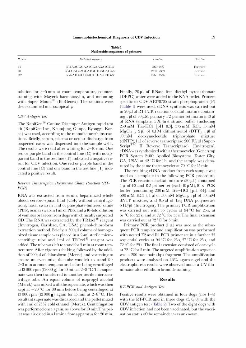
For antigen retrieval, autoclaving combined withmicrowave treatment yielded better labelling results(Fig.1) than did microwave treatment only (Fig. 2).Thenon-biotin HRP detection system gave essentially simi-lar labelling regardless of the substrate (AEC or DAB),but AEC labelling was preferable for small, ¢ne antigenparticles and for contrast. Immunolabelling producedby the non-biotin HRP detection system in CNS tissuesdid not di¡er signi¢cantly from that produced by theSuper SensitiveTMLink-Label detection system.All eight cases of suspected CD were con¢rmed
by the combination of methods used (RT-PCR and
CDV antigen test [Table 2], IHC [Table 3] andhistopathology [including and the demonstration ofintranuclear or cytoplasmic inclusion bodies]). Inclu-sionbodies were observed in seven cases (87.5%), albeitnot readily, their prevalence varying in di¡erenttissues. The most common sites were the periphery ofthe splenic central arteries, macrophages and lympho-cytes in the lymph nodes and urinary bladder epithe-lium, pulmonary macrophages and bronchiolarepithelium, ependymal cells lining the 4th ventricle,renal pelvis and cerebellar white matter astrocytes.Eosinophilic cytoplasmic inclusion bodies were occa-sionally seen in the renal pelvis and urinary bladderepithelium. Immunolabelling of the CDV antigen by
ARTICLE IN PRESS
Table 2
Details of the dogs with CDV infection
Clinical signs
Dog no. Breed Age (months) CNS GI Re CDVantigen
test
Positive RT-PCR Vaccination
record
1 Labrador
retriever
2 + + NA Blood, CSFand
urine
No
2 Chow chow 3 + + NA Blood NA
3 Shiba 3 + + NA Nasal swab,
ocular swab
NA
4 Dachshund 1 + + NA Blood, stool,
vomitus
No
5 Mongrel 3 + + NA NA
6 Shih tzu 2 + + + NA NA
7 Pomeranian 60 + NA NA NA
8 Miniature
pinscher
60 + + NA NA
CNS, central nervous system signs (seizure, ataxia, circling andmyoclonus); GI, gastrointestinal signs (diarrhoea, vomiting, depression andanorexia);
Re, respiratory signs including nasal and ocular discharge, cough, dyspnoea and sneezing; NA, not available.
Fig.1. Di¡use CDVimmunolabelling in the cerebellar whitematteris visible in the astrocytes (arrow). Non-biotin HRP (AEC)
with haematoxylin counterstain; autoclave with microwave
pretreatment. Bar, 50 mm.
Fig. 2. Same area as in Fig.1, showing CDV labelling in the cerebel-lar white matter, located focally in the astrocytes (arrow).
Non- biotin HRP (AEC) with haematoxylin counterstain;
microwave pretreatment only. Bar, 50 mm.
C.T. Liang et al.60
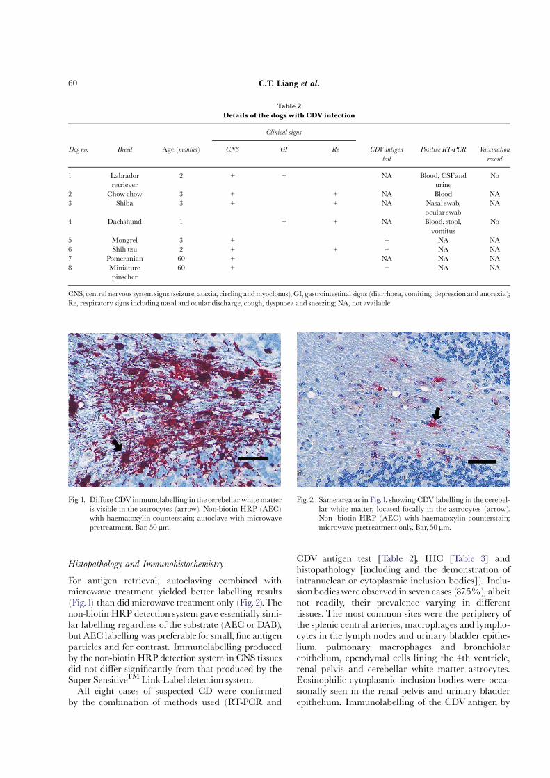
the non-biotin HRP method was observed in sevencases (Table 3).The brain lesions in the infected dogs consisted of de-
myelination and glial cell in¢ltration in the cerebel-lum, cerebellar peduncles and brain stem. CDVimmunolabelling in the cerebral cortex appeared dif-fusely or focally in the dendrites of large pyramidalcells (Fig. 3). It was particularly striking at the periph-ery of the 4th ventricle (Fig. 4) and in ependymal cells,but was present in only a few Purkinje cells. In somecases with cerebellar demyelinating areas, only a fewintranuclear inclusion bodies and neuropils were im-munolabelled. In more severely a¡ected cases, astro-cytes in the demyelinating white matter and brainstem showed immunolabelling with a spidery and
ARTICLE IN PRESSTable
3
Non-biotinHRPim
munohistochemicallabellingofCDVantigenin
tissuesofeightinfecteddogs
Resultsindogsno.
System
Tissues
12
34
56
78
Total
Respiratory
Lung
+,ecs
+,ecs
+,macrophages
ÿ+,ecsand
macrophages
+,ecsandsepta
+,ecsand
macrophages
+,ecsandsepta
7/8
Digestive
Stomach
++
+ÿ
++
++
7/8
Smallintestine
++
+ÿ
++,focal
++
7/8
Largeintestine
++
+ÿ
++
++
7/8
Nervous
Cerebrum
++
+ÿ
++
++
7/8
Cerebellum
++
+ÿ
++
++
7/8
Brain
stem
++
+ÿ
++
++
7/8
Lymphoid
Spleen
++
+ÿ
++
++
7/8
Lymphnode
++
NA
ÿ+
++
+6/7
Urinary
Urinary
bladder
++
NA
ÿ+
+,ecs
+,ecs
+,ecs
6/7
Kidney
++
+ÿ
++,pelvis,ecs
+,pelvis,ecs
+,pelvis,ecs
7/8
Others
Skin
++
+ÿ
++
++
7/8
Salivary
gland
+NA
+ÿ
++
++
6/7
NA,notavailable;ecs,epithelialcells.
Fig. 3. Di¡use CDV labelling in the cerebral cortex is seen in the
dendrites of large pyramidal cells (arrow). Non-biotin HRP(AEC) with haematoxylin counterstain; autoclave with mi-
crowave pretreatment. Bar, 50 mm.
Fig. 4. Extensive immunoreactivity with CDVantigen is exhibited
in the ependymal cells lining the 4th ventricle. Non-biotinHRP (AEC) with haematoxylin counterstain; autoclave
with microwave pretreatment. Bar,100 mm.
Immunohistochemical Diagnosis of CDV Infection 61
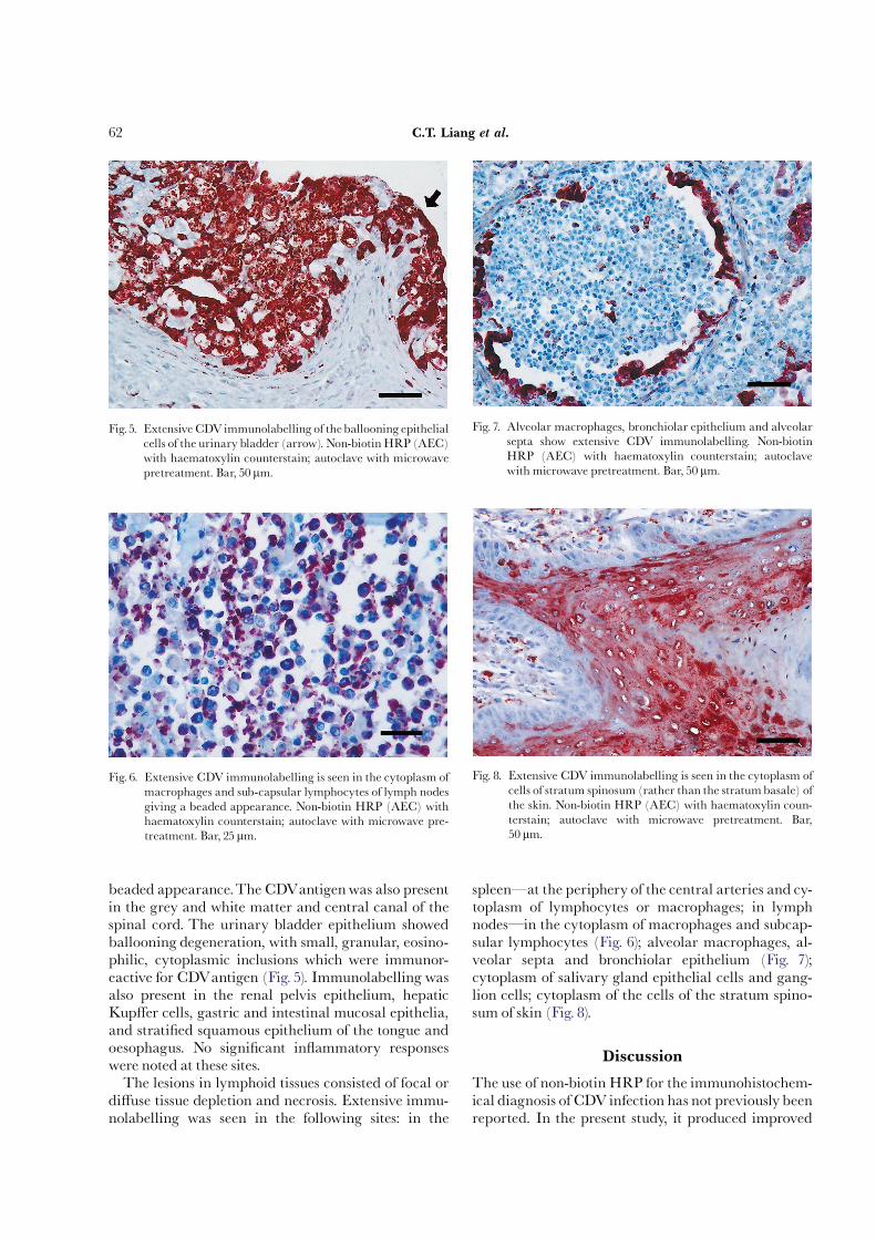
beaded appearance.The CDVantigenwas also presentin the grey and white matter and central canal of thespinal cord. The urinary bladder epithelium showedballooning degeneration, with small, granular, eosino-philic, cytoplasmic inclusions which were immunor-eactive for CDVantigen (Fig. 5). Immunolabelling wasalso present in the renal pelvis epithelium, hepaticKup¡er cells, gastric and intestinal mucosal epithelia,and strati¢ed squamous epithelium of the tongue andoesophagus. No signi¢cant in£ammatory responseswere noted at these sites.The lesions in lymphoid tissues consisted of focal or
di¡use tissue depletion and necrosis. Extensive immu-nolabelling was seen in the following sites: in the
spleençat the periphery of the central arteries and cy-toplasm of lymphocytes or macrophages; in lymphnodesçin the cytoplasm of macrophages and subcap-sular lymphocytes (Fig. 6); alveolar macrophages, al-veolar septa and bronchiolar epithelium (Fig. 7);cytoplasm of salivary gland epithelial cells and gang-lion cells; cytoplasm of the cells of the stratum spino-sum of skin (Fig. 8).
Discussion
The use of non-biotin HRP for the immunohistochem-ical diagnosis of CDVinfection has not previously beenreported. In the present study, it produced improved
ARTICLE IN PRESS
Fig.5. Extensive CDVimmunolabelling of theballooning epithelialcells of the urinary bladder (arrow). Non-biotin HRP (AEC)
with haematoxylin counterstain; autoclave with microwave
pretreatment. Bar, 50 mm.
Fig. 6. Extensive CDV immunolabelling is seen in the cytoplasm of
macrophages and sub-capsular lymphocytes of lymph nodes
giving a beaded appearance. Non-biotin HRP (AEC) withhaematoxylin counterstain; autoclave with microwave pre-
treatment. Bar, 25 mm.
Fig.7. Alveolar macrophages, bronchiolar epithelium and alveolarsepta show extensive CDV immunolabelling. Non-biotin
HRP (AEC) with haematoxylin counterstain; autoclave
with microwave pretreatment. Bar, 50 mm.
Fig. 8. Extensive CDV immunolabelling is seen in the cytoplasm of
cells of stratum spinosum (rather than the stratumbasale) of
the skin. Non-biotin HRP (AEC) with haematoxylin coun-
terstain; autoclave with microwave pretreatment. Bar,50 mm.
C.T. Liang et al.62
contrast against the background, making the detectionof CDVantigen easier than the histological detection ofintranuclear inclusion bodies. The cloned DV2-12 pri-mary antibody used required pretreatment of forma-lin-¢xed, wax-embedded sections by steaming,proteinase K pretreament having been shown byRamos-Vara and Beissenherz (2000) to be withoute¡ect for this antibody.The use of di¡erent ¢xatives, with or without micro-
wave unmasking, may in£uence the immunolabellingintensity, depending on the type of antibody (Lianget al.,2000). An alkaline phosphatase (AP) substrate de-tection system may be used in immunohistochemicaldiagnosis of infectious diseases (Liang et al., 2004) butwill not produce optimal labelling of ¢bres, processesor terminals in neural tissues. Unfortunately, the tar-geting of the CNS by CDV makes an AP system lesssuitable for use in its diagnosis. We tried to overcomethese problems by using a microwave unmasking andAP substrate detection system in CDV diagnosis, butthe results were unsatisfactory.Seven of the eight cases examined were acute to sub-
acute, with no signi¢cant associated in£ammatory re-sponse. Only one case (no. 4) had no CNS lesions. In aprevious study, the microscopical lesions of CDVence-phalopathy were divided into four types, namely acute,acute with necrosis, subacute, and chronic (Headleyet al., 2001). Acute distemper white matter lesions werecharacterized by vacuolar changes, mild microgliosiswith some reduction of myelin stain, and lack of peri-vascular mononuclear in£ammation. Subacutechanges were characterized by a ‘‘moth-eaten’’appear-ance,moderate to severe demyelination, foci of malaciawith Gitter cell in¢ltration, varying degrees of astrocy-tic hypertrophy and hyperplasia, a few multinucleatedastrocytes and gemistocytes, and minimal to severeperivascular lymphoplasmacytic cu¡s. Chronic lesionswere characterized by loosely or densely arranged as-trocytic proliferations and demyelinationwith or with-out perivascular lymphoplasmacytic cu¡s.There wereno hypertrophic astrocytes or macrophages in suchlesions (Baumgartner et al., 1989). The predominanceof early distemper encephalitis over late lesions in thepresent study was similar to that reported previously(Frisk et al.,1999; Headley et al., 2001).A higher percentage of cases had eosinophilic intra-
nuclear or cytoplasmic inclusion bodies in this study(87.5%) than in previous studies (17^72%) (Palmeret al., 1990; Haines et al., 1999;Wu et al., 2000; Stantonet al., 2002), but this discrepancy may be attributableto the small sample size. An explanation for the discre-pancybetween the detection of inclusionbodies and theimmunohistochemical ¢ndings may be that the forma-tion of intranuclear inclusion bodies should be viewedas paradoxical for an RNAvirus, which replicates en-
tirely in the cytoplasm (Summers and Appel, 1994).Bellini et al. (1994) presumed that intranuclear inclu-sions were composed of RNA surrounded only by theviral nucleocapsid (N) protein and appeared to be de-void of any other viral antigen. However, intranuclearinclusion bodies in the present study were more com-mon than cytoplasmic inclusion bodies.The eosinophi-lic inclusion bodies were found in gastric (dogs 1, 2 and8; data not shown) or urinary epithelial cells in thisstudy, despite the absence of obvious histopathologicalchanges. However, a previous study revealed gastroin-testinal lesions and inclusion bodies in the epithelialcells of the intestines or urinary epithelial cells only incases with enteritis (Okita et al., 1997). In the presentstudy, only two cases had digestive symptoms. In sevenout of the eight cases CDVantigenwas identi¢ed in thedigestive tract mucosa.In previous studies, CDV infection was con¢rmed
immunohistochemically in 58^76% of suspected cases(Frisk et al., 1999; Stanton et al., 2002). In these earlierstudies, CDV RNAwas detected by RT-PCR in 86%of serum samples and 88% of whole-blood and CSFsamples from dogs with immunohistochemically con-¢rmed distemper. There was no evidence of inhibitionof Taq DNA polymerase by haemoglobin (Frisk et al.,1999). However, with the aid of RT-PCR, CDwas diag-nosed in 17 cases (81%). The positive results given bydi¡erent organs varied from 71.4% (cerebellum), to33.3% (whole-blood).The speci¢c amplicons consistedof phosphoprotein (P) gene, which is unable to identifyfalse-positive results due to vaccination, and no immu-nohistochemical comparisons were made (Wu et al.,2000).Two dogs (1 and 4) in this study had not been vacci-
nated but still gave positive RT-PCR results in varioustissues. In addition, Kim et al. (2001) reported thatCDVantigen, as detected by nested PCR, disappeared14 days after vaccination. Dog 4 gave positive RT-PCRresults in blood, faeces and vomitus but was inclusionbody-negative and IHC-negative. Dog 4, aged only 1month, had peracute broncho-interstitial pneumoniaand splenic lymphoid depletion (data not shown), giv-ing no time for the accumulation of CDVantigen. Stan-ton et al. (2002), who studied 12 dogs with CD, founddiscrepancies between the positive results given byRT-PCR (83%), inclusion bodies detection (33%),immuno£uorescent labelling (50%) and immunohis-tochemical labelling (58%). Frisk et al. (1999) reportedthat in the chronic stage of infection, immunolabellingof CDV antigen was found mainly in the CNSbut rarely elsewhere.The immunohistochemical meth-od described provides a more sensitive method ofdiagnosing CD encephalitis than methods based ondemonstration of inclusion bodies or syncytial cells.Disadvantages include the relatively high cost of
ARTICLE IN PRESS
Immunohistochemical Diagnosis of CDV Infection 63
non-biotin kits and the limited life span of slides; more-over, AEC, if used, is soluble in organic solvents, neces-sitating the use of a water-based mounting medium(Haines and Chelack,1991).
References
Baumgartner,W., Orvell, C. andReinacher,M. (1989). Natu-rally occurring canine distemper virus encephalitis: dis-tribution and expression of viral polypeptides in nervoustissues. Acta Neuropathologica, 78, 504^512.
Bellini, V. J., Rota, J. S. and Rota, P. A. (1994). Virology ofmeasles virus. Journal of Infectious Diseases, 170, S15^S23.
Frisk, A. L., Konig, M., Motitz, A. and Baumgartner, W.(1999). Detection of canine distemper virus nucleoproteinRNA by reverse transcription-PCR using serum, wholeblood, and cerebrospinal £uid from dogs with distemper.Journal of ClinicalMicrobiology, 37, 3634^3643.
Haines, D. M. and Chelack, B. J. (1991).Technical considera-tions for developing enzyme immunohistochemical stain-ing procedures on formalin-¢xed para⁄n-embeddedtissues for diagnostic pathology. Journal ofVeterinary Diag-nostic Investigation, 11, 396^399.
Haines, D.M.,Martin, K.M., Chelack, B. J., Sargent, R. A.,Outerbridge, C. A. andClark, E. G. (1999). Immunohisto-chemical detection of canine distemper virus in hairedskin, nasal mucosa, and footpad epithelium: a methodfor antemortem diagnosis of infection. Journal ofVeterinaryDiagnostic Investigation, 11, 396^399.
Headley, S. A., Soares, I. C. and Graca, D. L. (2001). Glial¢brillary acidic protein (GFAP)çimmunoreactive astro-cytes in dogs infectedwith canine distemper virus. Journalof Comparative Pathology, 125, 90^97.
Iwatsuki, K., Okita, M., Ochikubo, F., Gemma,T., Shin,Y.S., Miyashita, N., Mikami,T. and Kai, C. (1995). Immu-nohistochemical analysis of the lymphoid organs of dogsnaturally infected with canine distemper virus. Journal ofComparative Pathology, 95, 425^435.
Kim,Y.H., Cho,K.W.,Youn,H.Y.,Yoo,H. S. andHan,H.R.(2001). Detection of canine distemper virus (CDV)through one step RT-PCR combined with nested PCR.Journal ofVeterinary Sciences, 2, 59^63.
Koutinas, A. F., Baumgartner,W., Tontis, D., Polizopoulou,Z., Saridomichelakis, M. N. and Lekkas, S. (2004). Histo-pathology and immunohistochemistry of canine distem-per virus-induced footpad hyperkeratosis (hard paddisease) in dogs with natural canine distemper.VeterinaryPathology, 41, 2^9.
Liang, C.T.,Wang, H. C. and Liu, C. H. (2000). A modi¢edimmunohistochemical staining method employed in for-malin or alcohol-¢xed, para⁄n-embedded porcine tissuesections. Journal of the Chinese Society ofVeterinary Science, 26,213^222.
Liang, C.T.,Wu, S. C., Huang,Y.T., Lin,Y. C., Chang,W. J.,Chou, J.Y., Liang, S. C. and Liu, C. H. (2004). Immuno-histochemical diagnosis of mouse hepatitis virus andMy-coplasmapulmonis infectionwithmurine antiserum. Journalof Comparative Pathology, 131, 214^220.
Masuoka, J., Guthrie, L. N. and Hazen, K. C. (2002). Com-plications in cell-surface labelingby biotinylation of Cana-da albicans due to avidin conjugate binding to cell-wallproteins.Microbiology, 148,1073^1079.
Mitchell, W. J., Summers, B. A. and Appel, M. J. G. (1991).Viral expression in experimental canine distemper de-myelinating encephalitis. Journal of Comparative Pathology,104,77^88.
Okita,M.,Yanai,T., Ochikubo, F., Gemma,T.,Mori,T.,Ma-seki, T.,Yamanouchi, K., Mikami,T. and Kai, C. (1997).Histopathological features of canine distemper recentlyobserved in Japan. Journal of Comparative Pathology, 116,403^408.
Palmer, D. G., Huxtable, C. R. andThomas, J. B. (1990). Im-munohistochemical demonstration of canine distempervirus antigen as an aid in the diagnosis of canine distem-per encephalomyelitis. Research in Veterinary Science, 49,177^181.
Ramos-Vara, J. A. (2005).Technical aspects of immunohisto-chemistry.Veterinary Pathology, 42, 405^426.
Ramos-Vara, J. A. and Beissenherz, M. E. (2000). Optimiza-tion of immunohistochemical methods using two di¡er-ent antigen retrieval methods on formalin-¢xed,para⁄n-embedded tissues: experience with 63 markers.Journal ofVeterinary Diagnostic Investigation, 12, 307^311.
Shi, S. R., Cote, R. J. andTaylor, C. R. (2001). Antigen retrie-val techniques: current perspectives. Journal of Histochem-istry and Cytochemistry, 49, 931^937.
Stanton, J. B., Poet, S., Frasca, S., Bienzle, D. and Brown, C.C. (2002). Development of a semi-nested reverse tran-scription polymerase chain reaction assay for the retro-spective diagnosis of canine distemper virus infection.Journal ofVeterinary Diagnostic Investigation, 14, 47^52.
Summers, B. A. and Appel, M. J. G. (1985). Syncytia forma-tion: an aid in the diagnosis of canine distemper encepha-lomyelitis. Journal of Comparative Pathology, 95, 425^435.
Summers, B. A. and Appel, M. J. G. (1994). Aspects of caninedistemper virus and measles virus encephalomyelitis.Neuropathology and Applied Neurobiology, 20, 525^534.
Summers, B. A., Greisen, H. A. and Appel, M. J. G. (1984).Canine distemper encephalomyelitis variation with virusstrain. Journal of Comparative Pathology, 94, 65^75.
True, L. D. (1990). Principles of immunohistochemistry. In:Atlas of Diagnostic Immunohistopathology,1st Edit., L.D.TrueandJ. Rosai, Eds, J.B. Lippincott, NewYork, NY, pp.1^34.
Wu, S. C., Chueh, L. L., Pang,V. F., Jeng, C. R., Lee, C. C.and Liu, C. H. (2000). Pathology and RT-PCR employedin the identi¢cation of canine distemper virus infection inTaiwan. Journal of the Chinese Society ofVeterinary Science, 26,328^338.
Yu, C. L., Chueh, L. L., Chen, H. C. and Liu, C. H. (2001).The detection of dural infection of canine distemper virusandcanine adenovirus by histopathology andpolymerasechain reaction. Journal of the Chinese Society of VeterinaryScience, 27, 288^294.
Received,March 27th, 2006
Accepted, November 6th, 2006
� �
ARTICLE IN PRESS
C.T. Liang et al.64