2. Breast Imaging Lexicon - · PDF fileBreast Imaging Lexicon Mammographic Pathology and...
18
2/9/2015 1 Breast Imaging Lexicon Mammographic Pathology and Assessment Categories DeborahThames, R.T.(R)(M)(QM) The Advanced Health Education Center Mass • A space occupying lesion seen in two different projections. If a potential mass is seen in only a single projection isi should be called a “density” until three‐ dimensionality is confirmed. Circumscribed (well‐defined or sharply‐defined) margins • The margins are sharply demarcated with an abrupt transition between the lesion and the surrounding tissue. Without additional modifiers there is nothing to suggest infiltration. Indistinct (ill‐defined) margins • The poor definition of the margins raises concern that there may be infiltration by the lesion and this is not likely due to superimposed normal breast tissue. Spiculated Margins • The lesion is characterized by lines radiating from the margins of a mass. Architectural Distortion • The normal architecture is distorted with no definite mass visible. This includes spiculations radiating from a point, and focal retraction or distortion of the edge of the parenchyma. Architectural distortion can also be an associated finding.
Transcript of 2. Breast Imaging Lexicon - · PDF fileBreast Imaging Lexicon Mammographic Pathology and...

2/9/2015
1
Breast Imaging LexiconMammographic Pathology and Assessment
Categories
Deborah Thames, R.T.(R)(M)(QM)
The Advanced Health Education Center
Mass
• A space occupying lesion seen in two different projections. If a potential mass is seen in only a single projection isi should be called a “density” until three‐dimensionality is confirmed.
Circumscribed (well‐defined or sharply‐defined) margins
• The margins are sharply demarcated with an abrupt transition between the lesion and the surrounding tissue. Without additional modifiers there is nothing to suggest infiltration.
Indistinct (ill‐defined) margins
• The poor definition of the margins raises concern that there may be infiltration by the lesion and this is not likely due to superimposed normal breast tissue.
Spiculated Margins
• The lesion is characterized by lines radiating from the margins of a mass.
Architectural Distortion
• The normal architecture is distorted with no definite mass visible. This includes spiculations radiating from a point, and focal retraction or distortion of the edge of the parenchyma. Architectural distortion can also be an associated finding.
2/9/2015
2
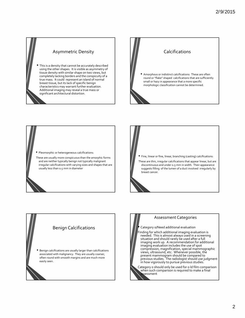
Asymmetric Density
• This is a density that cannot be accurately described using the other shapes. It is visible as asymmetry of tissue density with similar shape on two views, but completely lacking borders and the conspicuity of a true mass. It could represent an island of normal breast tissue, but its lack of specific benign characteristics may warrant further evaluation. Additional imaging may reveal a true mass or significant architectural distortion.
Calcifications
• Amorphous or indistinct calcifications: These are often round or “flake” shaped calcifications that are sufficiently small or hazy in appearance that a more specific morphologic classification cannot be determined.
• Pleomorphic or heterogeneous calcifications:
These are usually more conspicuous than the amorphic forms and are neither typically benign not typically malignant irregular calcifications with varying sizes and shapes that are usually less than 0.5 mm in diameter
• Fine, linear or fine, linear, branching (casting) calcifications:
These are thin, irregular calcifications that appear linear, but are discontinuous and under 0.5 mm in width. Their appearance suggests filling of the lumen of a duct involved irregularly by breast cancer.
Benign Calcifications
• Benign calcifications are usually larger than calcifications associated with malignancy. They are usually coarser, often round with smooth margins and are much more easily seen.
Assessment Categories
• Category 0/Need additional evaluation
Finding for which additional imaging evaluation is needed. This is almost always used in a screening situation and should rarely be used after a full imaging work up. A recommendation for additional imaging evaluation includes the use of spot compression, magnification, special mammographic views, ultrasound, etc. Whenever possible, the present mammogram should be compared to previous studies. The radiologist should use judgment in how vigorously to pursue previous studies.
Category 0 should only be used for o ld film comparison when such comparison is required to make a final assessment
2/9/2015
3
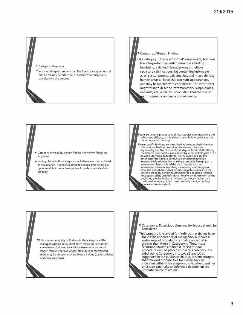
• Category 1/ Negative
There is nothing to comment on. The breasts are symmetrical and no masses, architectural disturbances or suspicious calcifications are present.
• Category 2/ Benign Finding
Like category 1, this is a “normal” assessment, but here the interpreter may wish to describe a finding. Involuting, calcified fibroadenomas, multiple secretory calcifications, fat containing lesions such as oil cysts, lipomas, galactoceles, and mixed density hamartomas all have characteristic appearances, and may be labeled with confidence. The interpreter might wish to describe intramammary lymph nodes, implants, etc. while still concluding that there is no mammographic evidence of malignancy.
• Category 3/ Probably benign finding‐short term follow‐up suggested
A finding placed in this category should have less than a 2% risk of malignancy. It is not expected to change over the follow‐up interval, but the radiologist would prefer to establish its stability.
There are several prospective clinical studies demonstrating the safety and efficacy of initial short‐term follow‐up for specific mammographic findings.
Three specific findings are described as being probably benign (the noncalcified circumscribed solid mass, the focal asymmetry and the cluster of round (punctate) calcifications; the latter is anecdotally considered by some radiologists to be an absolutely benign feature). All the published studies emphasize the need to conduct a complete diagnostic imaging evaluation before making a probably benign (cat 3) assessment; hence it is advisable to render such an assessment when interpreting a screening mammogram. Also, the published studies exclude palpable lesions, so the use of a probably benign assessment for a palpable lesion is not supported by scientific data. Finally, evidence from all the published studies indicate the need for biopsy rather than continued follow‐up when most probably benign findings increase in size or extent.
While the vast majority of findings in this category will be managed with an initial short‐term follow‐up (6 months) examination followed by additional examinations until longer‐term (2 years or longer) stability is demonstrated, there may be occasions where biopsy is done (patient wishes or clinical concerns).
• Category 4/ Suspicious abnormality‐biopsy should be considered
This category is reserved for findings that do not have the classic appearance of malignancy but have a wide range of probability of malignancy that is greater than those in Category 3. Thus, most recommendations of breast interventional procedures will be placed within this category. By subdividing Category 4 into 4A, 4B and 4C as suggested in the guidance chapter, it is encouraged that relevant probabilities for malignancy be indicated within this category so the patient and her physician can make an informed decision on the ultimate course of action.

2/9/2015
4
• Category 5/ Highly suggestive of malignancy‐appropriate action should be taken
These lesions have a high probability (> 95%) of being cancer. This category contains lesions for which one‐stage surgical treatment could be considered without preliminary biopsy. However, current oncologic management may require percutaneous tissue sampling as, for example, when sentinel node imaging is included in surgical treatment or when neoadjuvant chemotherapy is administered at the outset.
• Category 6/ Known biopsy‐proven malignancy‐appropriate action should be taken
This category is reserved for lesions identified on the imaging study with biopsy proof of malignancy prior to definitive therapy.
MassCircumscribedCalcificationBenign
BI-RADSCATEGORY:
2
Histology:
FA
Mass:(Circumscribed)
BI RADSCATEGORY:
3
Histology:
FA
Mass:(Circumscribed)
BI RADSCATEGORY:
3
Histology:
LN
Mass:(Circumscribed)
BI RADSCATEGORY:
3
Histology:
FA

2/9/2015
5
Mass:(Circumscribed)
BI RADSCATEGORY:
3
Histology:
FA
Mass:(Circumscribed)
BI RADSCATEGORY:
3
Histology:
FA
Mass:(Circumscribed)
BI RADSCATEGORY:
3
Histology:
LN
Mass:(Circumscribed)
BI RADSCATEGORY:
3
Histology:
FA
Mass:(Circumscribed)
BI RADSCATEGORY:
3
Histology:
FA
Mass:(Circumscribed)
BI RADSCATEGORY:
3
Histology:
C

2/9/2015
6
Mass:(Circumscribed)
BI RADSCATEGORY:
3
Histology:
FA
Mass:(Circumscribed)
BI RADSCATEGORY:
3
Histology:
FA
Mass:(Circumscribed)
BI RADSCATEGORY:
3
Histology:
LN
Mass:(Circumscribed)
BI RADSCATEGORY:
3
Histology:
FA
Mass:(Circumscribed)
BI RADSCATEGORY:
3
Histology:
FA
Mass:Indistinct
BI RADSCATEGORY:
4
Histology:
FA
2/9/2015
7
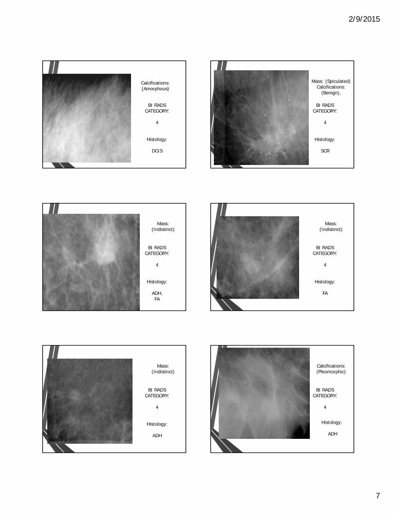
Calcifications:(Amorphous)
BI RADSCATEGORY:
4
Histology:
DCIS
Mass: (Spiculated)Calcifications:
(Benign),
BI RADSCATEGORY:
4
Histology:
SCR
Mass:(Indistinct)
BI RADSCATEGORY:
4
Histology:
ADH,FA
Mass:(Indistinct)
BI RADSCATEGORY:
4
Histology:
FA
Mass:(Indistinct)
BI RADSCATEGORY:
4
Histology:
ADH
Calcifications:(Pleomorphic)
BI RADSCATEGORY:
4
Histology:
ADH

2/9/2015
8
Mass:(Circumscribed)
BI RADSCATEGORY:
3
Histology:
FA
Calcifications:(Pleomorphic)
BI RADSCATEGORY:
4
Histology:
DCIS
Calcifications:(Pleomorphic)
BI RADSCATEGORY:
4
Histology:
DCIS
Mass:(Indistinct)
BI RADSCATEGORY:
4
Histology:
FA
Arch
BI RADSCATEGORY:
4
Histology:
LCIS
Mass:(Indistinct)
Calcifications(Pleomorphic)
BI RADSCATEGORY:
4
Histology:
DCIS

2/9/2015
9
Arch
Calcification:(Pleomorphic)
BI RADSCATEGORY:
4
Histology:
INVLC
Mass:(Indistinct)Calcification
(Benign)
BI RADSCATEGORY:
4
Histology:
FA
Mass:(Indistinct
BI RADSCATEGORY:
4
Histology:
FA
Mass:(Indistinct)
BI RADSCATEGORY:
4
Histology:
LN
Arch
Calcification(Benign)
BI RADSCATEGORY:
4
Histology:
DCIS
Asym
BI RADSCATEGORY:
4
Histology:
INVDC

2/9/2015
10
Calcifications:(Pleomorphic)
BI RADSCATEGORY:
4
Histology:
FA
Calcifications(Pleomorphic)
BI RADSCATEGORY:
4
Histology:
FA
Mass:(Circumscribed)
Calcification(Benign)
BI RADSCATEGORY:
4
Histology:
FA
Calficification(Linear)
BI RADSCATEGORY:
4
Histology:
SCR
Mass:(Indistinct)
BI RADSCATEGORY:
4
Histology:
C
Mass:(Indistinct)
BI RADSCATEGORY:
4
Histology:
INVDC

2/9/2015
11
Mass:(Indistinct)
BI RADSCATEGORY:
4
Histology:
FA
Calcifications(Pleomorphic)
BI RADSCATEGORY:
4
Histology:
DCIS
Mass:(Indistinct)
BI RADSCATEGORY:
4
Histology:
HARM
Mass:(Indistinct)
Calcification:(Benign)
BI RADSCATEGORY:
4
Histology:
FA
Mass:(Indistinct)
BI RADSCATEGORY:
4
Histology:
DH
Calcifications:(Pleomorphic)
BI RADSCATEGORY:
4
Histology:
DCIS
2/9/2015
12
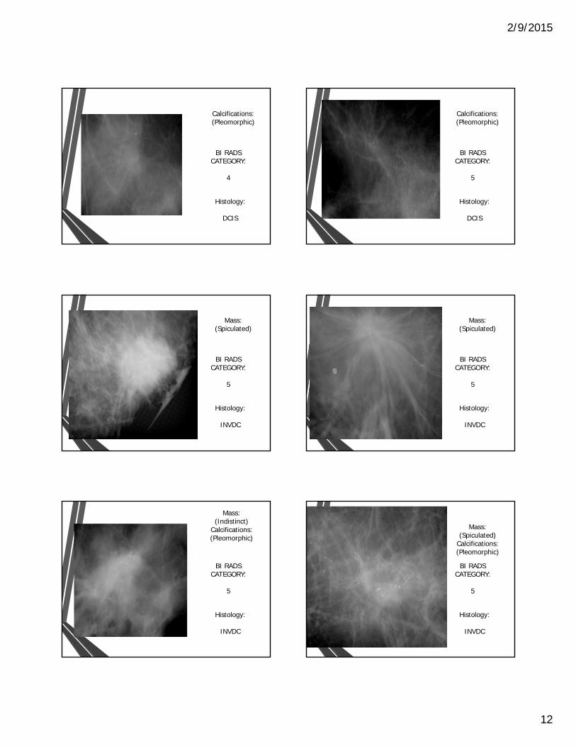
Calcifications:(Pleomorphic)
BI RADSCATEGORY:
4
Histology:
DCIS
Calcifications:(Pleomorphic)
BI RADSCATEGORY:
5
Histology:
DCIS
Mass:(Spiculated)
BI RADSCATEGORY:
5
Histology:
INVDC
Mass:(Spiculated)
BI RADSCATEGORY:
5
Histology:
INVDC
Mass:(Indistinct)
Calcifications:(Pleomorphic)
BI RADSCATEGORY:
5
Histology:
INVDC
Mass:(Spiculated)
Calcifications:(Pleomorphic)
BI RADSCATEGORY:
5
Histology:
INVDC
2/9/2015
13
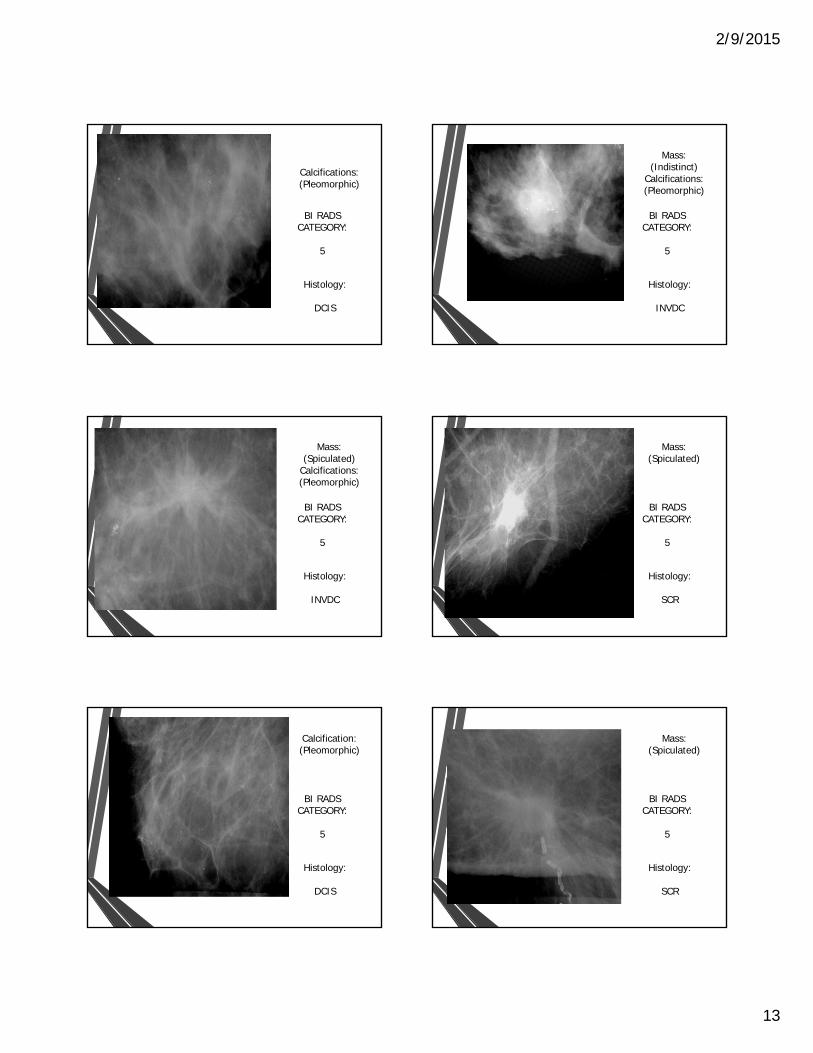
Calcifications:(Pleomorphic)
BI RADSCATEGORY:
5
Histology:
DCIS
Mass:(Indistinct)
Calcifications:(Pleomorphic)
BI RADSCATEGORY:
5
Histology:
INVDC
Mass:(Spiculated)
Calcifications:(Pleomorphic)
BI RADSCATEGORY:
5
Histology:
INVDC
Mass:(Spiculated)
BI RADSCATEGORY:
5
Histology:
SCR
Calcification:(Pleomorphic)
BI RADSCATEGORY:
5
Histology:
DCIS
Mass:(Spiculated)
BI RADSCATEGORY:
5
Histology:
SCR

2/9/2015
14
Calcifications:(Linear)
BI RADSCATEGORY:
5
Histology:
DCIS
Calcifications:(Linear)
BI RADSCATEGORY:
5
Histology:
DCIS
Mass:(Spiculated)
BI RADSCATEGORY:
5
Histology:
INVDC
Mass:(Spiculated)
BI RADSCATEGORY:
5
Histology:
INVDC
Calcifications:(Pleomorphic)
BI RADSCATEGORY:
5
Histology:
DCIS
Mass:(Spiculated)
BI RADSCATEGORY:
5
Histology:
INVDC

2/9/2015
15
Calcifications:(Linear)
BI RADSCATEGORY:
5
Histology:
DCIS
Calcifications:(Pleomorphic)
BI RADSCATEGORY:
5
Histology:
DCIS
Calcifications:(Pleomorphic)
BI RADSCATEGORY:
5
Histology:
DCIS
Calcifications:(Pleomorphic)
BI RADSCATEGORY:
5
Histology:
DCIS
Calcifications:(Linear)
BI RADSCATEGORY:
5
Histology:
INVDC
Calcifications:(Linear)
BI RADSCATEGORY:
5
Histology:
DCIS

2/9/2015
16
Mass:(Indistinct)
Arch
BI RADSCATEGORY:
5
Histology:
INVLC
Mass:(Spiculated)
BI RADSCATEGORY:
5
Histology:
SCR
Mass:(Spiculated)
BI RADSCATEGORY:
5
Histology:
INVDC
Mass:(Spiculated)
Calcifications:(Pleomorphic)
BI RADSCATEGORY:
5
Histology:
DCIS
Mass:(Spiculated)
BI RADSCATEGORY:
5
Histology:
INVDC
Mass:(Spiculated)
Calcifications:(Pleomorphic)
BI RADSCATEGORY:
5
Histology:
ADH

2/9/2015
17
Mass:(Spiculated)
BI RADSCATEGORY:
5
Histology:
INVLC
Mass:(Spiculated)
BI RADSCATEGORY:
5
Histology:
INVDC
Mass:(Spiculated)
BI RADSCATEGORY:
5
Histology:
INVDC
Mass:(Spiculated)
BI RADSCATEGORY:
5
Histology:
INVDC
Mass:(Spiculated)
BI RADSCATEGORY:
5
Histology:
INVDC
Mass:(Spiculated)
BI RADSCATEGORY:
5
Histology:
INVDC
2/9/2015
18
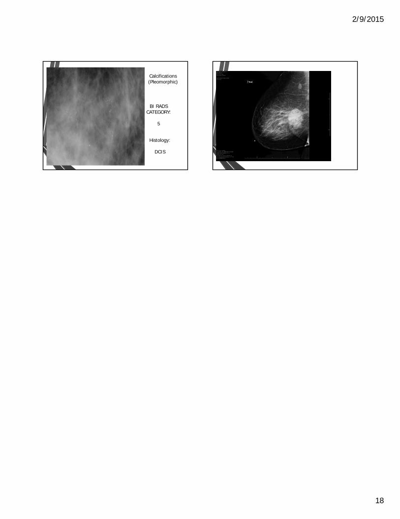
Calcifications(Pleomorphic)
BI RADSCATEGORY:
5
Histology:
DCIS