05 Hematology
31
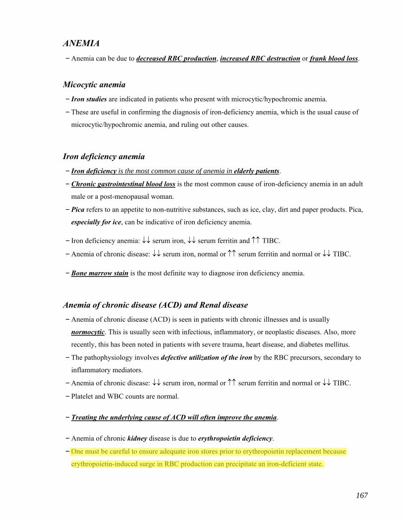
16 ANEMIA ‒ Anemia can be due to decreased RBC production , increased RBC destruction or frank blood loss . Micocytic anemia ‒ Iron studies are indicated in patients who present with microcytic/hypochromic anemia. ‒ These are useful in confirming the diagnosis of iron-deficiency anemia, which is the usual cause of microcytic/hypochromic anemia, and ruling out other causes. Iron deficiency anemia ‒ Iron deficiency is the most common cause of anemia in elderly patients . ‒ Chronic gastrointestinal blood loss is the most common cause of iron-deficiency anemia in an adult male or a post-menopausal woman. ‒ Pica refers to an appetite to non-nutritive substances, such as ice, clay, dirt and paper products. Pica, especially for ice, can be indicative of iron deficiency anemia. ‒ Iron deficiency anemia: ↓↓ serum iron, ↓↓ serum ferritin and ↑↑ TIBC. ‒ Anemia of chronic disease: ↓↓ serum iron, normal or ↑↑ serum ferritin and normal or ↓↓ TIBC. ‒ Bone marrow stain is the most definite way to diagnose iron deficiency anemia. Anemia of chronic disease (ACD) and Renal disease ‒ Anemia of chronic disease (ACD) is seen in patients with chronic illnesses and is usually normocytic . This is usually seen with infectious, inflammatory, or neoplastic diseases. Also, more recently, this has been noted in patients with severe trauma, heart disease, and diabetes mellitus. ‒ The pathophysiology involves defective utilization of the iron by the RBC precursors, secondary to inflammatory mediators. ‒ Anemia of chronic disease: ↓↓ serum iron, normal or ↑↑ serum ferritin and normal or ↓↓ TIBC. ‒ Platelet and WBC counts are normal. ‒ Treating the underlying cause of ACD will often improve the anemia . ‒ Anemia of chronic kidney disease is due to erythropoietin deficiency. ‒ One must be careful to ensure adequate iron stores prior to erythropoietin replacement because erythropoietin-induced surge in RBC production can precipitate an iron-deficient state. 7
description
UW Rankash notes USMLE STEP 2 CK
Transcript of 05 Hematology
16
ANEMIA ‒ Anemia can be due to decreased RBC production, increased RBC destruction or frank blood loss.
Micocytic anemia
‒ Iron studies are indicated in patients who present with microcytic/hypochromic anemia.
‒ These are useful in confirming the diagnosis of iron-deficiency anemia, which is the usual cause of
microcytic/hypochromic anemia, and ruling out other causes.
Iron deficiency anemia
‒ Iron deficiency is the most common cause of anemia in elderly patients.
‒ Chronic gastrointestinal blood loss is the most common cause of iron-deficiency anemia in an adult
male or a post-menopausal woman.
‒ Pica refers to an appetite to non-nutritive substances, such as ice, clay, dirt and paper products. Pica,
especially for ice, can be indicative of iron deficiency anemia.
‒ Iron deficiency anemia: ↓↓ serum iron, ↓↓ serum ferritin and ↑↑ TIBC.
‒ Anemia of chronic disease: ↓↓ serum iron, normal or ↑↑ serum ferritin and normal or ↓↓ TIBC.
‒ Bone marrow stain is the most definite way to diagnose iron deficiency anemia.
Anemia of chronic disease (ACD) and Renal disease ‒ Anemia of chronic disease (ACD) is seen in patients with chronic illnesses and is usually
normocytic. This is usually seen with infectious, inflammatory, or neoplastic diseases. Also, more
recently, this has been noted in patients with severe trauma, heart disease, and diabetes mellitus.
‒ The pathophysiology involves defective utilization of the iron by the RBC precursors, secondary to
inflammatory mediators.
‒ Anemia of chronic disease: ↓↓ serum iron, normal or ↑↑ serum ferritin and normal or ↓↓ TIBC.
‒ Platelet and WBC counts are normal.
‒ Treating the underlying cause of ACD will often improve the anemia.
‒ Anemia of chronic kidney disease is due to erythropoietin deficiency.
‒ One must be careful to ensure adequate iron stores prior to erythropoietin replacement because
erythropoietin-induced surge in RBC production can precipitate an iron-deficient state.
7
sun
Highlight
168
Lead poisoning
‒ Basophilic stippling and microcytic hypochromic anemia in a child are important clues to the
diagnosis of lead poisoning.
Macrovascular traumatic hemolysis
‒ In which intravascular fragmentation of red blood cells occur and leads to microcytic anemia
‒ It is common in patients with artificial heart valves or severely calcified aortic valves.
‒ Schistocytes (helmet cells) are the classic finding on peripheral blood smear.
Sideroblastic anemia ‒ Sideroblastic anemia is seen in inherited or acquired defects affecting the biosynthesis of heme
within red cell precursors.
‒ Hereditary form is due to defect in aminolevulinic acid synthase or abnormality in vitamin B6
(Pyridoxine) metabolism.
‒ Sideroblastic anemia is characterized by↑ serum iron levels and ↑ TIBC.
‒ Usually two groups of RBC can be demonstrated on microscopy hypochromic and normochromic
(“dimorphic” RBC population).
‒ It can progress to acute myelogenous leukemia (AML).
‒ Prussian blue stain of RBCs in the marrow reveals ringed sideroblasts.
‒ In patients with an identifiable cause of vitamin B6 (Pyridoxine) deficiency, (alcoholism,
chloramphenicol, isoniazid), the administration of pyridoxine can easily correct the problem.
sun
Highlight
sun
Typewriter
dec
169
THALASSEMIA
‒ Hereditary underproduction of either alpha or beta globulin chains of the hemoglobin molecule
resulting in microcytic hypochromic anemia.
Alpha thalassemia
‒ Alpha thalassemia is caused by abnormalities in the synthesis of alpha chains of hemoglobin.
‒ It is usually a hypochromic, microcytic anemia, and a positive family history is usually present.
‒ Peripheral blood smear shows hypochromic, microcytic anemia, and target cells.
‒ If the patient’s anemia is not severe (as evidenced by the hemoglobin level, adulthood diagnosis, and
lack of severe symptoms), it is probably alpha-thalassemia minor. The treatment of choice is
reassurance and follow-up monitoring.
β-thalassemia
‒ β-thalassemia minor occurs in people heterozygous for the β-hemoglobin chain this results in
reduced hemoglobin synthesis and eventually hypochromic microcytic anemia due to poor
hemoglobinization of RBCs (hemoglobinopathy).
‒ A patient with microcytic anemia non-responsive to iron supplementation is most likely to be β-
thalassemia minor (particularly in someone of Mediterranean origin).
‒ Peripheral blood smear shows hypochromic, microcytic anemia, and target cells.
‒ If the patient’s anemia is not severe (as evidenced by the hemoglobin level, adulthood diagnosis, and
lack of severe symptoms), it is probably beta-thalassemia minor. The treatment of choice is
reassurance and follow-up monitoring.
Beta Thalassemia Major (Cooley Anemia)
‒ β-thalassemia major occurs when both β-hemoglobin genes are defective, this results in severe
anemia and transfusion dependant at an early age (6-12 months of age).
‒ Peripheral blood smear shows hypochromic, microcytic anemia, and target cells.
‒ Growth failure, hepatosplenomegaly, jaundice, boney deformities, hemochromatosis, cirrhosis,
congestive heart failure and chronic anemia.
‒ Treatment includes: blood transfusions once or twice a month, Deferoxamine, splenectomy, and
bone marrow transplantation.
170
Diamond-Blackfan Anemia “Congenital Pure Red-Cell Anemia”
‒ The majority of cases are sporadic, although dominant and recessive inheritance is found in 15
percent of cases.
‒ The primary pathology is an intrinsic defect of erythroid progenitor cells which results in increased
apoptosis (programmed cell death).
‒ The condition often presents with pallor in the neonatal period.
‒ Congenital anomalies (triphalangeal thumbs and craniofacial deformities) are present in over 50
percent of cases.
‒ The macrocytic anemia of DBS is distinct from that of megaloblastic anemia because there is no
hypersegmentation of the nucleus in neutrophils and other blood cells in the former.
‒ Very low reticulocyte count.
‒ Increased RBC adenosine deaminase (ADA).
‒ Electrophoresis reveals elevated fetal Hb levels.
‒ Bone marrow biopsy shows significant decrease in RBCs precursors.
‒ Therapy is mainly corticosteroids.
‒ For unresponsive patients, transfusion and deferoxamine therapy is indicated.
‒ Definitive treatment is stem cell transplant.
Fanconi Anemia
‒ Fanconi's anemia is an autosomal recessive (chromosomal breaks) disorder marked by progressive
bone marrow failure characterized by progressive pancytopenia and macrocytosis (↑ MCV).
‒ The average age at diagnosis is 8 years.
‒ Associated deformities include cafe-au-lait spots (hypopigmentation of the skin), microcephaly,
microphthalmia, short stature, horseshoe kidneys, hypogonadism and absent thumbs.
‒ Bone marrow hypoplasia.
‒ Complications: increased risk of leukemia (AML) and other cancers.
‒ Therapy is mainly corticosteroids and androgens.
‒ Definitive treatment is bone marrow transplant.
sun
Highlight
sun
Highlight
sun
Highlight
sun
Highlight
sun
Highlight
sun
Highlight
sun
Highlight
sun
Highlight
sun
Highlight
171
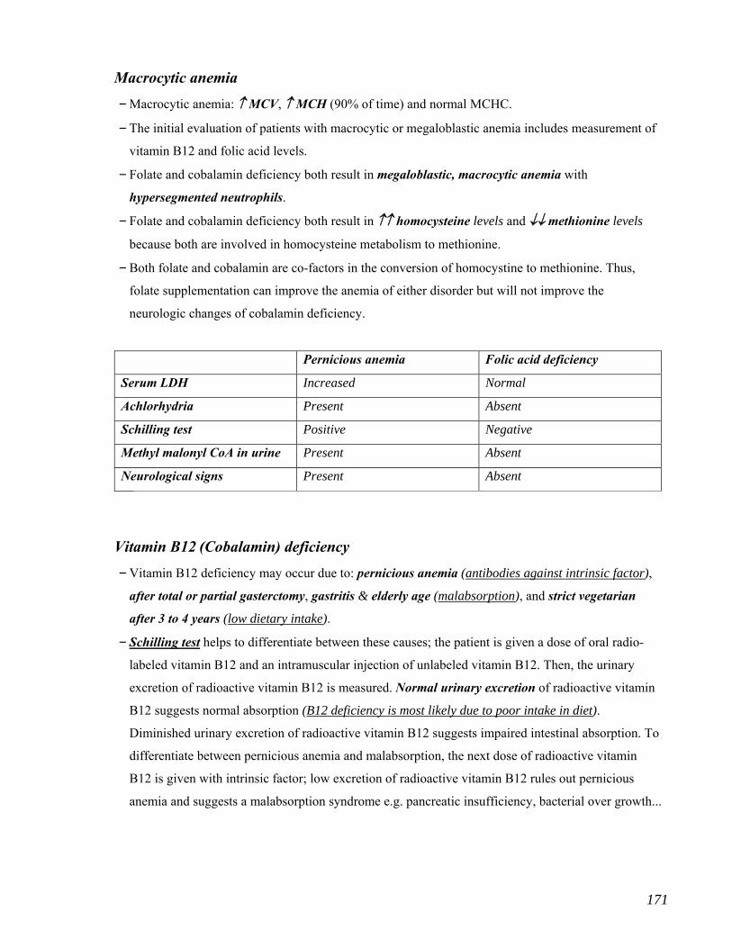
Macrocytic anemia
‒ Macrocytic anemia: ↑ MCV, ↑ MCH (90% of time) and normal MCHC.
‒ The initial evaluation of patients with macrocytic or megaloblastic anemia includes measurement of
vitamin B12 and folic acid levels.
‒ Folate and cobalamin deficiency both result in megaloblastic, macrocytic anemia with
hypersegmented neutrophils.
‒ Folate and cobalamin deficiency both result in ↑↑ homocysteine levels and ↓↓ methionine levels
because both are involved in homocysteine metabolism to methionine.
‒ Both folate and cobalamin are co-factors in the conversion of homocystine to methionine. Thus,
folate supplementation can improve the anemia of either disorder but will not improve the
neurologic changes of cobalamin deficiency.
Pernicious anemia Folic acid deficiency
Serum LDH Increased Normal
Achlorhydria Present Absent
Schilling test Positive Negative
Methyl malonyl CoA in urine Present Absent
Neurological signs Present Absent
Ass
Vitamin B12 (Cobalamin) deficiency
‒ Vitamin B12 deficiency may occur due to: pernicious anemia (antibodies against intrinsic factor),
after total or partial gasterctomy, gastritis & elderly age (malabsorption), and strict vegetarian
after 3 to 4 years (low dietary intake).
‒ Schilling test helps to differentiate between these causes; the patient is given a dose of oral radio-
labeled vitamin B12 and an intramuscular injection of unlabeled vitamin B12. Then, the urinary
excretion of radioactive vitamin B12 is measured. Normal urinary excretion of radioactive vitamin
B12 suggests normal absorption (B12 deficiency is most likely due to poor intake in diet).
Diminished urinary excretion of radioactive vitamin B12 suggests impaired intestinal absorption. To
differentiate between pernicious anemia and malabsorption, the next dose of radioactive vitamin
B12 is given with intrinsic factor; low excretion of radioactive vitamin B12 rules out pernicious
anemia and suggests a malabsorption syndrome e.g. pancreatic insufficiency, bacterial over growth...
172
‒ Vitamin B12 is a necessary co-factor in the pathway leading to purine synthesis.
‒ Vitamin B12 deficiency decreases DNA synthesis and erythropoiesis → megaloblastic anemia.
‒ Vitamin B12 deficiency presents with glossitis, neurologic changes & megaloblastic anemia.
‒ Cobalamin (vitamin B12) deficiency can result in peripheral neuropathy or posterior column defects
due to defective myelin synthesis.
‒ Pernicious anemia (PA) is the most common cause of megaloblastic anemia (common in elderly).
‒ In PA, Vitamin B12 deficiency is due to reduced intrinsic factor (IF) secondary to gastric atrophy.
‒ In the majority of cases, antibodies to parietal cells have been reported.
‒ The diagnosis of PA is confirmed by achlorhydria, dcreased serum Vitamin B12, positive IF
antibodies, and extremely elevated LDH.
‒ Pernicious anemia is associated with increase risk of gastric cancer.
Folic acid deficiency
‒ The most common cause of folic acid deficiency is nutritional due to poor diet and/or alcoholism.
‒ Alcohol abuse is the most common cause of folate deficiency in USA.
‒ Some drugs impair the absorption of folic acid (e.g. phenytoin) and some drugs antagonize its
physiologic effects (e.g. methotrexate, trimethoprim).
173
HEMOLYTIC ANEMIA
Any hemolysis → ↑ LDH, ↑ Bilirubin, ↑ Reticulocytes and ↓ Haptoglobin
Sickle cell disease
‒ Sickle cell disease is an autosomal recessive disorder.
‒ Sickle cell disease is characterized by chronic hemolysis of sickled cells, leading to a high RBC
turnover and anemia.
‒ Hemolysis is mainly extravascular and leads to ↑ LDH, ↑ Bilirubin, ↑ Reticulocytes and ↓
Haptoglobin
‒ The hematocrit is generally 20-30 percent, owing to the decreased RBC volume.
‒ The presence of hemoglobin F protects the infants from sickling during the first 4-6 months of life.
‒ Painful crises are the most common manifestation of sickle cell anemia.
‒ Dactylitis may be the initial presentation that warrants further work-up for sickle cell disease.
‒ Sickle cell disease can cause childhood stroke.
‒ Osteonecrosis is a common complication of sickle cell anemia due to vaso-occlusion of the bone. It
causes significant joint pain and functional limitation. The humerus and femur are the most
frequently affected bones.
‒ Vasoocclusive crisis are one of the complications of sickle cell disease, which may manifest as
stroke, priapism, or intractable pain. Whenever such acute vasoocclusive crisis occurs, Exchange
transfusion is indicated.
‒ Folic acid deficiency is much more common than vitamin B12 deficiency in sickle cell anemia.
‒ Because of chronic hemolysis and compensatory reticylocytosis, increased demand for folic acid can
lead to folic acid deficiency. For this reason, patients with sickle cell anemia should be on folic acid
supplementation.
‒ Folic acid supplementation is recommended in all patients with sickle cell anemia to prevent the
occurrence of aplastic crisis.
‒ Patients at an early age become functionally asplenic; thus they are susceptible to infection with
encapsulated organisms such as S. pneumoniae, H. influenzae, and N. meningitidis.
‒ Post-splenectomy patients or patients with auto-splenectomy are at increased risk for sepsis from
encapsulated organisms due to impaired antibody-mediated opsonization in phagocytosis.
sun
Highlight
sun
Highlight
174
‒ Start penicillin prophylaxis at 2 months age.
‒ Immunize the patient with regular vaccinations plus Pneumococcal vaccine, Polysaccharide
meningococcal vaccine H. influenzae type B vaccine, and hepatitis B virus vaccine.
‒ Howell-Jolly bodies are nuclear remnants within RBCs typically removed by the spleen. They
appear in blood smear as single, round blue inclusions on Wright stain. Their presence stongly
suggests physical or functional hyposplenism.
‒ Hyposthenuria is common in patients with sickle cell anemia and sickle cell trait. In which there is
an impairment of kidney’s ability to concentrate urine leads to nocturia.
Autoimmune hemolytic anemia
‒ Autoimmune hemolytic disease is extravascular hemolytic anemia; it is acquired.
‒ A negative family history and positive Coombs' test, suggestive of autoimmune hemolytic anemia.
‒ The peripheral blood smear may show spherocytes.
‒ Autoimmune hemolytic: may be associated with various tumors.
‒ Coombs' test or micro- Coombs' test is positive in such cases. Platelet and WBC counts are normal.
‒ Hemolytic anemia in a patient with a malignant lymphoproliferative disorder is likely to be of the
warm autoimmune type, caused by anti-red blood cell IgG antibodies. If prednisone therapy is
ineffective, splenectomy is usually indicated.
‒ Autoimmune hemolytic disease and hereditary spherocytosis are both extravascular hemolytic
anemias. However, Autoimmune hemolytic disease is acquired and hereditary spherocytosis has an
autosomal dominant transmission. A negative family history and positive Coombs' test are thus
suggestive of autoimmune hemolytic disease, whereas a positive family history is more suggestive
of hereditary spherocytosis. The peripheral blood smear in both conditions may show spherocytes.
sun
Highlight
175
Hereditary spherocytosis (HS)
‒ HS is an autosomal dominant trait and is the most common hereditary hemolytic anemia in white
population.
‒ There is congenital RBC membrane defect (spectrin) in HS leading to increased RBC membrane
osmotic fragility resulting in extravascular hemolysis occurring only in the presence of spleen.
‒ It is characterized by positive family history, splenomegaly, anemia, spherocytosis, jaundice and
can cause cholecystitis due to pigmented gallstones.
‒ Chronic leg ulcers may complicate the disease.
‒ Patients are complicated by episodes of aplastic crisis (in which erythropoiesis is suppressed and
hemolytic process continues), usually because of Parovirus B19 infection
‒ Severe anemia may also occur due to decreased intake of folate deficiency.
‒ Peripheral blood smear demonstrate spherocytes with polychromatophilia.
‒ Normal or ↓↓ MCV, normal MCH, but the MCHC is generally greater than 36%.
‒ Increased reticulocyte count, increased bilirubin, negative Coombs test.
‒ The osmotic fragility test is the diagnostic test for hereditary spherocytosis.
‒ The treatment for most patients involves supportive care with oral folic acid for life and blood
transfusion during periods of extreme anemia.
‒ Splenectomy is considered if the patients have moderate to sphere spherocytosis, or are refractory to
medical management.
‒ Studies have shown that the risk of sepsis is present up to 30 years and probably longer after
splenectomy. Current recommendations state that patients should receive anti-pneumococcal,
Haemophilus, and meningococcal vaccines several weeks before the operation and daily oral
penicillin prophylaxis for three to five years following splenectomy.

17
Paroxysmal nocturnal hemoglobinuria (PNH)
‒ PNH is an acquired disorder of hematopoietic cells.
‒ It is characterized by an intravascular hemolytic anemia, a hypercoagulable state, and bone
marrow aplasia.
‒ A red cell membrane defect (in PIG-A) causes increased binding of complement to the red cell
leading to increased intravascular hemolysis resulting in marked anemia.
‒ These cells are more susceptible to lysis in an acidic environment. Due to relative hypoventilation at
night, there is mild acidosis. So the hemolysis is more at night and hemosiderinuria and hematuria
is common in the first morning urine.
‒ Hemolysis is typically paroxysmal and complement mediated.
‒ PNH may cause pancytopenia, and should be considered in all patients when accompanied by
reticulocytosis and hemolytic anemia.
‒ Loss of iron in the urine may result in iron deficiency anemia.
‒ The other characteristic feature of PNH is unusually high incidence of venous thrombosis in the
hepatic and portal veins (Budd-Chiari syndrome).
‒ Lab tests show ↑↑ LDH, ↑↑ bilirubin, ↑↑ reticulocyte count and ↓↓ haptoglobin levels.
‒ Low Leukocyte Alkaline Phosphatase is seen in paroxysmal nocturnal hemoglobinuria (PNH).
‒ Test specific to PNH are sugar-water test and the acidified-hemolysis (Ham) test, which determines
the increased susceptibility of cells to lysis by complement.
‒ Decay Activating Factor (DAF) is diminished in PNH.
‒ Bone marrow examination shows hypocellular marrow.
‒ Flow cytometry is screening and confirmatory test, it is simple and has high sensitivity and
specificity. The expression of the GPI-anchored proteins CD55 and CD59 can be analyzed using
monoclonal antibodies and flow cytometry.
‒ Paroxysmal nocturnal hemoglobinuria (PNH) should be considered in the following situations:
1) Pancytopenia accompanied by hemolytic anemia (increased reticulocyte count and LDH and low
haptoglobin levels)
2) Recurrent thrombosis at unusual sites e.g., portal vein thrombosis or Budd Chiari syndrome.
‒ Low Leukocyte Alkaline Phosphatase is seen in CML, hypophosphatemia and paroxysmal
nocturnal hemoglobinuria (PNH).
6
17
Glucose-6-phosphate dehydrogenase (G6PD) deficiency
‒ It is an X-linked disorder, so there is typically a positive family history (common in African
Americans) and it is the most common enzymatic disorder of red blood cells in humans.
‒ G6PD is an enzyme involved in creating NADPH, a cofactor required for creating glutathione and
prevent oxidation of hemoglobin.
‒ Without G6PD, hemoglobin becomes oxidized and denatures into Heinz bodies.
‒ Denatured hemoglobin disrupts red blood cell (RBC) membranes and causes hemolysis.
‒ Episodes of hemolysis occur only due to oxidative stress from infection, fava beans or drugs
(primaquine, nitrofurantoin or sulfa drugs like sulphamethoxazole).
‒ Variants of G6PD deficiency are G6PD A- (moderate enzyme deficiency) and G6PD Mediterranean
(severe enzyme deficiency), where hemolysis is precipitated by infection, drugs or fava beans.
‒ The typical peripheral blood smear reveals bite cells and Heinz bodies.
‒ Platelet and WBC counts are normal.
‒ G6PD levels are often normal during the hemolytic episode.
Pyruvate kinase deficiency
‒ Pyruvate kinase deficiency can also lead to a similar clinical picture of hemolytic anemia; however,
the hemolysis in such cases is not precipitated by sulfa drugs.
‒ Furthermore, the typical peripheral smear does not include bite cells.
Aplastic anemia ‒ Idiopathic aplastic anemia is an acquired disease that results in pancytopenia.
‒ It may be due to chemicals (e.g., benzene, phenylbutazone), drugs (e.g., chloramphenicol,
sulfonamides), infectious agents (e.g., viral hepatitis) or ionizing radiation.
‒ The peripheral blood smear does not show any abnormal morphology of cells.
‒ Red blood cells (RBCs) are normocytic or macrocytic.
‒ There is neither hemolysis of RBCs nor splenomegaly.
‒ Reticulocyte count will be very low.
‒ Aplastic anemia shows hypoplastic fat-filled marrow with no abnormal cells.
‒ Bone marrow transplantation is the definitive treatment.
7
178
TTP-HUS ‒ Unexplained hemolytic anemia, and thrombocytopenia in a patient with renal failure and
neurologic symptoms should raise strong suspicious for TTP-HUS.
‒ Hemolytic uremic syndrome (HUS) and TTP come under a spectrum of diseases.
‒ TTP-HUS is thought to be due to deficiency or autoantibody against a specific von Willbrand factor-
cleaving protease (ADAMTS-13) → accumulation of large von Willbrand factor multimers and
platelet aggregation.
‒ If the patient has more neurologic symptoms and less of renal failure, it is considered TTP.
‒ If the patient has significant renal failure and less neurologic symptoms it is considered HUS.
‒ Without prompt institution of appropriate therapy, TTP-HUS proves lethal in 80% of patients.
‒ Both conditions are very serious and require emergent plasmapheresis (plasma exchange).
‒ Rate of recovery usually defined as normalization of platelet count and LDH levels.
‒ Renal function impairment and peripheral blood smear schistocytes may persist for several weeks
following clinical recovery.
‒ Schistocytes (helmet cells) are fragmented erythrocytes. They are found in micoangipathic
hemolytic anemias (TTP, HUS & DIC) and due to RBC destruction by artificial heart valves or
severely calcified aortic valves (macrovascular traumatic hemolysis).
Thrombotic Thrombocytopenic Purpura (TTP)
‒ TTP is a serious disorder, which presents with the following classical pentad:
1) Severe thrombocytopenia
2) Microangiopathic hemolytic anemia (RBC fragments)
3) Renal failure
4) Fluctuating neurological signs
5) Fever
‒ HIV increases the risk for TTP.
‒ The patients with TTP generally presents with fever, pallor, petechial, and confusion.
‒ CBC: Anemia, thrombocytopenia.
‒ Normal PT/PTT, ↑ bleeding time.
‒ ↑ LDH, ↑ indirect bilirubin, ↑ Reticulocyte count (due to hemolysis), Coombs’ test .
‒ Blood smear shows schistocytes, helmet cells and RBC fragments.
‒ Dipyridamole may help to treat TTP by preventing platelet aggregation.
179
Hemolytic uremic syndrome (HUS)
‒ HUS is a serious disorder, which presents with the following classical triad:
1) Thrombocytopenia
2) Microangiopathic hemolytic anemia (RBC fragments)
3) Renal failure (BUN and creatinine levels are markedly elevated)
‒ Hemolytic uremic syndrome (HUS) is typically a disease of young children.
‒ HUS often follows mild viral illness (upper respiratory infection) or gastroenteritis (bloody
diarrhea) caused by E. coli O157:H7, Shigella, Salmonella, Yersinia or Campylobacter. In the past,
E. coli O157:H7 has been transmitted via the intake of undercooked hamburger meat from fast food
chains.
‒ CBC: Anemia, thrombocytopenia.
‒ Normal PT/PTT.
‒ ↑ LDH, ↑ indirect bilirubin, ↑ Reticulocyte count (due to hemolysis), Coombs’ test .
‒ Blood smear shows schistocytes and giant cells.
‒ Plasmapheresis is indicated in patients with hemolytic uremic syndrome.
ACUTE LEUKEMIA
Acute lymphoblastic leukemia (ALL)
‒ Acute lymphoblastic leukemia (ALL) is the predominant type of leukemia in children from ages 2-
10 years. It is common in Down syndrome.
‒ Approximately 30-50% of patients present with infections, and about half present with
lymphadenopathy and splenomegaly.
‒ Lymphoblasts are typically seen on the peripheral smear.
‒ Varying degrees of anemia, neutropenia, and thrombocytopenia have been noted.
‒ The presence of more than 25% lymphoblasts in the bone marrow is diagnostic.
‒ ALL is characterized by the presence of common ALL antigen (CALLA) and terminal
deoxynucleotidyltransferase (TdT).
‒ Lymphoblasts lack peroxidase positive granules but often contain cytoplasmic aggregates of
periodic acid Schiff (PAS) positive material.
180
Acute myeloid leukemia (AML)
‒ Acute myeloid leukemia (AML) is the predominant type of leukemia in adults.
‒ In acute myelocytic leukemia (AML), the predominant cells are of myeloid origin.
‒ AML is characterized by the presence of Auer rods (M3), myeloperoxidase & esterase.
Acute monocytic (FAB M5) leukemia ‒ FAB M5 comprises less than 15% of all leukemias.
‒ The onset is dramatic, with most chief complaints being headaches, fever, weight loss, and bleeding
from the gums or nose.
‒ Signs include gingival hyperplasia and occasional skin lesions.
‒ Peripheral smear reveals leukocytosis with a high proportion of blast forms (monoblasts,
promonocytes and Monocytes).
‒ Monoblasts lack Auer rods, and are peroxidase negative and nonspecific esterase positive.
‒ A positive alpha-naphthyl esterase test is characteristic.
Acute promyelocytic leukemia
‒ APML is characterized by many hypergranular promyelocytes.
‒ Each cell has many Auer rods.
‒ There is a high incidence of DIC in these patients.
181
Chronic Myelogenous leukemia (CML)
‒ CML is a disorder characterized by massive over production of myeloid cells.
‒ CML is one of the myeloproliferative syndromes seen normally in adults (mostly age of 50).
‒ It is characterized by leukocytosis, Basophilia, anemia and marked splenomegaly.
‒ Night sweats, fatigue, and fever associated with increased metabolism due to granulocytic cell turn
over may occur.
‒ Abdominal pain from massive enlargement of the spleen is common.
‒ Lymphadenopathy is not a feature of CML.
‒ Peripheral blood smear shows leukocytosis (predominantly of mature granulocytic forms such as
neutrophils and band forms with left sided myeloid series).
‒ Presence of Philadelphia chromosome (9 and 22) and the ↓↓ Leukocyte Alkaline Phosphatase
makes the CML more likely than the leukemoid reaction.
‒ Examination of a bone marrow biopsy specimen reveals hypercellularity with prominent
granulocytic hyperplasia.
‒ Imatinib has changed the prognosis of patients with CML. It is tyrosine kinase inhibitor and works
by blocking signals within cancer cells and preventing a series of chemical reactions that cause the
cancer cells to grow and divide.
‒ Elevated Leukocyte Alkaline Phosphatase is characteristic of Leukemoid Reaction.
‒ Low Leukocyte Alkaline Phosphatase is seen in CML, hypophosphatemia and paroxysmal
nocturnal hemoglobinuria (PNH).
Leukemoid reaction
‒ Leukemoid reaction is marked increase in leucocytes due to severe infection or inflammation.
‒ ↑↑ Leukocyte Alkaline Phosphatase score typical for this condition.
sun
Highlight
182
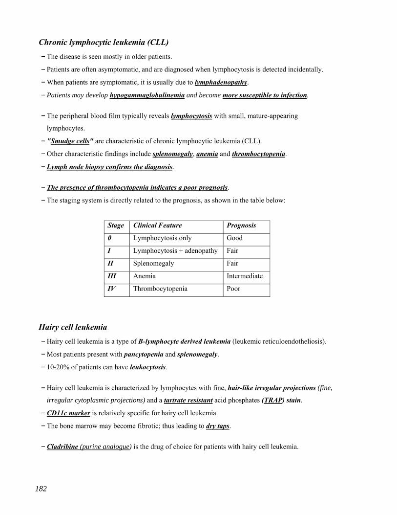
Chronic lymphocytic leukemia (CLL)
‒ The disease is seen mostly in older patients.
‒ Patients are often asymptomatic, and are diagnosed when lymphocytosis is detected incidentally.
‒ When patients are symptomatic, it is usually due to lymphadenopathy.
‒ Patients may develop hypogammaglobulinemia and become more susceptible to infection.
‒ The peripheral blood film typically reveals lymphocytosis with small, mature-appearing
lymphocytes.
‒ "Smudge cells" are characteristic of chronic lymphocytic leukemia (CLL).
‒ Other characteristic findings include splenomegaly, anemia and thrombocytopenia.
‒ Lymph node biopsy confirms the diagnosis.
‒ The presence of thrombocytopenia indicates a poor prognosis.
‒ The staging system is directly related to the prognosis, as shown in the table below:
Stage Clinical Feature Prognosis
0 Lymphocytosis only Good
I Lymphocytosis + adenopathy Fair
II Splenomegaly Fair
III Anemia Intermediate
IV Thrombocytopenia Poor
Hairy cell leukemia
‒ Hairy cell leukemia is a type of B-lymphocyte derived leukemia (leukemic reticuloendotheliosis).
‒ Most patients present with pancytopenia and splenomegaly.
‒ 10-20% of patients can have leukocytosis.
‒ Hairy cell leukemia is characterized by lymphocytes with fine, hair-like irregular projections (fine,
irregular cytoplasmic projections) and a tartrate resistant acid phosphates (TRAP) stain.
‒ CD11c marker is relatively specific for hairy cell leukemia.
‒ The bone marrow may become fibrotic; thus leading to dry taps.
‒ Cladribine (purine analogue) is the drug of choice for patients with hairy cell leukemia.
183
Multiple myeloma
‒ Multiple myeloma is a form of plasma cell leukemia, caused by the proliferation of a single
transformed plasma cell usually producing IgG.
‒ MM presents in old age.
‒ Classic tetrad of multiple myeloma “CRAB”: Calcium (hypercalcemia), Renal impairment (Urine
monoclonal proteins), Anemia (normocytic), and Bones (bone pain, bone lytic lesions, fractures).
‒ Back pain is the most common manifestation.
‒ Hypercalcemia is a common finding in a patient with multiple myeloma. Hypercalcemia may cause
severe constipation, anorexia, weakness, renal tubular dysfunction, and neurologic symptoms.
‒ IgG antibodies or paraproteins, produced by the myeloma cells can collect in the glomeruli, causing
renal failure or “myeloma kidney”.
‒ There is increased risk for infection due to total decrease in the functional antibodies and
leukopenia secondary to bone marrow crowiding with maligant plasma cells.
‒ Normally, there is a 3-4 g/dL difference separating the serum total protein and albumin
concentration, the gap is greater in patients with multiple myeloma.
‒ Laboratory findings include anemia, increased ESR (often more than 100), and Bence Jones
proteins in the urine (monoclonal proteins).
‒ RBC morphology is significant for a rouleaux appearance.
‒ X-rays reveal lytic lesions, especially in the axial skeleton, as well as osteoporosis and fractures.
‒ Bone marrow biopsy shows 10–20% plasma cells (normal is < 5%).
‒ Serum immunoelectrophoresis demonstrates abnormal M-spike due to excess IgG production.
‒ The complete work-up consists of CBC with differential and morphology, serum electrolytes,
kidney and liver screening profiles, skeletal survey, serum electrophoresis, and bone marrow biopsy.
‒ Complications include renal failure, hypercalcemia, and hyperviscosity syndrome.
‒ Bisphosphonates (e.g. Zoledronic acid) are the drugs of choice for mild to moderate hypercalcemia
due to malignancy.
sun
Highlight
184
Waldenstrom’s Macroglobulinemia (WM)
‒ It is a rare chronic plasma cell neoplasm.
‒ It is characterized by abnormal plasma cells which multiply out-of-control and invade the bone
marrow, lymph nodes and spleen.
‒ Typically, there is production of excessive amounts of IgM antibodies in the blood that causes
hyperviscosity (thickening) of the blood.
‒ Major Signs & Symptoms are:
1) Increased size of spleen, liver, lymph node
2) Tiredness, usually due to anemia(too few RBCs)
3) Tendency to bleed and bruise easily (little platelets)
4) Night sweats
5) Headache and dizziness
6) Visual problems (engorgement of the retinal veins caused by hyperviscosity of the blood)
7) Pain and numbness in extremities.
‒ In multiple Myeloma, there is IgA/G not IgM. Also, No hyperviscosity like in WM.
Monoclenal gammopathy of undetermined significance (MGUS)
‒ The characteristic laboratory findings of this condition include an M component (IgA, or IgG, or
IgM) < 3000 mg/dL, and fewer than 10 % plasma cells in the bone marrow.
‒ Patients are initially asymptomatic, and may remain so for years.
‒ In the early course of their disease, patients do not manifest with any lytic lesions, anemia,
hypercalcemia, or renal insufficiency (these are typically seen in patients with multiple myeloma).
The reason behind this is that the levels of M-proteins initially remain stable for years.
‒ Nevertheless, patients with MGUS have a 25 % risk of development of a serious disease, which is
usually a late event. In addition, the evolution from MGUS to multiple myeloma, which occurs at a
rate of 1 %/yr, may be abrupt.
‒ For these reasons, patients initially do not require any treatment, but proper education and
counseling are necessary.
‒ Regular follow-up visits are recommended, and all patients are instructed to promptly obtain
medical evaluation if any clinical symptoms occur.
185
Hodgkin's disease
‒ Neoplastic transformation of lymphocytes.
‒ It present with enlarged, painless, rubbery lymph nodes (mostly cervical and supraclavicular).
‒ Pruritus is common.
‒ Patients with Hodgkin's lymphoma usually have normal blood smears (eosinophilia is common).
‒ Reed Sternberg cells “Owl’s eye cells” are seen in the lymph node biopsy.
‒ Hypercalcemia in cases of Hodgkin's disease is almost always produced by calcitriol.
‒ Nephrotic syndrome is a well-known complication of Hodgkin's lymphoma, and is usually caused
by minimal change disease.
Non-Hodgkin lymphoma
‒ Non-Hodgkin lymphoma is a complication of Sjogren's syndrome.
‒ CHOP regimen is used for non-Hodgkin's lymphoma.
‒ Anemia of lymphoproliferative disorders is due to bone marrow infiltration with cancerous cells
replacing RBC progenitor cells.
Burkitt lymphoma ‒ Burkitt lymphoma is a neoplasm of mature B cells.
‒ It is associated with the Epstein-Barr virus infection.
‒ Most patients present with either a mass involving the mandible or abdominal viscera.
‒ High mitotic index is typical.
‒ Histological examination shows characteristic "starry sky appearance".
‒ It is a very aggressive tumor but responds well to the high dose chemotherapy.
‒ Tumors with high cell turnovers are frequently associated with tumor lysis syndrome.
‒ Tumor lysis syndrome is characterized by hyperphosphatemia, hypocalcemia, hyperkalemia, and
hyperuricemia.
‒ The tumors most often associated with this syndrome are the poorly differentiated lymphomas (e.g.,
Burkitt's lymphoma) and the leukemias (particularly ALL and less often AML). Although the
introduction of allopurinol has reduced the acute urate nephropathy to a large extent, the possibility
of tumor lysis syndrome remains.
sun
Highlight
sun
Highlight
18
Mucosa-associated lymphoid tissue (MALT) lymphoma
‒ There is an important role for H. pylori infection in the pathogenesis of low-grade gastric MALT
lymphoma.
‒ Such lymphomas regress after the eradication of H. pylori using antibiotic therapy.
‒ Antibiotic therapy against H. pylori is the most accepted and recommended management of gastric
MALT lymphoma without any metastasis.
‒ If antibiotic therapy fails to regress the lymphoma, chemotherapy is used (CHOP regimen or
CHOP+Bleomycin).
Idiopathic thrombocytopenia purpura (ITP) ‒ ITP is a diagnosis of exclusion
‒ It is characterized by thrombocytopenia and increased bleeding time.
‒ All other cell lines, PT, and PTT will be normal.
‒ Bone marrow biopsy shows megakaryocytes.
‒ Autoimmune platelet destruction is a common cause of thrombocytopenia and should be
suspected in patients with ecchmoses, petechiae, and mucosal bleeding without signs or symptoms
of TTP/HUS, pancytopenia, bone marrow failure or splenomegaly.
‒ ITP in children is typically acute and self-limited.
‒ ITP in adults tend to be of insidious and chronic course, and treated with immunosuppression by
steroids.
‒ Thrombocytopenia can be an initial manifestation of SLE, CMV, hepatitis or HIV.
‒ Isolated thrombocytopenia may be a presenting feature of SLE, especially if the patient is a young
female, thus we need to use antinuclear antibody test to diagnose SLE.
Von Willebrand's disease
‒ Von Willebrand's disease is the most frequently diagnosed inherited bleeding disorder in adults.
‒ Labs would show an increase in bleeding time with an increase in PTT.
‒ PT will be normal. Platelet count is normal.
‒ vWF Level is decreased in Von Willebrand disease.
‒ DDAVP increases the release of factor VIII:von Willebrand factor multimers from endothelial
storage sites.
6
sun
Highlight
sun
Highlight
18
Bernard-Soulier syndrome
‒ Bernard-Soulier syndrome is an autosomal recessive rare bleeding disorder.
‒ It is characterized by thrombocytopenia, giant platelets, and a bleeding tendency, which is typically
greater than expected bleeding for the degree of thrombocytopenia.
‒ There is decrease or abnormality in the membrane glycoprotein (GP Ib) so the platelets can not
adhere to the endothelium.
‒ Platelets from these patients do not aggregate in the presence of normal vWF and ristocetin because
of the decrease or abnormality in the membrane glycoprotein (GP Ib).
Hemophilia ‒ Recurrent hemarthroses in patients with coagulopathies lead to a joint injury called 'hemophilic
arthropathy'.
‒ Spontaneous hemarthrosis raises the suspicion for hemophilia, for which factor VIII assay is
diagnostic.
‒ Prolonged PTT, normal prothrombin time, normal bleeding time, normal fibrinogen level and low
serum factor VIII activity are the typical lab findings.
‒ The standard treatment for hemophilia is to replace the factor VIII.
‒ However, mild hemophilia may be treated with desmopressin (DDAVP), which causes release of
factor VIII from the endothelial cells.
Vitamin K deficiency
‒ The body gets vitamin K from two sources: exogenous from the food (absorbed in small intestine)
and endogenous from the bacterial production of vitamin K in the intestine (inhibited by antibiotics).
‒ A 30-day store of vitamin K is stored in normal liver; however, an acutely sick person (e.g.
alcoholic, hepatocellular carcinoma, liver cirrhosis etc…) will be vitamin K-deficient in 7-10 days.
‒ Cystic fibrosis → exocrine dysfunction of the pancreas → malabsorption of fat soluble vitamins →
deficiency of vitamin K → deficiency of Factors II, VII, IX and X as well as protein C and S →
prolonged PT.
‒ Vitamin K deficiency leads to fall in all the prothrombin complex proteins (Factors II, VII, IX, X
and protein C and S) → ↑ PT/INR levels, followed by ↑ PPT (↑ PT > ↑ PPT ).
7
188
‒ Regardless of the cause, the first step in such setting (without an evidence of active bleeding) is
empiric vitamin K administration to correct any underlying coagulopathy.
‒ Parenteral administration of vitamin K rapidly restores the stores in 8-12 hours.
‒ Fresh frozen plasma (FFP) is indicated if the patient is actively bleeding.
‒ Elevated PT/INR levels due to liver disease are not corrected by giving vitamin K; however, vitamin
K is still given first since such patients always have an underlying vitamin K deficiency due to
several co-morbidities.
‒ Vitamin K may cause hyperbilirubinemia in premature infants.
‒ Vitamin K reverses the action of warfarin, but takes 8-12 hours to be effective.
‒ Fresh frozen plasma is indicated if the patient is actively bleeding, or if the patient needs immediate
surgery or an invasive procedure.
Liver disease
‒ In chronic liver disease, factor VII is the first factor to be depleted.
‒ Prolonged PT, and PTT.
‒ Low platelets count.
‒ Elevated PT/INR levels due to liver disease are not corrected by giving vitamin K; however, vitamin
K is still given first since such patients always have an underlying vitamin K deficiency due to
several co-morbidities.
‒ Fresh frozen plasma is indicated if the patient is actively bleeding.
Chronic renal failure
‒ Platelet dysfunction is the most common cause of abnormal hemostasis in patients with CRF.
‒ PT, PTT, and platelet count are normal. BT is prolonged.
‒ DDAVP is usually the treatment of choice, if needed. DDAVP increases the release of factor
VIII:von Willebrand factor multimers from endothelial storage sites.
‒ Platelet transfusion is not indicated because the transfused platelets quickly become inactive.
sun
Highlight
189
Glanzmann's thrombasthenia
‒ It is a rare bleeding disorder
‒ It is an autosomal recessive disorder that results in deficient glycoproteins IIb-IIIa complex so
fibrinogen will not cross-connect.
‒ The patient presents with increased bleeding episodes for some time.
‒ Platelet counts may be normal but on the peripheral blood stream, platelets remain isolated and do
not exhibit clumping that is normally seen.
‒ The bleeding time (BT) is a measure of the interaction of platelets with the blood vessel wall.
Bleeding time is markedly increased and clot retraction is decreased.
‒ The diagnosis lies in qualitative platelet tests.
‒ Epinephrine, collagen and thrombin fail to induce aggregation.
‒ There is absence of primary wave of aggregation in response to ADP however platelet aggregation
studies with ristocetin are normal.
‒ Von Willebrand factor is also normal.
Disseminated intravascular coagulation (DIC)
‒ Causes: sepsis, transfusion reaction, neoplasia, trauma, and obstetric complications.
‒ Physical examination: Bleeding: Venipuncture sites, epistaxis. Thrombosis: Digital gangrene,
hypotension.
‒ CBC: Anemia, thrombocytopenia.
‒ ↑ PT/PTT, ↑ bleeding time.
‒ ↑ D-dimer, ↑ fibrin split products, ↓ fibrinogen.
‒ Blood smear shows schistocytes.
‒ Treat the underlying condition; transfuse with platelets and cryoprecipitate.
CHRONIC DIC
‒ Migratory thrombophlebitis and atypical venous thromboses are suggestive of chronic DIC, which
is most likely due to cancer.
‒ The typical laboratory findings of chronic DIC include mild prolongation of PT, low fibrinogen
levels and positive Fibrin split products (FSP).
‒ The most common causes are malignancies of the lung, pancreas, stomach and prostate.
sun
Highlight
190
‒ CT of the chest, abdomen and pelvis are thus indicated to identify and further evaluate the
underlying malignancy, along with age-appropriate cancer screening (e.g., digital rectal exam,
mammography, colonoscopy).
Deep vein thrombosis (DVT) ‒ Deep vein thrombosis occurs when Virchow’s triad of stasis, endothelial injury, and
hypercoabulability are present.
‒ Major surgery is a significant risk factor for DVTs.
‒ Patients with DVT should be treated acutely with a heparin product and warfarin for several
months (with a goal INR of 2-3).
‒ The goal of therapy is to prevent extension of the clot and development of future clots, not lysis of
the clot already present.
‒ Whereas superficial thromboses do not need anticoagulation.
DVT in the setting of increased homocysteine levels
‒ Homocysteine is a highly reactive amino acid. ↑↑ homocysteine levels predisposes to venous
thrombosis as well as atherosclerosis.
‒ Homocysteine can be metabolized to cysteine or demethylated to form methionine. If either of these
pathways is disrupted by an enzyme or co-factor deficiency, elevation of homocysteine level occurs.
‒ Homocysteine is catalyzed by → cystathionine β-synthase (using Pyridoxine as a cofactor) → to
form cysteine.
‒ Homocysteine is catalyzed by → methylenetetrahydrofolate reductase and methionine synthase
(with folic acid and cobalamin as essential cofactors) → to form methionine.
‒ Independent of the underlying cause, homocysteine levels can be normalized by administration of
pyridoxine (B6) and folic acid.
‒ Vitamin B12 should be added if a B12 deficiency is documented.
‒ Thus, DVT treatment should include an attempt to correct the homocysteine level.
191
Antiphospholipid antibody syndrome
‒ It is characterized by recurrent arterial or venous thrombosis, or recurrent fetal losses in the
presence of antiphospholipid antibodies.
‒ There are three types of antiphospholipid antibodies. The first type is responsible for false-positive
syphilis serology. The second type is lupus anticoagulant, and it falsely elevates the APTT level.
The third type is anticardiolipin antibody.
‒ Antiphospholipid antibody syndrome may occur as a primary condition, or it may be associated with
other autoimmune disorders such as SLE.
‒ In patients with SLE and venous thromboembolic disease, the “lupus anticoagulant”, or anti-
phospholipid antibody syndrome, must be suspected.
‒ Lupus anticoagulant is an IgM or IgG immunoglobulin prolongs PTT.
‒ This syndrome places a pregnant patient at increased risk for a first or second trimester abortion,
and the use of heparin and aspirin reduces this risk.
‒ Patients with arterial or venous thrombosis or fetal losses due to antiphospholipid antibody
syndrome is immediately treated with anticoagulant therapy.
‒ Acute thrombosis is treated with heparin, and factor anti-X-a activity is measured, since APTT is
not reliable in such cases. Subsequently, such patient is anticoagulated with warfarin (unless the
patient is pregnant).
SLE
‒ Pancytopenia (decreased RBCs, WBCs and Platelets) is common in patients with SLE. It occurs due
to formation of autoantibodies (warm IgG antibodies) against blood cells ( a form of type II
hypersensitivity reaction), leading to peripheral destruction of blood cells.
Warfarin
‒ Warfarin is a vitamin K antagonist used for anticoagulation in numerous clinical settings.
‒ Foods rich in vitamin K (e.g. dark green vegetables) will decrease its efficacy.
‒ Vitamin supplements, alcohol, vitamin E, garlic, ginkgo biloba, ginseng, St. John’s wort, and several
types of antibiotics increase its activity.
sun
Highlight
192
Warfarin-induced skin necrosis
‒ Warfarin is a vitamin K antagonist, inhibits the production of vitamin K-depebdant clotting factors
II, VII, IX, & X (half-lives around 60 hours). It also inhibits the blood natural anticoagulants,
Protein C and S (half-live is only 9 hours).
‒ Therefore, warfarin in the first days of treatment can lead to hypercoagulable state (Protein C and S
deficiency) and placing the patient at risk of thrombus formation and skin necrosis.
‒ Warfarin-induced skin necrosis presents with pain followed by bullae formation and skin necrosis.
‒ The breasts, buttocks, thighs, and abdomen are commonly involved.
‒ Vitamin K should be promptly administered in the early stages of the lesion, and warfarin is
discontinued if the lesion progresses.
‒ Heparin should be used to maintain anticoagulation until the necrotic lesions heal.
‒ Few patients require skin grafting.
Heparin
‒ Elevated PTT is a therapeutic effect of heparin.
‒ Thrombocytopenia (which manifests as prolonged bleeding from the venapuncture site), is
associated with paradoxical hypercoagulation (arterial/venous thrombosis), which may manifest as
an acute ischemic stroke, in patients receiving heparin therapy is highly suggestive of heparin-
induced thrombocytopenia (HIT) most likely caused by unfractioned heparin.
‒ Patients on heparin should be regularly followed up with platelet counts.
‒ There are two types of HIT:
• HIT I occurs in the first two days of heparin therapy due to the direct effect of heparin on platelet
activation.
• HIT II occurs within 4 to 10 days of heparin therapy and it is an autoimmune disorder
characterized by the formation of antibodies against heparin-platelet factor 4 complex and
resolution within 4 to 5 days of discontinuation of heparin.
‒ The first step in management of patient with HIT is immediate cessation of all exposure to heparin,
including low-molecular weight heparin (LMWH).
‒ Subsequently, an alternative means of anticoagulation is usually given; currently the two
recommended alternatives are danaparoid and direct thrombin inhibitor (e.g. lepirudin,
argatroban).
193
Blood transfusion
‒ Patients who received the equivalent of more than one blood volume of blood transfusion or packed
RBCs over 24 hours may develop elevated plasma levels of citrate (a substance added to stored
blood). Citrates cheleates calcium and magnesium and may reduce their plasma levels, causing
paresthesias.
‒ Calcium gluconate infusion is employed in rare cases of severe hypocalcaemia following massive
blood transfusion.
‒ Warming the blood is recommended only during rapid massive transfusion to prevent hypothermia.
‒ Individuals who received blood transfusions before 1992 should be screened for hepatitis C.
‒ Those who received blood transfusions before 1986 should be screened for hepatitis B.
‒ IgA deficiency significantly increases the risk of developing anaphylactic reaction to transfused
blood products. The risk of anaphylaxis may be reduced in susceptible patients by providing IgA-
deficient blood products or by performing additional washes on red blood cells or platelet products.
Anaphylaxis typically presents within minutes of starting transfusion and manifests as wheezing,
respiratory distress, tachycardia and hypotension. Similar mechanism seen in patients with
ahaptoglobinemia or anti-haptoglobin antibodies.
REACTION TO CYTOKINES ‒ It is the most common transfusion reaction leads to febrile non hemolytic reactions (fever, chills,
malaise).
‒ It is usually responds to NSAIDs and acetaminophen.
‒ During blood storage, leukocytes release cytokines, which when transfused cause transient fever,
chills, and malaise.
‒ Therefore, leukocyte depletion techniques (although not commonly employed due to high cost) can
reduce the probability of febrile transfusion reaction. These techniques are cell washing, use of
frozen deglycerolized red cells, use of leukocyte depletion RBC filters, etc.
sun
Highlight
194
ACUTE HEMOLYTIC TRANSFUSION REACTION
‒ In which donor erythrocytes are rapidly destroyed by preformed recipient antibodies. This is
typically due to ABO mismatching.
‒ It is classically presents as fever, chills, flank pain, and hemoglobinuria.
‒ Diagnosis is made by positive direct antiglobulin test (Coombs), plasma free hemoglobin showing
pink plasma with hemoglobin concentration > 25 mg/dl, urine analysis will also show the presence
of hemoglobin, and repeated typing & cross matching revealing a mismatch.
‒ Complications: DIC, ARF, cardiovascular collapse (shock) and even death.
‒ Stop the transfusion, & supportive treatment (e.g. NSAIDs, acetaminophen, IV fluids …).
FRESH FROZEN PLASMA
‒ FFP is the therapeutic agent of choice for coagulopathy in patients with liver failure.
‒ Warfarin treated patients should be given fresh frozen plasma instead of vitamin K when emergency
surgical procedure is to be performed.
‒ Patients who develop serious bleeding (e.g., intracerebral hemorrhage) due to excess anticoagulation
with warfarin should be given fresh frozen plasma (FFP) for the rapid reversal of anticoagulation.
‒ Vitamin K reverses the action of warfarin, but takes 8-12 hours to be effective.
‒ Patients with prosthetic valves usually tolerate cessation of oral anticoagulant therapy for about a
week without significant increase of the risk of thrombosis. So, reversing with FFP is a very good
option. It is difficult to anticoagulate after the operation if you give vitamin K in these people and it
takes several days to achieve therapeutic INR.
Carbon monoxide poisoning ‒ Always consider carbon monoxide poisoning in patients with environmental risk factors (e.g.
enclosed space) who present with nausea, headache and dizziness as well as polycythemia.
‒ CO binds hemoglobin → decrease blood’s oxygen-carrying capacity → decrease oxygen delivery →
body try to compensate by increasing red blood cell production → polycythemia.
195
Obstructive sleep apnea (OSA)
‒ Patients tend to be overweight or obese and have excessive daytime sleeping, snoring, morning
headaches, impotence and arterial hypertension.
‒ Recurrent transient obstruction of the upper airway due to pharyngeal collapse during sleeping →
short-term hypoxia → stimulate the kidney to increase erythropoietin production → creation of more
red blood cells → polycythemia.
‒ Treatment OSA causes the polycythemia to improve.
Polycythemia Vera (PV)
‒ PV is a myeloproliferative Disease characterized by increased production of all three blood lines,
especially RBCs (increase in RBC mass and total blood volume “↑ RDW”).
‒ A typical patient is an old plethoric male who may complain of pruritus after hot bathing (due to
histamine release from an increased number of circulating basophils).
‒ Up to 40% of patients with PCV suffer from gout.
‒ Myeloproliferative Disease (MPD) → ↑ catabolism and turnover of purines → over-production of
uric acid → ↑ serum uric acid → precipitation of gouty arthritis.
‒ Symptoms such as headache, dizziness, and paresthesias are frequently related to hyperviscosity.
They also have granulocytosis and thrombocytosis on peripheral blood smear, and splenomegaly.
‒ Both thrombosis and bleeding can occur due to elevated platelet count and impaired platelet
function.
‒ Reversible moderate hypertension frequently occurs as a result of expanded blood volume.
‒ Bone marrow is virtually always hypercellular.
‒ There is an elevated leukocyte alkaline phosphatase, normal oxygen saturation, and low
erythropoetin level.
‒ In polycythemia ESR will be very low or close to '0'
‒ Absence of measurable erythropoietin in Urine is an important diagnostic feature of PV.
sun
Highlight
19
Hereditary telangiectasia (Osler-Weber-Rendu syndrome)
‒ It is an autosomal dominant disorder characterized by diffuse telangiectasias (ruby-colored papules
that blanch partially with pressure), recurrent epistaxis, and widespread AV malformations
(AVMs).
‒ AVMs tend to occur in the mucous membranes, skin and gastrointestinal tract, but may be also
present in the liver, brain and lung.
‒ AVMs in the lungs may present with hemoptysis and can shunt blood from the right to the left side
of the heart, causing chronic hypoxemia (digital clubbing) and reactive polycythemia.
Anabolic steroids ‒ Anabolic steroids are commonly abused by athletes in an effort to improve performance.
‒ Side effects include: suppressed endogenous testicular function (resulting in infertility),
gynecomastia, erythrocytosis (↑↑ Hematocrit), hepatotoxicity, cardiac disease, dyslipidemia
(lowered HDL and elevated LDL), increased coagulation, and premature epiphseal fusion (which
shunts growth).
‒ Virilization is often seen in females who abuse steroids.
Infectious mononucleosis (IM) ‒ IM is an acute, benign and self-limiting lymphoproliferative condition caused by Epstein-Bar Virus
(EBV). EBV is transmitted primarily by close contact with infectious oropharyngeal secretions.
‒ Clinical manifestations include extreme fatigue, malaise, sore throat, fever, and a generalized
maculopapular rash.
‒ Posterior cervical lymphadenopathy and palatal petechiae can be present.
‒ Splenomegaly is also common.
‒ Contact sports should be avoided to prevent the chances of splenic rupture. When rupture occurs,
the mortality is significant.
‒ Hematological studies reveal leukocytosis with variant lymphocytes (atypical lymphocytes).
‒ Heterophile antibodies are very sensitive and specific, but may be negative early in the illness.
Repeating the test may be helpful. For this reason, a negative antibody test does not exclude the
diagnosis of IM.
6
19
Senile purpura
‒ Senile purpura is an ecchymotic lesion that occurs in areas susceptible to trauma in the elderly.
‒ Senile purpura occurs because of perivascular connective tissue atrophy as people age.
‒ The most commonly affected areas are dorsum of the hand and forearms.
‒ The lesions develop rapidly and resolve over several days, typically leaving a brownish
discoloration from hemosiderin deposition.
‒ Senile purpura is not a dangerous condition and requires no further investigation.
‒
‒
7