“ Heart Blocks” Why does AVB occur? Disease of the atrioventricular node A change in the...
18
“ “ Heart Blocks” Heart Blocks”
-
Upload
anissa-cobb -
Category
Documents
-
view
217 -
download
0
Transcript of “ Heart Blocks” Why does AVB occur? Disease of the atrioventricular node A change in the...
“ “ Heart Blocks”Heart Blocks”
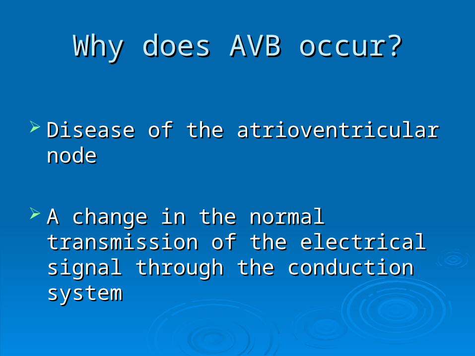
Why does AVB occur?Why does AVB occur?
Disease of the atrioventricular nodeDisease of the atrioventricular node
A change in the normal transmission of the A change in the normal transmission of the electrical signal through the conduction electrical signal through the conduction systemsystem

Types of Atrioventricular Blocks Types of Atrioventricular Blocks
1st Degree AV Block1st Degree AV Block
2nd Degree AV Block, Type I2nd Degree AV Block, Type I
2nd Degree AV Block, Type II2nd Degree AV Block, Type II
3rd Degree AV Block3rd Degree AV Block

First degree heart blockFirst degree heart block
Signal originates in SA nodeSignal originates in SA node
Signal conducted to ventriclesSignal conducted to ventricles
BUT there is a delay in the conduction BUT there is a delay in the conduction pathwaypathway
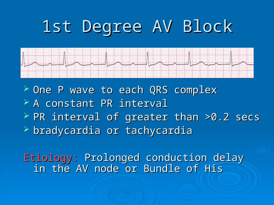
1st Degree AV Block1st Degree AV Block
One P wave to each QRS complex One P wave to each QRS complex A constant PR intervalA constant PR interval PR interval of greater than >0.2 secsPR interval of greater than >0.2 secs bradycardia or tachycardiabradycardia or tachycardia
Etiology:Etiology: Prolonged conduction delay in the AV Prolonged conduction delay in the AV node or Bundle of Hisnode or Bundle of His
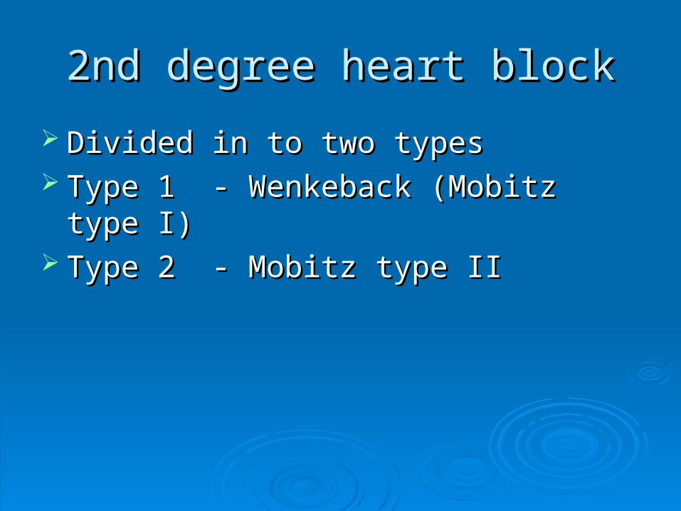
2nd degree heart block2nd degree heart block
Divided in to two types Divided in to two types Type 1 - Wenkeback (Mobitz type I)Type 1 - Wenkeback (Mobitz type I) Type 2 - Mobitz type IIType 2 - Mobitz type II
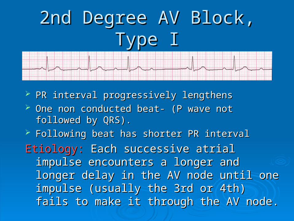
2nd Degree AV Block, Type I2nd Degree AV Block, Type I
PR interval progressively lengthensPR interval progressively lengthens One non conducted beat- One non conducted beat- (P wave not followed by (P wave not followed by
QRS).QRS). Following beat has shorter PR intervalFollowing beat has shorter PR interval
Etiology:Etiology: Each successive atrial impulse Each successive atrial impulse encounters a longer and longer delay in the AV encounters a longer and longer delay in the AV node until one impulse (usually the 3rd or 4th) node until one impulse (usually the 3rd or 4th) fails to make it through the AV node.fails to make it through the AV node.
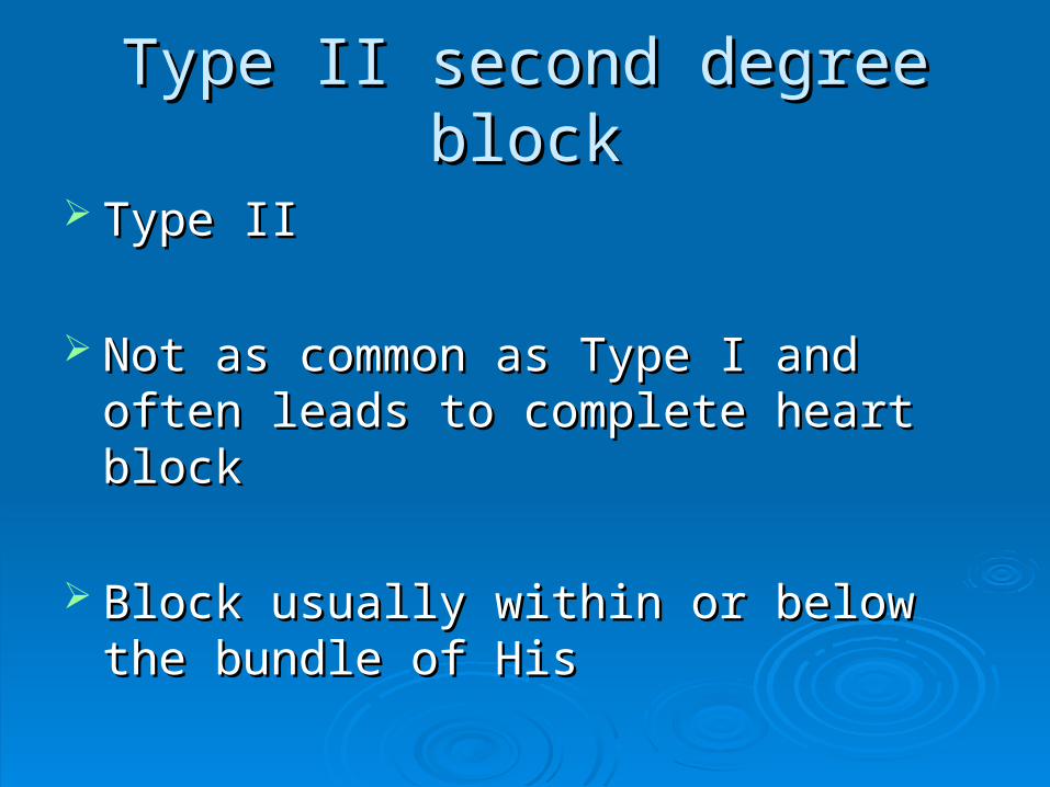
Type II second degree blockType II second degree block
Type IIType II
Not as common as Type I and often leads Not as common as Type I and often leads to complete heart blockto complete heart block
Block usually within or below the bundle of Block usually within or below the bundle of HisHis
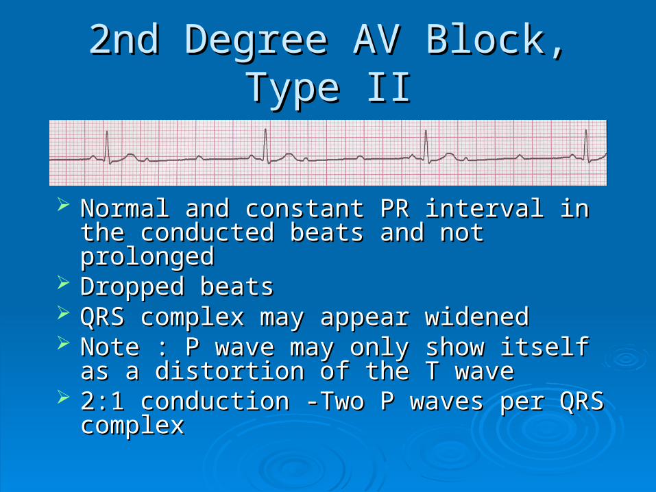
2nd Degree AV Block, Type II2nd Degree AV Block, Type II
Normal and constant PR interval in the Normal and constant PR interval in the conducted beats and not prolongedconducted beats and not prolonged
Dropped beatsDropped beats QRS complex may appear widenedQRS complex may appear widened Note : P wave may only show itself as a Note : P wave may only show itself as a
distortion of the T wavedistortion of the T wave 2:1 conduction -Two P waves per QRS 2:1 conduction -Two P waves per QRS
complexcomplex
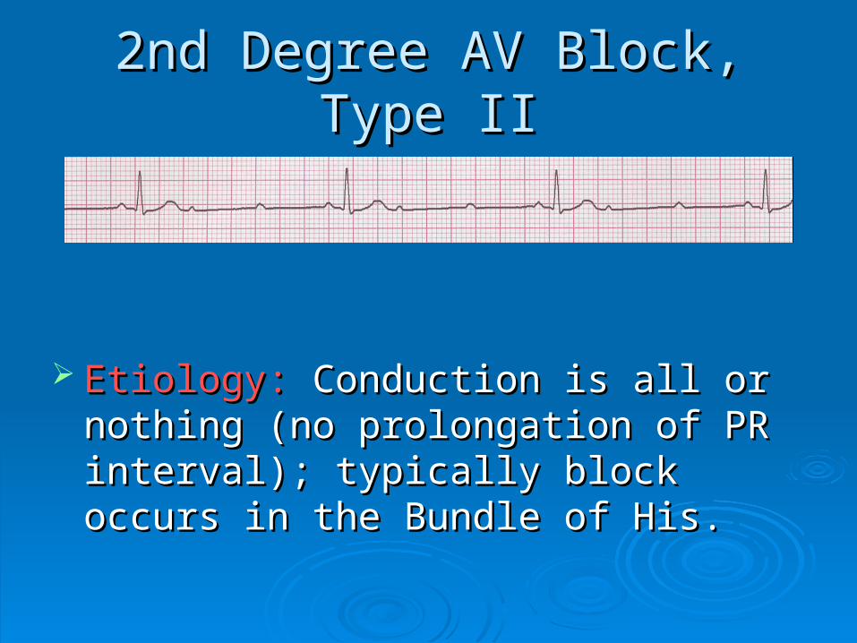
2nd Degree AV Block, Type II2nd Degree AV Block, Type II
Etiology:Etiology: Conduction is all or nothing (no Conduction is all or nothing (no prolongation of PR interval); typically block prolongation of PR interval); typically block occurs in the Bundle of His.occurs in the Bundle of His.

3rd Degree AV Block3rd Degree AV Block
The P-P interval and R-R interval will be The P-P interval and R-R interval will be regular and consistent .Atria will beat at regular and consistent .Atria will beat at intrinsic rate (60-80).Ventricles (20-40)intrinsic rate (60-80).Ventricles (20-40)
No relation between P and QRS complexNo relation between P and QRS complex Note QRS may be abnormal shape (P Note QRS may be abnormal shape (P
wave and abnormal spread of wave and abnormal spread of depolarisation)depolarisation)
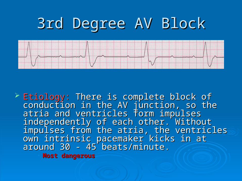
3rd Degree AV Block3rd Degree AV Block
Etiology:Etiology: There is complete block of conduction There is complete block of conduction in the AV junction, so the atria and ventricles in the AV junction, so the atria and ventricles form impulses independently of each other. form impulses independently of each other. Without impulses from the atria, the ventricles Without impulses from the atria, the ventricles own intrinsic pacemaker kicks in at around 30 - own intrinsic pacemaker kicks in at around 30 - 45 beats/minute.45 beats/minute.
• Most dangerousMost dangerous

Differentiating Atrioventricular Differentiating Atrioventricular BlockBlock
Examine Atrial rateExamine Atrial rate Examine ventricular rateExamine ventricular rate P wavesP waves PR intervalPR interval QRS complexQRS complex

RememberRemember
When an impulse originates in a ventricle, When an impulse originates in a ventricle, conduction through the ventricles will be conduction through the ventricles will be inefficient and the QRS will be wide and inefficient and the QRS will be wide and bizarre.bizarre.
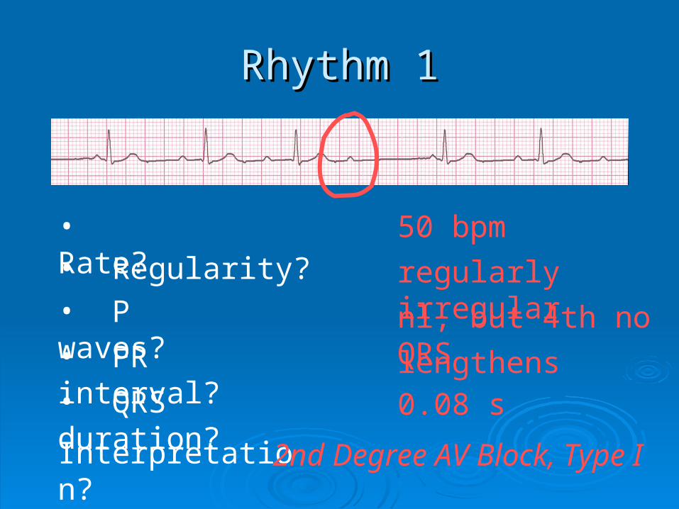
Rhythm 1Rhythm 1
50 bpm• Rate?• Regularity? regularly irregular
nl, but 4th no QRS
0.08 s
• P waves?
• PR interval? lengthens• QRS duration?
Interpretation? 2nd Degree AV Block, Type I
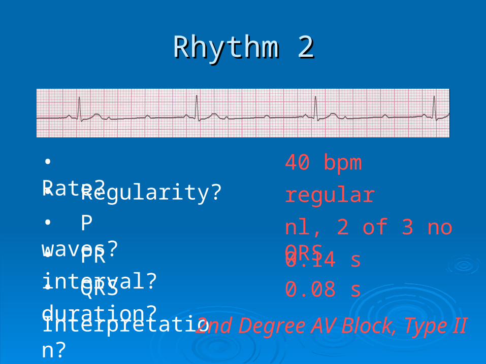
Rhythm 2Rhythm 2
40 bpm• Rate?• Regularity? regular
nl, 2 of 3 no QRS
0.08 s
• P waves?
• PR interval? 0.14 s• QRS duration?
Interpretation? 2nd Degree AV Block, Type II
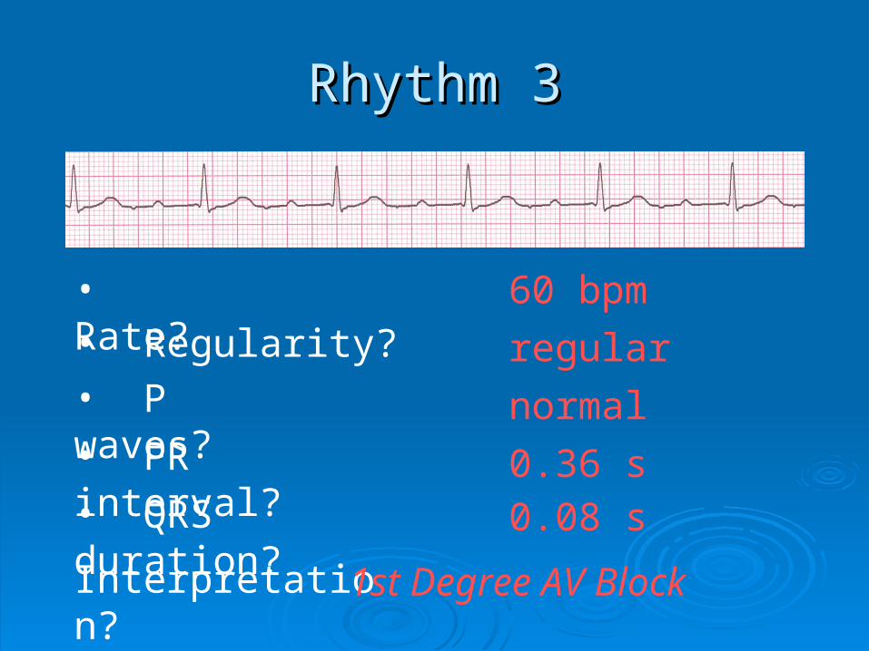
Rhythm 3Rhythm 3
60 bpm• Rate?• Regularity? regular
normal
0.08 s
• P waves?
• PR interval? 0.36 s• QRS duration?
Interpretation? 1st Degree AV Block
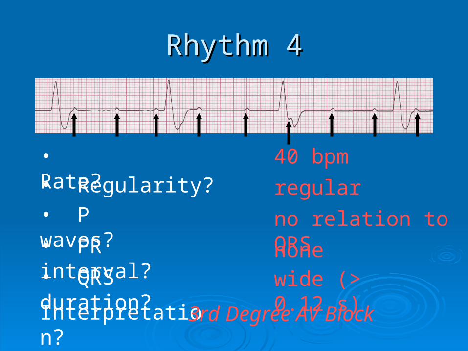
Rhythm 4Rhythm 4
40 bpm• Rate?• Regularity? regular
no relation to QRS
wide (> 0.12 s)
• P waves?
• PR interval? none• QRS duration?
Interpretation? 3rd Degree AV Block